Review
. 2023 Feb 16;24(4):4004.
doi: 10.3390/ijms24044004.

Fibrosis: Types, Effects, Markers, Mechanisms for Disease Progression, and Its Relation with Oxidative Stress, Immunity, and Inflammation

Affiliations
Review

Fibrosis: Types, Effects, Markers, Mechanisms for Disease Progression, and Its Relation with Oxidative Stress, Immunity, and Inflammation

Samar A Antar et al. Int J Mol Sci. .

Abstract

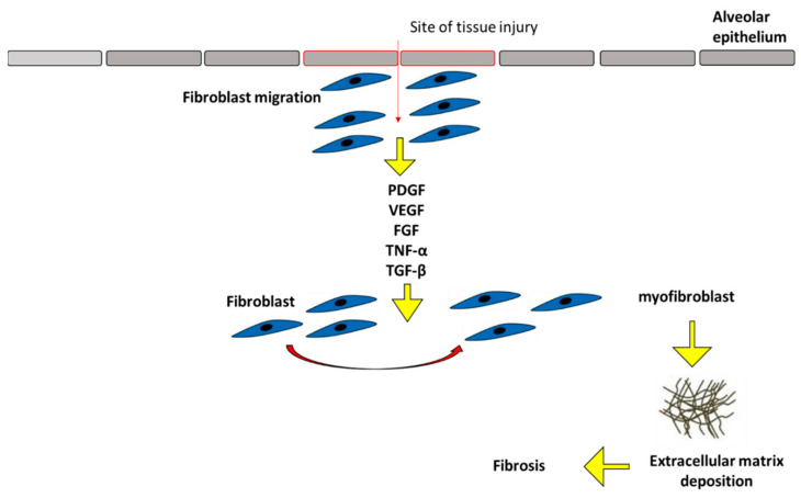
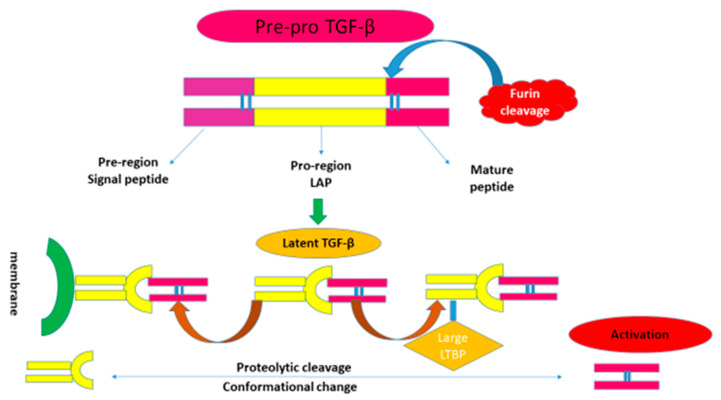
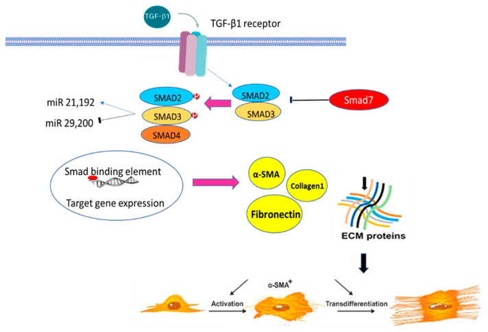
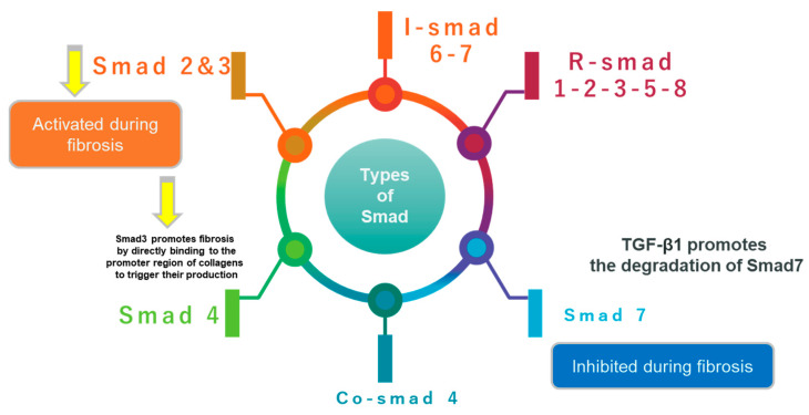
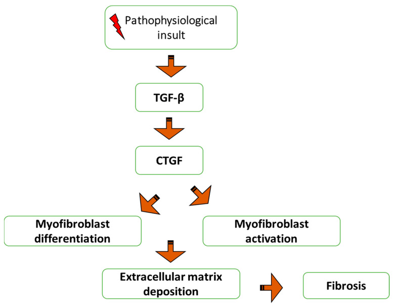
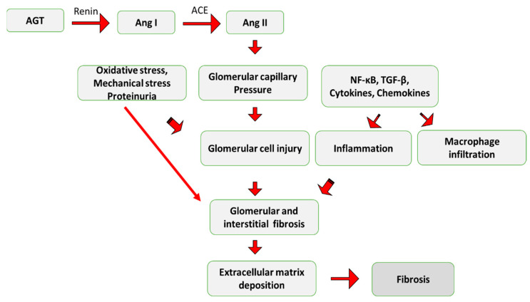
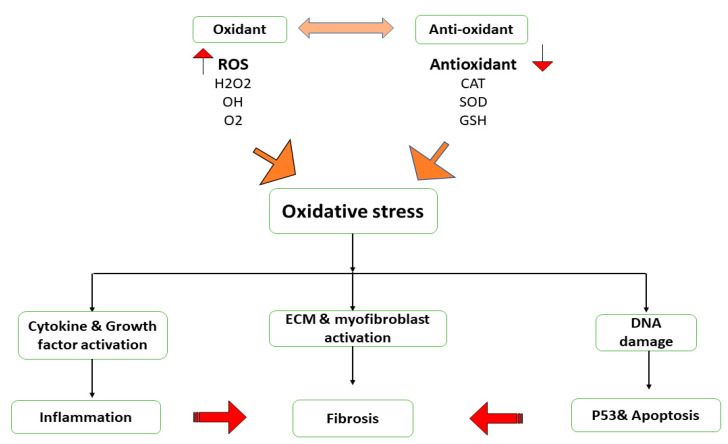
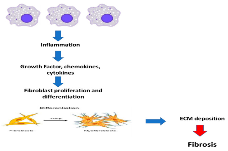
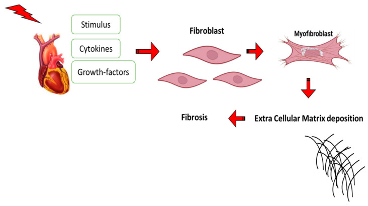
Most chronic inflammatory illnesses include fibrosis as a pathogenic characteristic. Extracellular matrix (ECM) components build up in excess to cause fibrosis or scarring. The fibrotic process finally results in organ malfunction and death if it is severely progressive. Fibrosis affects nearly all tissues of the body. The fibrosis process is associated with chronic inflammation, metabolic homeostasis, and transforming growth factor-β1 (TGF-β1) signaling, where the balance between the oxidant and antioxidant systems appears to be a key modulator in managing these processes. Virtually every organ system, including the lungs, heart, kidney, and liver, can be affected by fibrosis, which is characterized as an excessive accumulation of connective tissue components. Organ malfunction is frequently caused by fibrotic tissue remodeling, which is also frequently linked to high morbidity and mortality. Up to 45% of all fatalities in the industrialized world are caused by fibrosis, which can damage any organ. Long believed to be persistently progressing and irreversible, fibrosis has now been revealed to be a very dynamic process by preclinical models and clinical studies in a variety of organ systems. The pathways from tissue damage to inflammation, fibrosis, and/or malfunction are the main topics of this review. Furthermore, the fibrosis of different organs with their effects was discussed. Finally, we highlight many of the principal mechanisms of fibrosis. These pathways could be considered as promising targets for the development of potential therapies for a variety of important human diseases.

Keywords: ECM; TGF-β; anti-oxidant system; fibrosis; inflammation; organ malfunction.


Conflict of interest statement

The authors declare that they have no conflict of interest.

Figures

Figure 1
Figure 1
The major mechanisms of fibrosis. PDGF: Platelet-derived growth factor; VEGF: Vascular endothelial growth factor; FGF: Fibroblast growth factor; TNF-α Tumor necrosis factor-alpha; TGF-β: Transforming growth factor beta.
Figure 2
Figure 2
Schematic of the different forms of latent TGF-β. TGF-β: transforming growth factor-beta; LAP: latency-associated peptide. TGF-β is synthesized as an inactive form and cleaved by endopeptidase furin to generate a mature form which is still without biological activity. This is a literature review of the presence of latency-associated peptide (LAP) and latent TGF-β binding protein (LTBP). This large TGF-β associated complex is excreted into the extracellular matrix, cross-linked by tissue transglutaminase, and stored in the tissues in an inactive form. Once TGF-β releases from the latency-maintaining protein complex LAP/LTBP, it will display its powerful biological activity. The active form of TGF-β is a dimer stabilized by hydrophobic interactions and further strengthened by an inter-subunit disulfide bridge in most cases.
Figure 3
Figure 3
Activation of TGF-β; transforming growth factor-beta, α-SMA; alpha-smooth muscle actin plays a key role in fibrosis through activation of Smad, enhancing ECM deposition and the trans-differentiation of fibroblast to myofibroblast.
Figure 4
Figure 4
The schematic diagram illustrates the different types of Smad: receptor regulated Smad (R-Smad); common partner-Smad (co-Smad); and Inhibitory Smad (I-Smad).
Figure 5
Figure 5
The regulatory scheme for CTGF and its role in fibrosis. TGF-β; transforming growth factor-beta, CTGF; connective tissue growth factor.
Figure 6
Figure 6
A schematic illustration demonstrates how Angiotensin II (Ang II) contributes to fibrosis. AGT: Angiotensinogen; Ang I: Angiotensin I; ACE: Angiotensin Converting Enzyme.
Figure 7
Figure 7
In the pathogenesis of fibrosis, oxidative stress plays a crucial role by promoting inflammation by increasing the production of cytokines and growth factors, increasing myofibroblast differentiation and fibrogenesis, and as a result of DNA damage and p53 activation, ROS promotes apoptosis. These changes aid in the development of fibrosis. CAT; catalase, SOD; superoxide dismutase, GSH; glutathione, H2O2; Hydrogen peroxide, O2: Oxygen.
Figure 8
Figure 8
The schematic diagram illustrates how fibrosis is triggered by persistent inflammation. TFG-β; transforming growth factor.
Figure 9
Figure 9
The schematic diagram illustrates the role of Jun N terminal kinase (JNK) in activating the pro-fibrotic genes.
Figure 10
Figure 10
JAK/STAT signaling pathway; When JAK1; Janus kinase connects to TGF-β RI, STAT3; the signal transducer and activator of transcription is activated. Additionally, TGF-β-stimulates STAT3 in a SMAD-dependent way.
Figure 11
Figure 11
Wnt signaling pathway; Wnt protein interacts with a Frizzled family receptor to initiate Wnt signaling; Axin is taken out of the receptor complex and activates β-catenin to help with receptor activation; the transcription factor on DNA is bound by β-catenin as it travels into the nucleus, activating the transcription of the target genes.
Figure 12
Figure 12
The diagram illustrates cardiac fibrosis; the stimulus encourages the release of cytokines and growth factors, the differentiation of fibroblasts to myofibroblasts, and the deposition of the extracellular matrix.
Figure 13
Figure 13
The activation of the Smad2, 3, and 4 complexes by TGF-β cause pro-fibrogenic genes to be induced, which in turn causes the fibrosis process.
Figure 14
Figure 14
Liver fibrosis results from changes in cytokine release such as connective tissue growth factor (CTGF), transforming growth factor-beta (TGF-β), platelet-derived growth factor (PDGF), tumor necrosis factor-alpha (TNF-α), and the accumulation of extracellular matrix deposition.
Figure 15
Figure 15
The function of TGF-β in inducing pulmonary fibrosis.

References

    1. Conte E.J. Targeting monocytes/macrophages in fibrosis and cancer diseases: Therapeutic approaches. Pharmacol. Ther. 2021;234:108031. doi: 10.1016/j.pharmthera.2021.108031. - DOI - PubMed
    1. Davidson M.D., Burdick J.A., Wells R.G. Engineered biomaterial platforms to study fibrosis. Adv. Healthc. Mater. 2020;9:1901682. - PMC - PubMed
    1. Al-Hattab D.S., Chattopadhyaya S., Czubryt M.P. Canadian Contributions in Fibroblast Biology. Cells. 2022;11:2272. doi: 10.3390/cells11152272. - DOI - PMC - PubMed
    1. Schuster R., Rockel J.S., Kapoor M., Hinz B. The inflammatory speech of fibroblasts. Immunol. Rev. 2021;302:126–146. doi: 10.1111/imr.12971. - DOI - PubMed
    1. Lafuse W.P., Wozniak D.J., Rajaram M.V. Role of cardiac macrophages on cardiac inflammation, fibrosis and tissue repair. Cells. 2020;10:51. doi: 10.3390/cells10010051. - DOI - PMC - PubMed

Substances

LinkOut - more resources