Review
. 2023 Feb 17;24(4):4089.
doi: 10.3390/ijms24044089.

The Gut-Organ-Axis Concept: Advances the Application of Gut-on-Chip Technology

Affiliations
Review

The Gut-Organ-Axis Concept: Advances the Application of Gut-on-Chip Technology

Yuxi Guo et al. Int J Mol Sci. .

Abstract

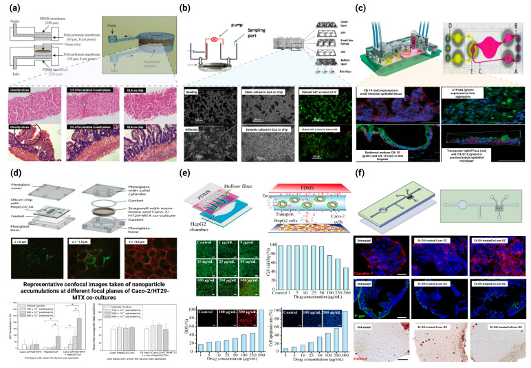
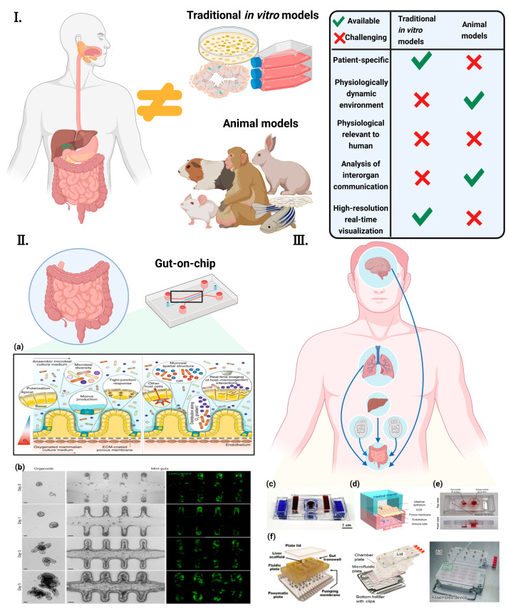
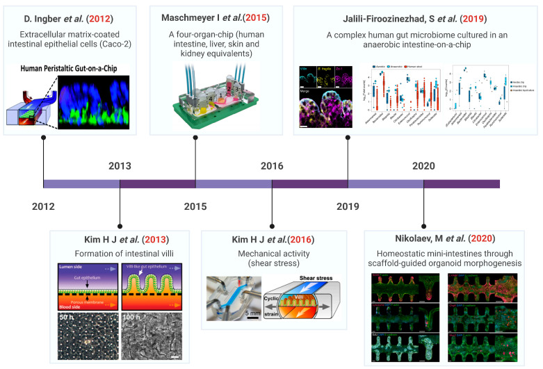
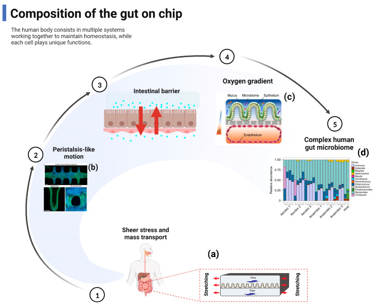
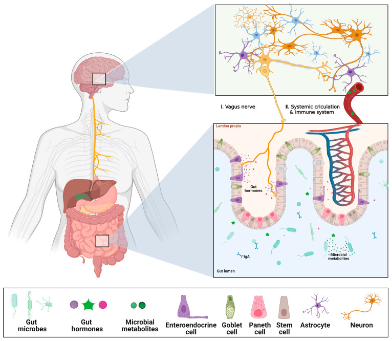
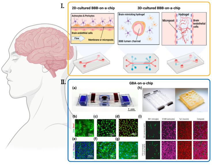
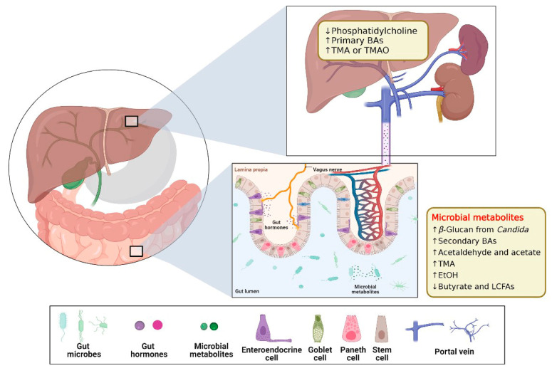
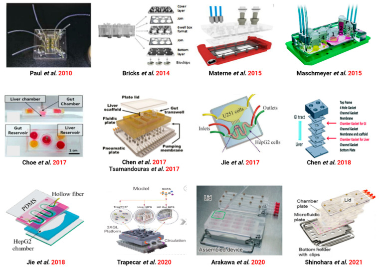
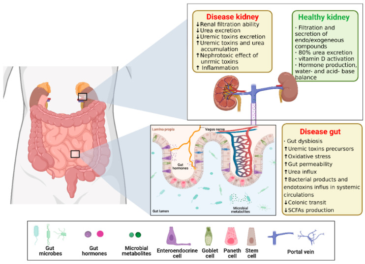
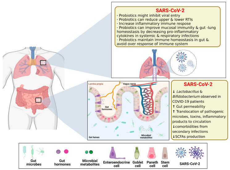
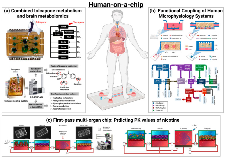
The intestine is considered to be a vital digestive organ to absorb nutrients and is the largest immune organ, while numerous microorganisms coexist with the host. It is well known that the complex interactions between the gut microbiota and the host's immune system inevitably affect the function of other organs, creating an "axis" between them. During the past few years, a new technique based mainly on microfluidics and cell biology has been developed to emulate the structure, function, and microenvironment of the human gut, called the "gut-on-chip". This microfluidic chip provides insight into key aspects of gut function in health and disease, such as the gut-brain axis, gut-liver axis, gut-kidney axis, and gut-lung axis. In this review, we first describe the basic theory of the gut axis and the various composition and parameter monitoring of the gut microarray systems, as well as summarize the development and emerging advances in the gut-organ-on-chip, with a focus on the host-gut flora and nutrient metabolism, and highlight their role in pathophysiological studies. In addition, this paper discusses the challenges and prospects for the current development and further use of the gut-organ-on-chip platform.

Keywords: bio-inspired microfluidic; disease; gut–organ-axis; gut–organ-on-chip; organ-on-chip.


Conflict of interest statement

The authors declare no conflict of interest.

Figures

Figure 8
Figure 8
Application of GLA-on-chip in drug experiments and disease models. (a) A microfluidic approach for in vitro assessment of inter-organ interactions in drug metabolism using GLA-on-chip. Top: Schematic illustration and photograph of the PDMS biochip. Bottom: Morphological evaluation of liver and intestinal slices directly after slicing, after 3 h of incubation in well plates, and in the biochip. (Magnification: 100×) [88] ©Copyright 2010, Royal Soc Chemistry. (b) Development of a new microfluidic platform integrating co-cultures of intestinal and liver cell lines. Top: Principle, design of GLA-on-chip. Bottom: Microscopic analysis of HepG2 C3A integrity in the microchips [89] ©Copyright 2014, Elsevier. (c) A four-organ-chip for interconnected long-term co-culture of human intestine, liver, skin, and kidney equivalents. Top: The microfluidic four-organ-chip device at a glance. Bottom: Performance of human tissues in the 4 °C after 28 days of co-culture [58] ©Copyright 2015, Royal Soc Chemistry. (d) Body-on-a-chip simulation with gastrointestinal (GI) tract and liver tissues suggests that ingested nanoparticles have the potential to cause liver injury. Top: Schematic of the silicon chip and GI tract module of the body-on-a-chip system. Middle: Representative confocal images taken of nanoparticle accumulations at different focal planes of Caco-2/HT29-MTX co-cultures. Bottom: Mean concentrations of aspartate aminotransferase (AST) and the percent area of on-chip liver chambers that was covered with viable HepG2/C3A cells after 24 h of exposure to 50 nm carboxylate polystyrene nanoparticles at varying concentrations [97] ©Copyright 2014, Royal Soc Chemistry. (e) GLA-on-chip for multiple drugs absorption and metabolism behavior simulations. Top: Schematic illustration of the double-layer microchip. Bottom: Influence of genistein and dacarbazine combination influence on HepG2 cell viability and apoptosis in the intestine–liver model [94] ©Copyright 2018, Science Press. (f) GLA-on-chip reveals the intestinal protective role on hepatic damage by emulating ethanol first-pass metabolism. Top: Schematic representation of the InLiver-OC and CFD study. Bottom: Protective role of 3D-HIM on Et-OH-induced liver cytotoxicity [98] ©Copyright 2020, Frontiers. * represents significance analysis, * p < 0.05.
Figure 1
Figure 1
Concept of “gut-axis” organ-on-a-chip (I) Conventional in vitro models and animal models are physiologically different from the human body. They also hinder the understanding of human diseases and the development of new therapeutic strategies [27] ©Copyright 2021, MDPI. (II) The concept of the GoC simulates the dynamic 3D microenvironment of an organ on a small scale. (a) Human-gut-microbiome on a chip [28] ©Copyright 2019, Nature.; (b) Establishment of long-term homeostatic culture of tubular mini-guts [29] ©Copyright 2020, Nature. (III) “Gut-axis” organ-on-a-chip. (c) Gut–brain axis on chip [30] ©Copyright 2021, Elsevier. (d) Gut–lung axis on chip [31] (e) Gut–kidney axis on chip [32] ©Copyright 2021, Elsevier. (f) Gut–liver axis on chip [33] ©Copyright 2017, Wiley [34] ©Copyright 2021, Nature. [35] ©Copyright 2020, Royal Soc Chemistry.
Figure 2
Figure 2
Development timeline of the gut-on-chip technology. Ref. [18] ©Copyright 2012 Nature. Ref. [42] ©Copyright 2013, Royal Soc Chemistry. Ref. [58] ©Copyright 2015, Royal Soc Chemistry. Ref. [40] ©Copyright 2016, Natl Acad Sciences. Ref. [44] ©Copyright 2019, Nature. Ref. [29] ©Copyright 2020, Nature.
Figure 3
Figure 3
The gut-on-chip reproduces the composition and function of the gut. Main relevant reproduced include shear stress and mass transport, peristalsis-like motion, intestinal barrier, and oxygen gradient. (a) Gut-on-chip reveals mechanical forces impacting Shigella infection [49] ©Copyright 2019, Cell Press; (b) Bioengineering intestinal stem cell epithelia with a tubular, in vivo-like architecture [29] ©Copyright 2020, Nature; (c) A schematic of the two-channel gut-on-chip device with an oxygen gradient [44] ©Copyright 2019, Nature; (d) Differences in microbial abundance between gut-on-chip samples and a human microbiome stool sample from the Human Microbiome Project [44] ©Copyright 2019, Nature.
Figure 4
Figure 4
Schematics of Gut–Brain Axis (GBA). The gut luminal environment, including the gut microbiome, affects the physiology and behavior of the brain via multiple routes. (I) The vague nerve senses gut hormones and microbial metabolites from the gut environment and delivers the signals to the brain. (II) Microbial products can cross the gut epithelial barrier, which makes them eventually enter systemic circulation. Additionally, some gut hormones are secreted into the bloodstream, stimulating the immune system, or traveling to the BBB, which affects changes in the physiology and behavior of the BBB and brain. Created with BioRender.com. The reproduction of this image has been licensed from BioRender.
Figure 5
Figure 5
Various BBBs-on-a-chip and GBA-on-chip. (I) Strategies for reconstructing the in vitro BBB in an OOC platform are diverse. Cells can be cultured in a 2D environment under fluidic flow or 3D-cultured with different approaches, such as seeding in hollow hydrogel or the angio/vasculogenesis approach [27] ©Copyright 2020, MDPI. (II) Various GBA-on-chip. (a) The picture of assembled GBA chip [30] ©Copyright 2020, Elsevier; (bg) Fluorescent images of cells seeded in the chip [30] ©Copyright 2020, Elsevier: (b) Live/Dead Images of Caco-2, (c) bEnd.3, and (d) hBMECs when co-cultured (green = live, red = dead). F-actin/nucleus stain images of (e) Caco-2 cells, (f) bEnd.3 cells and (g) hBMECs when co-cultured (blue = nucleus, green = F-actin); (h) Left: pneumatic plates machined in acrylic; Right: mesofluidic plate machined from monolithic polysulfone [82] ©Copyright 2021, Amer Assoc Advancement Science; (i) Representative, 3D rendered confocal images of the PD (top) and control PD-C (bottom) cerebral MPSs composed of hiPSC-derived microglia (green), astrocytes (purple), and neurons (red) cocultured on 0.4-μm microporous 24-well Transwells [82] ©Copyright 2021, Amer Assoc Advancement Science.
Figure 6
Figure 6
Schematics of Gut–liver Axis (GLA). The gut luminal environment, including the gut microbiome, affects the physiology and behavior of the liver via multiple routes. Created with BioRender.com. The reproduction of this image has been licensed from BioRender.
Figure 7
Figure 7
Various GLA-on-chip from 2010 to 2021. Ref. [88] ©Copyright 2010, Royal Soc Chemistry. Ref. [89] ©Copyright 2014, Pergamon-Elsevier Science Ltd. Ref. [90] ©Copyright 2015, Amer Chemical Soc. Ref. [58] ©Copyright 2015, Royal Soc Chemistry. Ref. [56] ©Copyright 2017, Springer. Ref. [33] ©Copyright 2017, Wiley. Ref. [91] ©Copyright 2017, Springer. Ref. [92] ©Copyright 2017, Royal Soc Chemistry. Ref. [93] ©Copyright 2018, Royal Soc Chemistry. Ref. [94] ©Copyright 2018, Royal Soc Chemistry. Ref. [86] ©Copyright 2020, Cell Press. Ref. [35] ©Copyright 2020, Royal Soc Chemistry. Ref. [34] ©Copyright 2021, Science.
Figure 9
Figure 9
Schematics of Gut–kidney Axis (GKA). The gut luminal environment, including the gut microbiome, affects the physiology and behavior of the kidney via multiple routes. Created with BioRender.com. The ↑ and ↓ arrows in the graph represent an increase or decrease in content, performance, and/or activity, respectively. The reproduction of this image has been licensed from BioRender.
Figure 10
Figure 10
GKA on chip for studying effects of antibiotics on risk of hemolytic uremic syndrome by Shiga toxin-producing Escherichia coli. (a) Assembly of a main body in four layers with modules. (b) View of completed GKA on chip showing gut and kidney modules and two reservoirs. (c) Gravity-induced perfusion by periodically tilting the chip 10 degrees (0.1 degree/s) every 10 min. (d) View of the tilting machine inducing gravity-driven perfusion of cell culture medium in GKA on chip. (e) LIVE/DEAD stained images and (f) EZ-CytoX assay of Caco-2 and HKC-8 cells either mono-cultured or co-cultured for 3 days. (g) Immunostaining of occludin in Caco-2 cells at day 7. (h) Viabilities of Caco-2 and HKC-8 cells to Stx2 in GKA on chip. Only the gut module was treated with the toxin at different concentrations (0–21.2 nM) for 72 h. (i) Simulation of Stx2 transport from the gut module to the kidney module in the Experimental Section. (j) TEER Value of Caco-2 cells to Stx2 at 21.2 nM in Transwell for 72 h. (k) TEER values of HKC-8 cells after treatment with 21.2 nM of Stx2 in the gut module for 72 h. Sample number n= 3, Student’s t-test. NS; not significant, * p < 0.05, ** p < 0.01, *** p < 0.001. (l,m) LIVE/DEAD stained images from 106 and 107 CFU per module and (n,o) cell viability of Caco-2 and HKC-8 cells being treated with O157 lysed by either CIP or GEN in the chip for 72 h. The relative viability was calculated as the number of viable cells divided by the number of viable cells in the control (no infection and no antibiotics) from LIVE/DEAD stained images. (p) TEER values of HKC-8 cells in module of the chip when the gut module was infected with O157 at 107 CFU and treated with either CIP or GEN for 72 h. n = 3, Student’s t-test, NS; not significant, * p < 0.05, ** p < 0.01, *** p < 0.001. [32] ©Copyright 2021, MDPI.
Figure 11
Figure 11
Schematics of Gut–lung Axis (GLAx). The gut luminal environment, including the gut microbiome, affects the physiology and behavior of the lung via multiple routes. Created with BioRender.com. The ↑ and ↓ arrows in the graph represent an increase or decrease in content, performance, relative abundance, and/or activity, respectively. The reproduction of this image has been licensed from BioRender.
Figure 12
Figure 12
SARS-CoV-2-induced intestinal responses with a GLA-on-chip. (a) The configuration of the multilayered intestine on the chip device infected with SARS-CoV-2. (b) Confocal micrographs of the intestinal epithelial barrier on the chip visualized by the expression of an adhesion junction (E-cadherin) and tight junction markers (ZO-1). The intestinal villus-like structures with high levels of ZO-1 expression are indicated by white dashed lines. (c) Confocal micrographs of the vascular endothelium identified by the expression of an adhesion junction protein (VEcadherin) and ZO-1. (d) DIC image of an intestinal villus-like structure with clumps of cells (indicated by white dashed lines). (e) Immunostaining of a mucin marker (MUC2) in intestinal epithelial cells. Scale bars: 50 lm. (f,h) The 3D reconstructed confocal image and side view of the human intestinal epithelium (E-cadherin) and endothelium (VE-cadherin). (g,i) The 3D reconstructed confocal image and side view of the intestinal epithelium, endothelium, and intestinal villus-like structures (indicated by white arrows). (j) Confocal micrographs of SARS-CoV-2 infection (Spike protein) on the intestinal epithelium (E-cadherin) and intestinal villus-like structures (indicated by yellow dashed lines) at day 3 post-infection. Scale bars: 50 lm. (k) The 3D reconstructed confocal image and side view of a mock-infected gut-on-chip. The 3D reconstructed confocal image and side view of the virus-infected intestinal model. SARS-CoV-2 infection was identified in the epithelial layer by the expression of the viral Spike protein. (l) Confocal micrographs of SARS-CoV-2 infection (Spike protein) and MUC2 expression in the intestinal epithelium at day 3 post-infection. Scale bars: 50 lm. (m) Confocal micrographs of viral infection (Spike protein) in the vascular endothelium (VE-cadherin). Scale bars: 50 lm. (n,o) Quantification of endothelial cell density and size for mock- and SARS-CoV-2-infected chips. Four chips were counted for cell density quantification in each group, and 100 cells were counted for cell size quantification in each group. Data are presented as the mean ± SD and were analyzed by Student’s t-test (***, p < 0.001) [31] ©Copyright 2021, Elsevier.
Figure 13
Figure 13
Schematics of human-on-chip. (a) Analysis of an Integrated Human Multi-organ Microphysiological System for Combined Tolcapone Metabolism and Brain Metabolomics [114] ©Copyright 2019, Amer Chemical Soc. (b) Functional Coupling of Human Microphysiology Systems: Intestine, Liver, Kidney Proximal Tubule, Blood–Brain Barrier and Skeletal Muscle [55] ©Copyright 2017, Nature Research. (c) Quantitative prediction of human pharmacokinetic responses to drugs via fluidically coupled vascularized organ chips [113] ©Copyright 2020, Nature Research.

References

    1. Cryan J.F., O’Riordan K.J., Sandhu K., Peterson V., Dinan T.G. The gut microbiome in neurological disorders. Lancet Neurol. 2020;19:179–194. doi: 10.1016/S1474-4422(19)30356-4. - DOI - PubMed
    1. Koh A., Backhed F. From association to causality: The role of the gut microbiota and its functional products on host metabolism. Mol. Cell. 2020;78:584–596. doi: 10.1016/j.molcel.2020.03.005. - DOI - PubMed
    1. Compare D., Coccoli P., Rocco A., Nardone O.M., De Maria S., Carteni M., Nardone G. Gut-liver axis: The impact of gut microbiota on non alcoholic fatty liver disease. Nutr. Metab. Cardiovasc. Dis. 2012;22:471–476. doi: 10.1016/j.numecd.2012.02.007. - DOI - PubMed
    1. Lyte M. Microbial endocrinology in the microbiome-gut-brain axis: How bacterial production and utilization of neurochemicals influence behavior. PLoS Pathog. 2013;9:230–239. doi: 10.1371/journal.ppat.1003726. - DOI - PMC - PubMed
    1. Moser G., Fournier C., Peter J. Intestinal microbiome-gut-brain axis and irritable bowel syndrome. Wien. Med. Wochenschr. 2018;168:62–66. doi: 10.1007/s10354-017-0592-0. - DOI - PMC - PubMed

LinkOut - more resources