. 2023 Feb 18;24(4):4131.
doi: 10.3390/ijms24044131.

Spermidine-Eugenol Supplement Preserved Inflammation-Challenged Intestinal Cells by Stimulating Autophagy

Affiliations

Spermidine-Eugenol Supplement Preserved Inflammation-Challenged Intestinal Cells by Stimulating Autophagy

Francesca Truzzi et al. Int J Mol Sci. .

Abstract

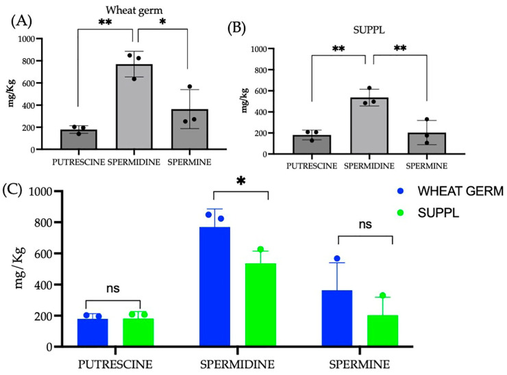
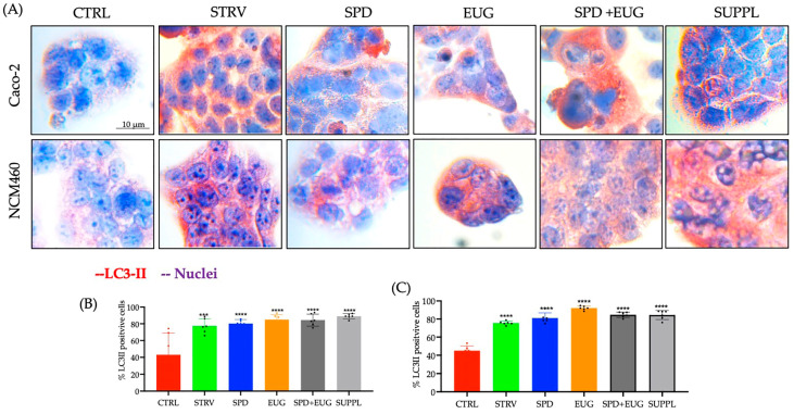
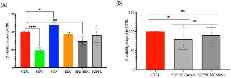
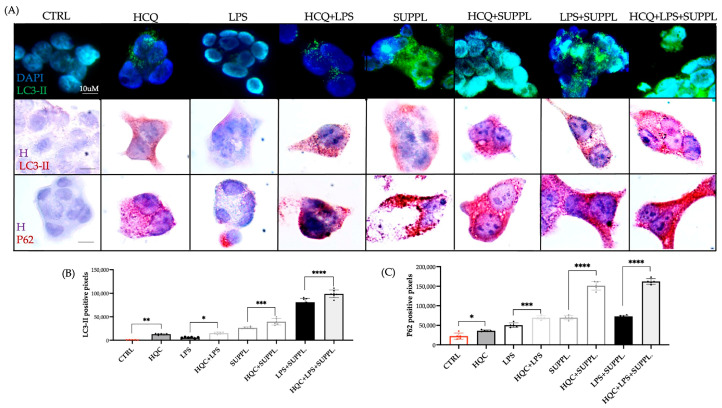
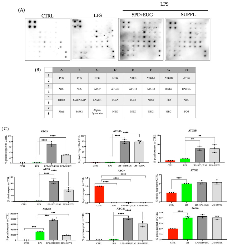
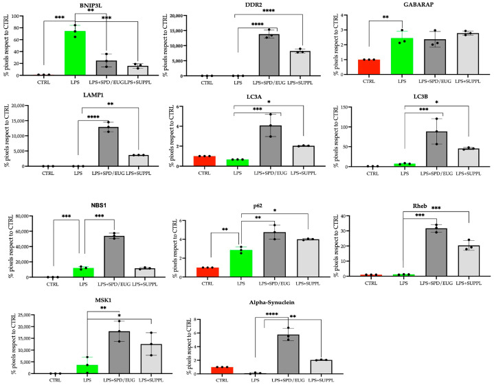
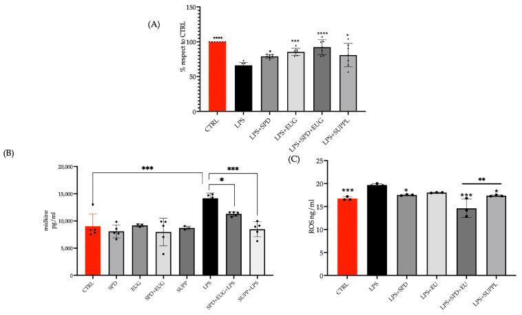
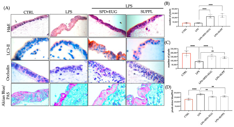
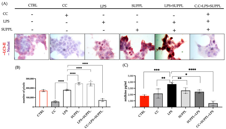
Increases in non-communicable and auto-immune diseases, with a shared etiology of defective autophagy and chronic inflammation, have motivated research both on natural products in drug discovery fields and on the interrelationship between autophagy and inflammation. Within this framework, the tolerability and protective effects of a wheat-germ spermidine (SPD) and clove eugenol (EUG) combination supplement (SUPPL) were investigated on inflammation status (after the administration of lipopolysaccharide (LPS)) and on autophagy using human Caco-2 and NCM460 cell lines. In comparison to the LPS treatment alone, the SUPPL + LPS significantly attenuated ROS levels and midkine expression in monocultures, as well as occludin expression and mucus production in reconstituted intestinal equivalents. Over a timeline of 2-4 h, the SUPPL and SUPPL + LPS treatments stimulated autophagy LC3-11 steady state expression and turnover, as well as P62 turnover. After completely blocking autophagy with dorsomorphin, inflammatory midkine was significantly reduced in the SUPPL + LPS treatment in a non-autophagy-dependent manner. After a 24 h timeline, preliminary results showed that mitophagy receptor BNIP3L expression was significantly downregulated in the SUPPL + LPS treatment compared to the LPS alone, whereas conventional autophagy protein expression was significantly higher. The SUPPL shows promise in reducing inflammation and increasing autophagy to improve intestinal health.

Keywords: Caco-2 cells; LPS; NCM460 cells; autophagy; compound C; eugenol; inflammation; spermidine.


Conflict of interest statement

The authors declare no conflict of interest.

Figures

Figure 1
Figure 1
Polyamine content (putrescine, spermidine and spermine) in both the pressed wheat germ source (A) and the supplement (B) with a statistical comparison between the pressed wheat germ and supplement (C). Significant differences were represented as follows: ns p ≥ 0.05, * 0.01 < p < 0.05, ** 0.001 < p < 0.01. The black dots indicate the positioning of the individual replicates within the bar for each sample.
Figure 2
Figure 2
The effect of 1.2 µM spermidine (SPD) and 0.2 mM eugenol (EUG) alone, in combination (1.2 µM SPD + 0.2 mM EUG), and in supplement (SUPPL) form (1.2 µM SPD + 92 µM EUG), respectively, on LC3-II activation in Caco-2 (A,B) and NCM460 (A,C) cell lines compared to the untreated control (CTRL). (AC) LC3-II expression in the CTRL, the starvation (STRV)-induced control, and all treatments was detected with red chromogen staining after 4 h and visualized at ×40 magnification. (B,C) Statistical analysis of the LC3-II positive cells stained with red chromogen was performed with significant differences represented as follows: *** 0.0001 < p < 0.001, **** p < 0.0001. The black dots indicate the positioning of the individual replicates within the bar for each sample.
Figure 3
Figure 3
(A) The effect of 1.2 µM spermidine (SPD) and 0.2 mM eugenol (EUG) alone, in combination (1.2 µM SPD + 0.2 mM EUG), and in supplement (SUPPL) form (1.2 µM SPD + 92 µM EUG), respectively, on cell viability (MTT assay) in NCM460 cells compared to the untreated control (CTRL). (B) The effect of the SUPPL on cell viability in Caco-2 and NCM460 cells compared to the respective controls. Significant differences were represented as follows: ns p ≥ 0.05, * 0.01 < p < 0.05, ** 0.001 < p < 0.01, **** p < 0.0001. The black dots indicate the positioning of the individual replicates within the bar for each sample.
Figure 4
Figure 4
(A) The effect of the supplement (SUPPL, 1.2 µM SPD + 92 µM EUG) alone or in combination with ng/mL lipopolysaccharide (LPS) for 2 h on fluorescent green (FG)-stained LC3-II puncta, chromogen red-stained LC3-II and chromogen red-stained P62 in comparison to the untreated control and LPS alone (magnification ×60). All treatments were performed in the presence and absence of 20 µM hydroxychloroquine (HCQ). (B) Quantification of the LC3-II steady state and turnover levels. (C) Quantification of the P62 steady state and turnover levels. Significant differences were represented as follows: * 0.01 < p < 0.05, ** 0.001 < p < 0.01, *** 0.0001 < p < 0.001, **** p < 0.0001. The black dots indicate the positioning of the individual replicates within the bar for each sample. DAPI = nuclear staining in FG imaging: H = hematoxylin.
Figure 5
Figure 5
Expression (A) of 20 individual human autophagy proteins in NCM460 cells, identified according to the (B) position location on the micro-array. Protein expression (A) was performed in the untreated control (CTRL) and lipopolysaccharide (LPS)-treated cells (1 ng/mL LPS) after 24 h and compared to cells pre-incubated for 1 h with 1.2 µM spermidine (SPD) + 0.2 mM eugenol (EUG) and in supplement (SUPPL) form (1.2 µM SPD + 92 µM EUG) prior to LPS addition. (C) Protein expression for each of the 20 proteins was quantified for each treatment. Significant differences were represented as follows: * 0.01 < p < 0.05, ** 0.001 < p < 0.01, *** 0.0001 < p < 0.001, **** p < 0.0001. The black dots indicate the positioning of the individual replicates within the bar for each sample.
Figure 5
Figure 5
Expression (A) of 20 individual human autophagy proteins in NCM460 cells, identified according to the (B) position location on the micro-array. Protein expression (A) was performed in the untreated control (CTRL) and lipopolysaccharide (LPS)-treated cells (1 ng/mL LPS) after 24 h and compared to cells pre-incubated for 1 h with 1.2 µM spermidine (SPD) + 0.2 mM eugenol (EUG) and in supplement (SUPPL) form (1.2 µM SPD + 92 µM EUG) prior to LPS addition. (C) Protein expression for each of the 20 proteins was quantified for each treatment. Significant differences were represented as follows: * 0.01 < p < 0.05, ** 0.001 < p < 0.01, *** 0.0001 < p < 0.001, **** p < 0.0001. The black dots indicate the positioning of the individual replicates within the bar for each sample.
Figure 6
Figure 6
The effect of a pretreatment (1 h) of 1.2 µM spermidine (SPD) and 0.2 mM eugenol (EUG) alone, in combination (1.2 µM SPD + 0.2 mM EUG), and in supplement (SUPPL) form (1.2 µM SPD + 92 µM EUG) prior to the addition of 1 ng/mL lipopolysaccharide (LPS) for 24 h on (A) cell viability, (B) Midkine levels, and (C) ROS levels in NCM460 cells compared to the untreated control (CTRL) and LPS treatment alone. Significant differences were represented as follows: * 0.01 < p < 0.05, ** 0.001 < p < 0.01, *** 0.0001 < p < 0.001, **** p < 0.0001. The black dots indicate the positioning of the individual replicates within the bar for each sample.
Figure 7
Figure 7
(A) Hematoxylin and eosin (H&E) staining, and LC3-II, occludin, and mucus expression of 3D intestinal equivalents (Caco-2/U937/L929 co-cultures) exposed to a 24 h pretreatment of either 1.2 spermidine (SPD) and 0.2 mM eugenol (EUG) or supplement (1.2 µM SPD + 92 µM EUG) prior to the addition of lipopolysaccharide (LPS, 1 ng/mL) for a further 24 h compared to the untreated control (CTRL) and LPS treatments alone. The magnification was ×60. (B) Quantification of LC3-II-red chromogen staining, (C) occludin–red chromogen staining (D) mucus expression from Alcian Blue/Periodic Acid-Schiff Stain (PAS) staining of the Caco-2 cells. Significant differences were represented as follows: ** 0.001 < p < 0.01, **** p < 0.0001. The black dots indicate the positioning of the individual replicates within the bar for each sample.
Figure 8
Figure 8
(A) Exposure of NCM460 cells to the supplement (SUPPL, 1.2 µM spermidine (SPD)) + 92 µM eugenol (EUG), lipopolysaccharide (LPS, 1 ng/mL) and Compound C (CC, 5 µM) either alone or in combination compared to the untreated control (CTRL). (A) Red chromogen-stained LC3-II expression at ×40 mag with (B) LC3-II expression quantification and (C) comparison to midkine levels. Compound C was administered 30 min prior to the addition of the SUPPL and SUPPL pretreatments were for 1 h prior to LPS exposure for 4 h. Significant differences were represented as follows: * 0.01 < p < 0.05, ** 0.001 < p < 0.01, *** 0.0001 < p < 0.001, **** p < 0.0001. The black dots indicate the positioning of the individual replicates within the bar for each sample.

References

    1. Mizushima N., Komatsu M. Autophagy: Renovation of cells and tissues. Cell. 2011;147:728–741. doi: 10.1016/j.cell.2011.10.026. - DOI - PubMed
    1. Netea-Maier R.T., Plantinga T.S., van de Veerdonk F.L., Smit J.W., Netea M.G. Modulation of inflammation by autophagy: Consequences for human disease. Autophagy. 2016;12:245–260. doi: 10.1080/15548627.2015.1071759. - DOI - PMC - PubMed
    1. Naveja J.J., Madariaga-Mazón A., Flores-Murrieta F.J., Granados-Montiel J., Maradiaga-Ceceña M.A., Alaniz V.D., Maldonado-Rodríguez M.R., García-Morales J., Senosiain-Peláez J.P., Martínez-Mayorga K. Union is strength: Antiviral and anti-inflammatory drugs for COVID-19. Drug Discov. Today. 2021;26:29–239. doi: 10.1016/j.drudis.2020.10.018. - DOI - PMC - PubMed
    1. Pereira G., Leão A., Erustes A.G., Morais I., Vrechi T., Zamarioli L., Pereira C., Marchioro L.O., Sperandio L.P., Lins Í., et al. Pharmacological Modulators of Autophagy as a Potential Strategy for the Treatment of COVID-19. Int. J. Mol. Sci. 2021;22:4067. doi: 10.3390/ijms22084067. - DOI - PMC - PubMed
    1. Seiler K., Tschan M.P.P. Autophagy and coronavirus infection—A Trojan horse or Achilles’ heel? Swiss Med. Wkly. 2021;151:w20468. doi: 10.4414/smw.2021.20468. - DOI - PubMed

Grants and funding

LinkOut - more resources