Review
. 2023 Feb 20;24(4):4177.
doi: 10.3390/ijms24044177.

Placental Mitochondrial Function and Dysfunction in Preeclampsia

Affiliations
Review

Placental Mitochondrial Function and Dysfunction in Preeclampsia

Fahmida Jahan et al. Int J Mol Sci. .

Abstract

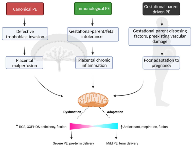
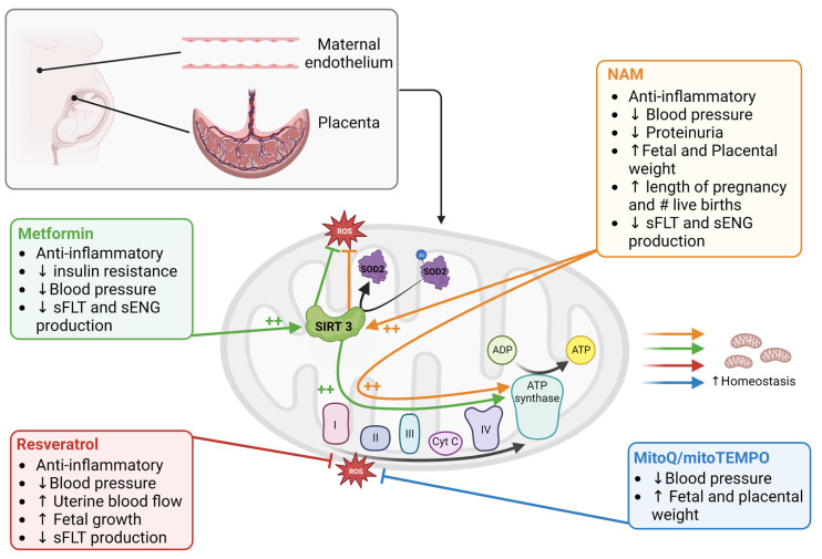
The placenta is a vital organ of pregnancy, regulating adaptation to pregnancy, gestational parent/fetal exchange, and ultimately, fetal development and growth. Not surprisingly, in cases of placental dysfunction-where aspects of placental development or function become compromised-adverse pregnancy outcomes can result. One common placenta-mediated disorder of pregnancy is preeclampsia (PE), a hypertensive disorder of pregnancy with a highly heterogeneous clinical presentation. The wide array of clinical characteristics observed in pregnant individuals and neonates of a PE pregnancy are likely the result of distinct forms of placental pathology underlying the PE diagnosis, explaining why no one common intervention has proven effective in the prevention or treatment of PE. The historical paradigm of placental pathology in PE highlights an important role for utero-placental malperfusion, placental hypoxia and oxidative stress, and a critical role for placental mitochondrial dysfunction in the pathogenesis and progression of the disease. In the current review, the evidence of placental mitochondrial dysfunction in the context of PE will be summarized, highlighting how altered mitochondrial function may be a common feature across distinct PE subtypes. Further, advances in this field of study and therapeutic targeting of mitochondria as a promising intervention for PE will be discussed.

Keywords: disease subclasses; hypertension; mitochondria; placenta; preeclampsia; pregnancy; reactive oxygen species; therapies.


Conflict of interest statement

The authors declare no conflict of interest.

Figures

Figure 1
Figure 1
Mitochondrial dysfunction as a common feature across all PE subclasses. PE disease driven by either placental malperfusion, chronic inflammation at the utero–placental interface and/or poor gestational parent adaptation, likely all demonstrate altered mitochondrial respiration, increased ROS production and mitochondrial fragmentation within the placenta and across numerous gestational parent tissues. The inherent ability of the mitochondria to adapt during pregnancy to various pathological insults may in part dictate the clinical outcome of the pregnancy, with high degrees of mitochondrial adaptation associated with a milder clinical presentation of the disease, prolonged gestation, and improved fetal growth profiles. The “↑” arrow indicates an increase. Figure was created with Biorender.com, accessed on 24 January 2023.
Figure 2
Figure 2
Mitochondria targeting treatments in preeclampsia. Among the drugs and nutraceuticals tested in PE studies, the most promising ones that target mitochondria are metformin, resveratrol, nicotinamide (NAM), and mitoQ/mitoTEMPO. Metformin, resveratrol, and NAM function on multiple pathways that improve mitochondrial biogenesis, function, quality control (by mitophagy), and reduce ROS; thereby establishing mitochondrial homeostasis under pathological conditions. MitoQ/mitoTEMPO act by reducing mitochondrial ROS which eventually improve mitochondrial homeostasis. The symbol ++ indicates increased activation, # indicates number, ↑ indicates increase and ↓ indicates decrease. Figure was created with Biorender.com, accessed on 13 February 2023.

References

    1. Bauer M.K., Harding J.E., Bassett N.S., Breier B.H., Oliver M.H., Gallaher B.H., Evans P.C., Woodall S.M., Gluckman P.D. Fetal growth and placental function. Mol. Cell. Endocrinol. 1998;140:115–120. doi: 10.1016/S0303-7207(98)00039-2. - DOI - PubMed
    1. Gude N.M., Roberts C.T., Kalionis B., King R.G. Growth and function of the normal human placenta. Thromb. Res. 2004;114:397–407. doi: 10.1016/j.thromres.2004.06.038. - DOI - PubMed
    1. Rana S., Lemoine E., Granger J.P., Karumanchi S.A. Preeclampsia: Pathophysiology, Challenges, and Perspectives. Circ. Res. 2019;124:1094–1112. doi: 10.1161/CIRCRESAHA.118.313276. - DOI - PubMed
    1. Leavey K., Benton S.J., Grynspan D., Kingdom J.C., Bainbridge S.A., Cox B.J. Unsupervised Placental Gene Expression Profiling Identifies Clinically Relevant Subclasses of Human Preeclampsia. Hypertension. 2016;68:137–147. doi: 10.1161/HYPERTENSIONAHA.116.07293. - DOI - PubMed
    1. Griffiths S.K., Campbell J.P. Placental structure, function and drug transfer. Contin. Educ. Anaesth. Crit. Care Pain. 2014;15:84–89. doi: 10.1093/bjaceaccp/mku013. - DOI