

Post-Traumatic Atlanto-Axial Instability: A Combined Clinical and Radiological Approach for the Diagnosis of Pathological Rotational Movement in the Upper Cervical Spine
- PMID: 36836004
- PMCID: PMC9964642
- DOI: 10.3390/jcm12041469
Post-Traumatic Atlanto-Axial Instability: A Combined Clinical and Radiological Approach for the Diagnosis of Pathological Rotational Movement in the Upper Cervical Spine
Abstract
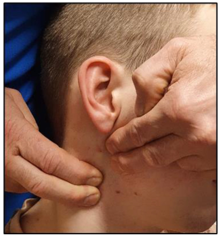
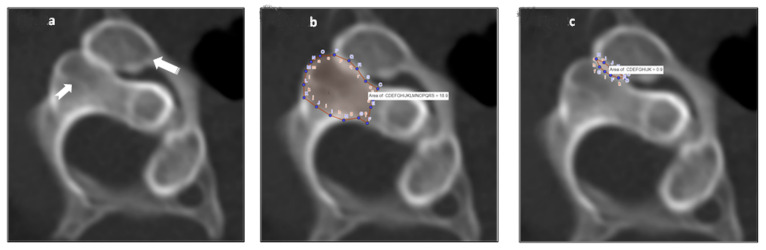
Post-traumatic rotational instability at the atlanto-axial (C1-2) joint is difficult to assess, much less quantify, due to the orientation and motion plane of the joint. Prior investigations have demonstrated that a dynamic axial CT scan, during which the patient maximally rotates the head right and left, can be used to evaluate and quantify the amount of residual overlap between the inferior articulating facet of C1 and the superior facet of C2, as an index of ligamentous laxity at the joint. We have previously demonstrated that a novel orthopedic test of rotational instability, the atlas-axis rotational test (A-ART), may have utility in identifying patients with imaging evidence of upper cervical ligament injury. In the present investigation, we assessed the correlation between a positive A-ART and a CT scan assessment of the relative quantity of residual C1-2 overlap, as a percent of the superior articulating facet surface area of C2. A retrospective review was conducted of the records of consecutive patients presenting to a physical therapy and rehabilitation clinic, over a 5-year period (2015-20) for chronic head and neck pain after whiplash trauma. The primary inclusion criteria were that the patient had undergone both a clinical evaluation with A-ART and a dynamic axial CT to evaluate for C1-2 residual facet overlap at maximum rotation. The records for a total of 57 patients (44 female/13 male) were identified who fit the selection criteria, and among these, there were 43 with a positive A-ART (i.e., "cases") and 14 with a negative A-ART (i.e., "controls). The analysis demonstrated that a positive A-ART was highly predictive of decreased residual C1-2 facet overlap: the average overlap area among the cases was approximately one-third that of the control group (on the left, 10.7% versus 29.1%, and 13.6% versus 31.0% on the right). These results suggest that a positive A-ART is a reliable indicator of underlying rotational instability at C1-2 in patients with chronic head and neck symptoms following whiplash trauma.
Keywords: CT scan; atlas-axis rotational test (A-ART); upper cervical instability; whiplash trauma.
Conflict of interest statement
M.D.F. provides expert medicolegal consultation. No conflicts are declared for the remaining authors.
Figures
Similar articles
-
CT scan study of atlantoaxial rotatory mobility in asymptomatic adult subjects: a basis for better understanding C1-C2 rotatory fixation and subluxation.Spine (Phila Pa 1976). 2009 May 20;34(12):1292-5. doi: 10.1097/BRS.0b013e3181a4e4e9. Spine (Phila Pa 1976). 2009. PMID: 19412141
-
[In vivo measurement of three-dimensional motion of the upper cervical spine using CT three-dimensional reconstruction].Zhongguo Gu Shang. 2019 Jul 25;32(7):658-665. doi: 10.3969/j.issn.1003-0034.2019.07.014. Zhongguo Gu Shang. 2019. PMID: 31382726 Chinese.
-
Chronic neck pain: making the connection between capsular ligament laxity and cervical instability.Open Orthop J. 2014 Oct 1;8:326-45. doi: 10.2174/1874325001408010326. eCollection 2014. Open Orthop J. 2014. PMID: 25328557 Free PMC article.
-
Pediatric cervical kyphosis in the MRI era (1984-2008) with long-term follow up: literature review.Childs Nerv Syst. 2022 Feb;38(2):361-377. doi: 10.1007/s00381-021-05409-z. Epub 2021 Nov 22. Childs Nerv Syst. 2022. PMID: 34806157 Review.
-
Atlantoaxial dislocation.Neurol India. 2012 Jan-Feb;60(1):9-17. doi: 10.4103/0028-3886.93582. Neurol India. 2012. PMID: 22406773 Review.
Cited by
-
Don't Throw the 'Bio' out of the Bio-Psycho-Social Model: Editorial for Spine Rehabilitation in 2022 and Beyond.J Clin Med. 2023 Aug 28;12(17):5602. doi: 10.3390/jcm12175602. J Clin Med. 2023. PMID: 37685669 Free PMC article.
-
Chronic Widespread Spinal Pain (CWSP) Alleviated With Chiropractic Biophysics®: A Case Report With Three-Year Follow-Up.Cureus. 2024 Jan 3;16(1):e51620. doi: 10.7759/cureus.51620. eCollection 2024 Jan. Cureus. 2024. PMID: 38179324 Free PMC article.
-
Documenting Cervical Spine Injuries Following Negative MRI Findings: Clinical and Medico-Legal Overview of Dynamic Imaging.Cureus. 2025 Jul 16;17(7):e88121. doi: 10.7759/cureus.88121. eCollection 2025 Jul. Cureus. 2025. PMID: 40687407 Free PMC article. Review.
References
-
- Mönckeberg J.E., Tomé C.V., Matías A., Alonso A., Vásquez J., Zubieta J.L. CT Scan Study of Atlantoaxial Rotatory Mobility in Asymptomatic Adult Subjects: A Basis for Better Understanding C1-C2 Rotatory Fixation and Subluxation. Spine. 2009;34:1292–1295. doi: 10.1097/BRS.0b013e3181a4e4e9. - DOI - PubMed
LinkOut - more resources
Full Text Sources
Miscellaneous
