

Corneal Optical Densitometry in the Evaluation of 2-Year Graft Function Following Endothelial Keratoplasty
- PMID: 36836087
- PMCID: PMC9963363
- DOI: 10.3390/jcm12041552
Corneal Optical Densitometry in the Evaluation of 2-Year Graft Function Following Endothelial Keratoplasty
Abstract
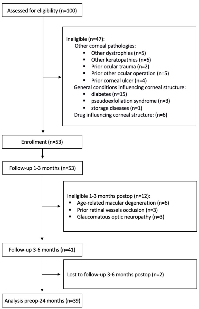
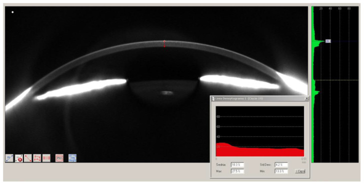
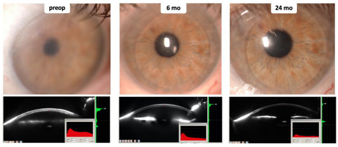
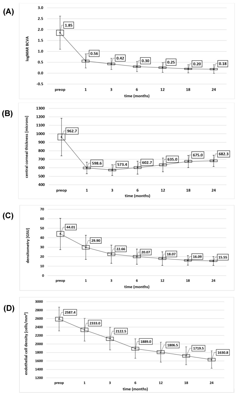
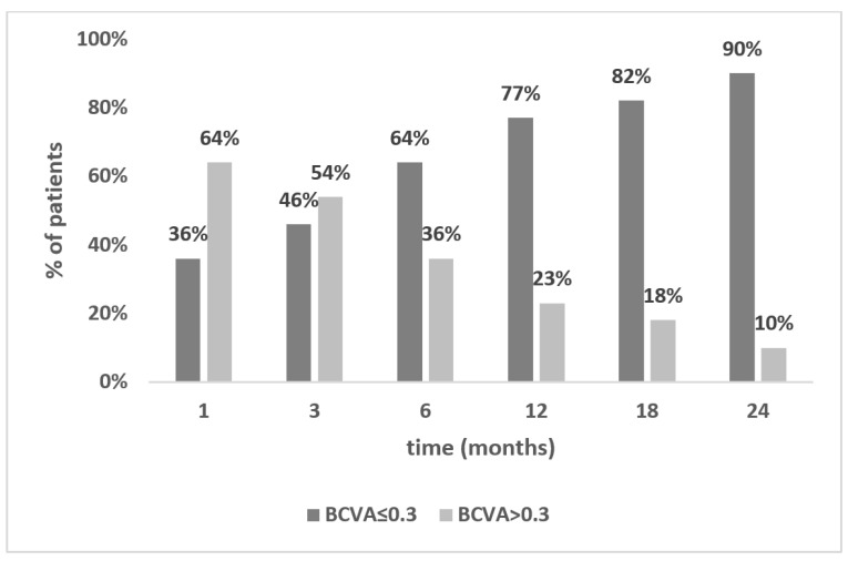
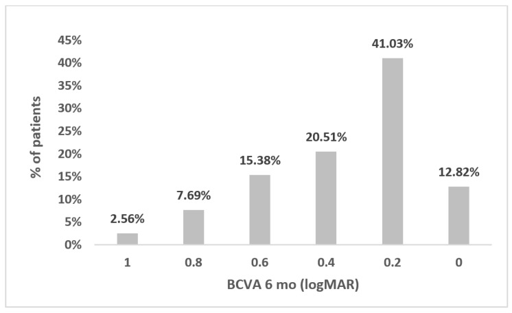
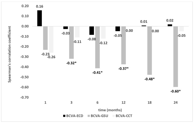
This study aimed to assess clinical application of the Scheimpflug corneal tomography for objective evaluation of corneal optical density in eyes undergoing Descemet's stripping endothelial keratoplasty (DSEK). In this prospective study, 39 pseudophakic eyes with bullous keratopathy were enrolled. All eyes underwent primary DSEK. Ophthalmic examination included best corrected visual acuity (BCVA) measurement, biomicroscopy, Scheimpflug tomography, pachymetry, and endothelial cell count. All measurements were taken preoperatively and within a 2-year follow-up period. Gradual BCVA improvement was observed in all patients. After two years, the mean and median BCVA values were 0.18 logMAR. A decrease in central corneal thickness was noted only during the first 3 months postoperatively and was followed by a gradual increase. Corneal densitometry decreased constantly and most significantly in the first 3 months postoperatively. The consecutive decrease in endothelial cell count of the transplanted cornea was most significant during the first 6 months postoperatively. Six months postoperatively, the strongest correlation (Spearman's r = -0.41) with BCVA was found for densitometry. This tendency was maintained throughout the entire follow-up period. Corneal densitometry is applicable for objective monitoring of early and late outcomes of endothelial keratoplasty, showing a higher correlation with visual acuity than pachymetry and endothelial cell density.
Keywords: Descemet stripping endothelial keratoplasty; Scheimpflug system; corneal densitometry; corneal optical density; corneal transparency.
Conflict of interest statement
The authors declare no conflict of interest.
Figures
Similar articles
-
A Comprehensive Investigation Into the Outcomes of Descemet's Stripping Endothelial Keratoplasty (DSEK) as a Treatment for Corneal Endothelial Disorders.Cureus. 2023 Sep 27;15(9):e46076. doi: 10.7759/cureus.46076. eCollection 2023 Sep. Cureus. 2023. PMID: 37900453 Free PMC article.
-
Early postoperative results of Descemet's stripping endothelial keratoplasty in six dogs with corneal endothelial dystrophy.Vet Ophthalmol. 2019 Nov;22(6):879-890. doi: 10.1111/vop.12666. Epub 2019 Mar 21. Vet Ophthalmol. 2019. PMID: 30895742
-
Femtosecond laser-assisted Descemet's stripping endothelial keratoplasty: a prospective study of 6-month visual outcomes, corneal thickness and endothelial cell loss.Int Ophthalmol. 2020 Aug;40(8):2065-2075. doi: 10.1007/s10792-020-01383-8. Epub 2020 Apr 21. Int Ophthalmol. 2020. PMID: 32318937
-
Infectious interface keratitis (IIK) following lamellar keratoplasty: A literature review.Ocul Surf. 2019 Oct;17(4):635-643. doi: 10.1016/j.jtos.2019.08.001. Epub 2019 Aug 12. Ocul Surf. 2019. PMID: 31415815
-
Corneal optical density: Structural basis, measurements, influencing factors, and roles in refractive surgery.Front Bioeng Biotechnol. 2023 Apr 6;11:1144455. doi: 10.3389/fbioe.2023.1144455. eCollection 2023. Front Bioeng Biotechnol. 2023. PMID: 37091331 Free PMC article. Review.
Cited by
-
Corneal stromal changes following simple limbal epithelial transplantation on Scheimpflug densitometry: Early results.Indian J Ophthalmol. 2025 Jan 1;73(1):77-82. doi: 10.4103/IJO.IJO_105_24. Epub 2024 Aug 14. Indian J Ophthalmol. 2025. PMID: 39186626 Free PMC article.
References
-
- Edelhauser H.F., Ubels J.L., Hejny C. The cornea and the sclera. In: Kaufman P.L., Alm A., editors. Adler’s Physiology of the Eye. 10th ed. Mosby; St. Louis, MO, USA: 2003. pp. 47–114.
LinkOut - more resources
Full Text Sources
