. 2023 Jan 21;9(2):145.
doi: 10.3390/jof9020145.

FgAP1σ Is Critical for Vegetative Growth, Conidiation, Virulence, and DON Biosynthesis in Fusarium graminearum

Affiliations

FgAP1σ Is Critical for Vegetative Growth, Conidiation, Virulence, and DON Biosynthesis in Fusarium graminearum

Congxian Wu et al. J Fungi (Basel). .

Abstract

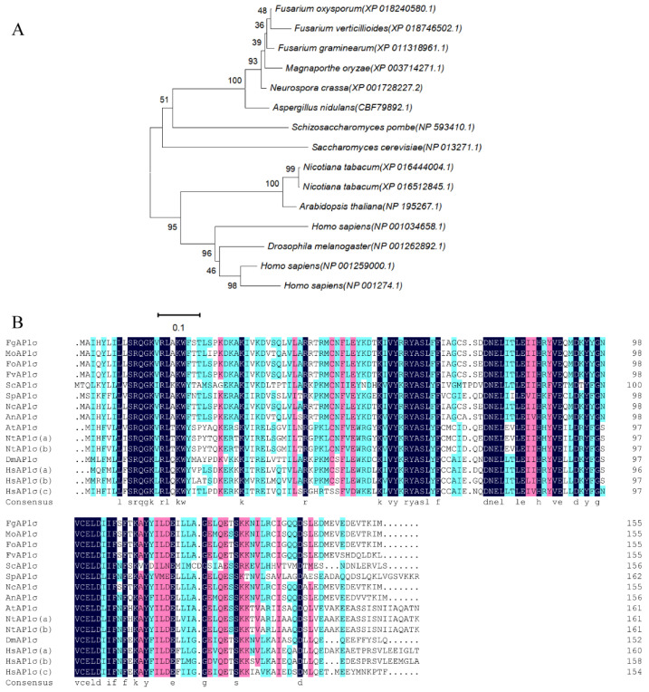
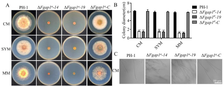
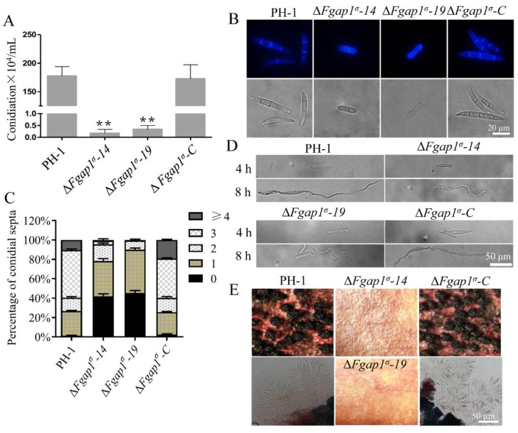
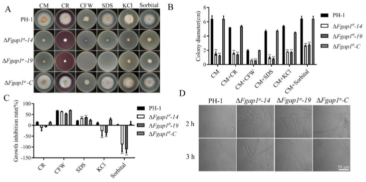
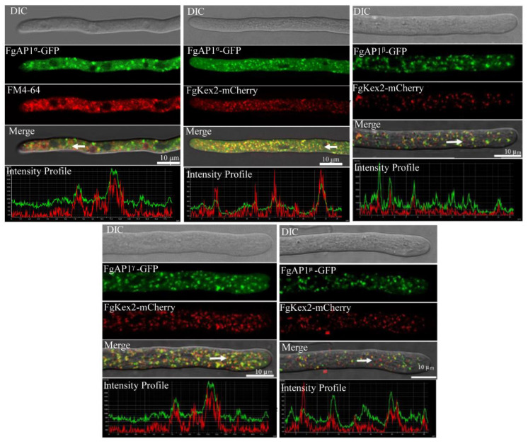
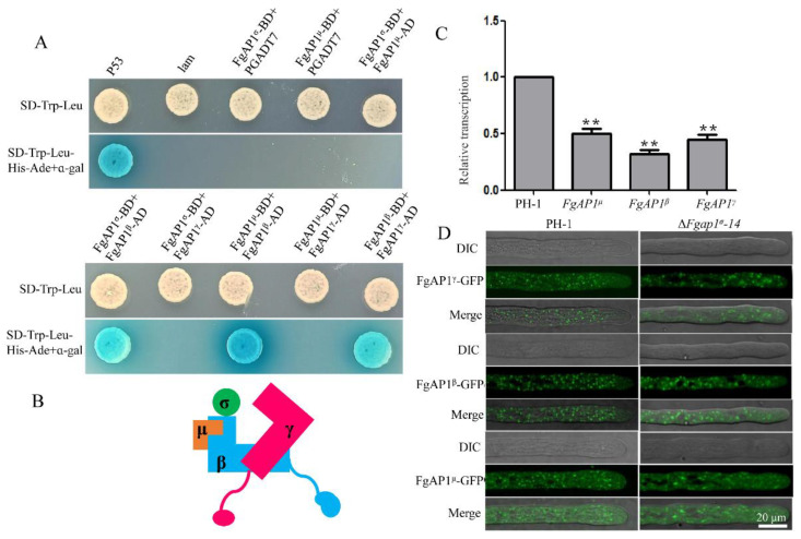
The AP1 complex is a highly conserved clathrin adaptor that plays important roles in regulating cargo protein sorting and intracellular vesicle trafficking in eukaryotes. However, the functions of the AP1 complex in the plant pathogenic fungi including the devastating wheat pathogen Fusarium graminearum are still unclear. In this study, we investigated the biological functions of FgAP1σ, a subunit of the AP1 complex in F. graminearum. Disruption of FgAP1σ causes seriously impaired fungal vegetative growth, conidiogenesis, sexual development, pathogenesis, and deoxynivalenol (DON) production. The ΔFgap1σ mutants were found to be less sensitive to KCl- and sorbitol-induced osmotic stresses but more sensitive to SDS-induced stress than the wild-type PH-1. Although the growth inhibition rate of the ΔFgap1σ mutants was not significantly changed under calcofluor white (CFW) and Congo red (CR) stresses, the protoplasts released from ΔFgap1σ hyphae were decreased compared with the wild-type PH-1, suggesting that FgAP1σ is necessary for cell wall integrity and osmotic stresses in F. graminearum. Subcellular localization assays showed that FgAP1σ was predominantly localized to endosomes and the Golgi apparatus. In addition, FgAP1β-GFP, FgAP1γ-GFP, and FgAP1μ-GFP also localize to the Golgi apparatus. FgAP1β interacts with FgAP1σ, FgAP1γ, and FgAP1μ, while FgAP1σ regulates the expression of FgAP1β, FgAP1γ, and FgAP1μ in F. graminearum. Furthermore, the loss of FgAP1σ blocks the transportation of the v-SNARE protein FgSnc1 from the Golgi to the plasma membrane and delays the internalization of FM4-64 dye into the vacuole. Taken together, our results demonstrate that FgAP1σ plays vital roles in vegetative growth, conidiogenesis, sexual reproduction, DON production, pathogenicity, cell wall integrity, osmotic stress, exocytosis, and endocytosis in F. graminearum. These findings unveil the functions of the AP1 complex in filamentous fungi, most notably in F. graminearum, and lay solid foundations for effective prevention and control of Fusarium head blight (FHB).

Keywords: AP1 complex; FgAP1σ; Fusarium graminearum; wheat scab.


Conflict of interest statement

The authors have no conflict of interest to declare.

Figures

Figure 1
Figure 1
Bioinformatics analyses of FgAP1σ homologues. (A) Phylogenetic relationship of FgAP1σ protein from different organisms. The gene names are from the Broad Institute genome annotation: Fusarium graminearum (XP_011318961.1), Magnaporthe oryzae (XP_003714271.1), Fusarium oxysporum (XP_018240580.1), Fusarium verticillioides (XP_018746502.1), Saccharomyces cerevisiae (NP_013271.1), Schizosaccharomyces pombe (NP_593410.1), Neurospora crassa (XP_001728227.2), Aspergillus nidulans (CBF79892.1), Arabidopsis thaliana (NP_195267.1), Nicotiana tabacum (XP_016444004.1), Nicotiana tabacum (XP_016512845.1), Homo sapiens (NP_001259000.1), Homo sapiens (NP_001274.1), Homo sapiens (NP_001034658.1), Drosophila melanogaster (NP_001262892.1). (B) Multiple alignment of amino acid sequences of FgAP1σ homologues.
Figure 2
Figure 2
FgAP1σ is required for the vegetative growth of F. graminearum. (A) Vegetative growth of the wild-type PH-1, ΔFgap1σ mutants, and the complemented strain ΔFgap1σ-C after growth on complete media (CM), starch yeast medium (SYM), and minimal medium (MM) agar at 28 °C for 3 days. (B) Statistical analysis of the colony diameters of the PH-1, ΔFgap1σ, and ΔFgap1σ-C strains on CM, SYM, and MM media after 3 days. Level of significance was measured using an unpaired t-test (** p < 0.01). (C) Mycelial morphologies of PH-1, ΔFgap1σ, and ΔFgap1σ-C strains on CM medium. Hyperbranching was observed in the ΔFgap1σ mutants. Bar = 50 μm.
Figure 3
Figure 3
FgAP1σ is involved in the conidiation and sexual development of F. graminearum. (A) Conidiation of the wild-type PH-1, ΔFgap1σ, and ΔFgap1σ-C strains in liquid CMC media for 3 days. Level of significance was measured using an unpaired t-test (** p < 0.01). (B) Conidial morphology was observed under a light microscope after the PH-1, ΔFgap1σ, and ΔFgap1σ-C strains were cultured in liquid CMC media for 3 days. Bar = 20 μm. (C) The number of septa in the conidia produced by the indicated strains. (D) Germination of the indicated strains in CM media after 4 h and 8 h of inoculation. Bar = 50 μm. (E) Images of the perithecia and ascospores produced by the PH-1, ΔFgap1σ, and ΔFgap1σ-C strains after 2 weeks of inoculation on carrot agar plates. Bar = 50 μm.
Figure 4
Figure 4
FgAP1σ is required for virulence. (A) Pathogenicity of ΔFgap1σ mutants on flowering wheat heads was significantly reduced compared with the wild-type PH-1 and ΔFgap1σ-C strains. Mycelia plugs from the various strains were inoculated on flowering wheat heads under moist conditions, and disease symptoms were recorded after two weeks. (B) The pathogenicity of ΔFgap1σ mutants on wheat coleoptiles was abolished. (C) Level of deoxynivalenol (DON) production by ΔFgap1σ mutants. Mycelia of the indicated strains were dried and weighed to quantify the fungal biomass. Level of significance was measured using an unpaired t-test (** p < 0.01). (D) The expression levels of some trichothecene biosynthetic genes FgTRI were significantly reduced in the ΔFgap1σ mutants. Level of significance was measured using an unpaired t-test (* p < 0.05, ** p < 0.01).
Figure 5
Figure 5
Deletion of FgAP1σ resulted in cell wall integrity and osmotic pressure defects. (A) The PH-1, ΔFgap1σ, and ΔFgap1σ-C strains were incubated on CM plates supplemented with Congo red (CR, 200 μg/mL), calcofluor white (CFW, 200 μg/mL), SDS (0.01 %), sorbitol (1 M), and KCl (1 M) at 28 °C for 3 days. (B) Colony diameter of the indicated strains under cell wall and osmotic stresses. Level of significance was measured using an unpaired t-test (** p < 0.01). (C) Growth inhibition rate of the strains due to cell wall and osmotic stresses induced by the indicated agents. Level of significance was measured using an unpaired t-test (** p < 0.01). (D) Protoplast releasing potential of the PH-1 and ΔFgap1σ mutants after treatment with cell-wall-degrading enzymes for 2 h and 3 h. Bar = 50 μm.
Figure 6
Figure 6
The localization of AP1 complex in F. graminearum. Colocalization of FgAP1σ-GFP with FM4-64 and trans-Golgi network (TGN, FgKex2-mCherry) as well as that of FgAP1β-GFP, FgAP1γ-GFP, and FgAP1μ-GFP with FgKex2-mCherry in F. graminearum. Images were captured from laser scanning confocal microscope. Bar = 10 μm.
Figure 7
Figure 7
The relationship between FgAP1 complex subunits and the localizations of the various subunits in ΔFgap1σ mutant. (A) Yeast two-hybrid assay showing the interactions of the AP1 complex subunits. There were positive interactions between FgAP1β and FgAP1σ and between FgAP1γ and FgAP1μ. The interaction between pGBKT7-Lam and pGADT7-T was used as a negative control while that between pGBKT7-53 and pGADT7-T served as a positive control. (B) The interaction model of FgAP1σ with the other subunits of FgAP1 complex. FgAP1σ interacts with FgAP1β, and FgAP1β interacts with FgAP1γ and FgAP1μ, which indicates that FgAP1σ, FgAP1β, FgAP1γ, and FgAP1μ function as a complex in F. graminearum. (C) The expression levels of FgAP1β, FgAP1γ, and FgAP1μ were significantly downregulated in ΔFgap1σ mutant. Level of significance was measured using an unpaired t-test (** p < 0.01). (D) The localizations of FgAP1β-GFP, FgAP1γ-GFP, and FgAP1μ-GFP were not affected in ΔFgap1σ mutant. Bar = 20 μm.
Figure 8
Figure 8
FgAP1σ is involved in exocytosis and endocytosis processes. (A) The plasma membrane localization of GFP-FgSnc1 is disrupted in the ΔFgap1σ mutant, as indicated by red circles. Bar = 10 μm. (B) FgAP1σ delays internalization of FM4-64 into the vacuole membrane. Hyphae of the wild-type PH-1, ΔFgap1σ-14, ΔFgap1σ-19, and complement strain ΔFgap1σ-C were cultured in liquid CM for 24 h and then stained with FM4-64 and observed at different time points using a fluorescence confocal microscope. Bar = 5 μm.

Similar articles

Cited by

References

    1. Schmid S.L. Clathrin-coated vesicle formation and protein sorting: An integrated process. Annu. Rev. Biochem. 1997;66:511–548. doi: 10.1146/annurev.biochem.66.1.511. - DOI - PubMed
    1. Hirst J., Robinson M.S. Clathrin and adaptors. Biochim. Biophys. Acta. 1998;14:173–193. doi: 10.1016/S0167-4889(98)00056-1. - DOI - PubMed
    1. Owen D.J., Collins B.M., Evans P.R. Adaptors for clathrin coats: Structure and function. Annu. Rev. Cell Dev. Biol. 2004;20:153–191. doi: 10.1146/annurev.cellbio.20.010403.104543. - DOI - PubMed
    1. Ohno H. Clathrin-associated adaptor protein complexes. J. Cell Sci. 2006;119:3719–3721. doi: 10.1242/jcs.03085. - DOI - PubMed
    1. Popova N.V., Deyev I.E., Petrenko A.G. Clathrin-mediated endocytosis and adaptor proteins. Acta Nat. 2013;5:62–73. doi: 10.32607/20758251-2013-5-3-62-73. - DOI - PMC - PubMed