. 2023 Jan 17;13(2):259.
doi: 10.3390/life13020259.

IGFBP-6 Alters Mesenchymal Stromal Cell Phenotype Driving Dasatinib Resistance in Chronic Myeloid Leukemia

Affiliations

IGFBP-6 Alters Mesenchymal Stromal Cell Phenotype Driving Dasatinib Resistance in Chronic Myeloid Leukemia

Daniela Cambria et al. Life (Basel). .

Abstract

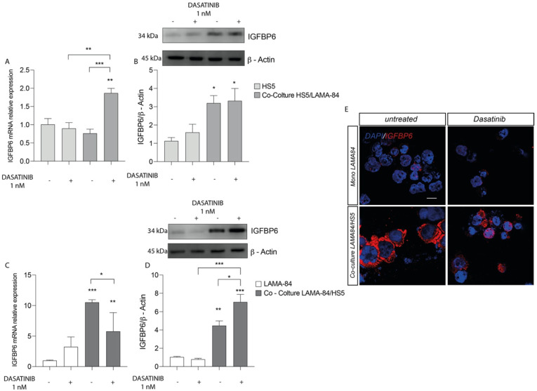
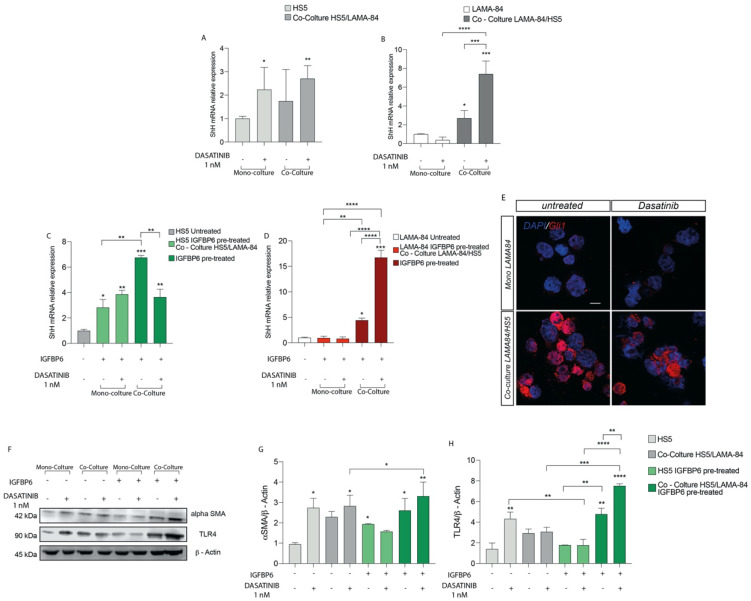
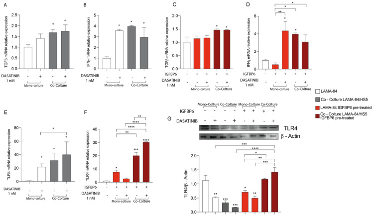
Chronic myeloid leukemia (CML), BCR-ABL1-positive, is classified as a myeloproliferative characterized by Philadelphia chromosome/translocation t(9;22) and proliferating granulocytes. Despite the clinical success of tyrosine kinase inhibitors (TKi) agents in the treatment of CML, most patients have minimal residual disease contained in the bone marrow microenvironment, within which stromal cells assume a pro-inflammatory phenotype that determines their transformation in cancer-associated fibroblasts (CAF) which, in turn can play a fundamental role in resistance to therapy. Insulin-like Growth Factor Binding Protein-6 (IGFBP-6) is expressed during tumor development, and is involved in immune-escape and inflammation as well, providing a potential additional target for CML therapy. Here, we aimed at investigating the role of IGFBP-6/SHH/TLR4 axis in TKi response. We used a CML cell line, LAMA84-s, and healthy bone marrow stromal cells, HS-5, in mono- or co-culture. The two cell lines were treated with Dasatinib and/or IGFBP-6, and the expression of inflammatory markers was tested by qRT-PCR; furthermore, expression of IGFBP-6, TLR4 and Gli1 were evaluated by Western blot analysis and immumocytochemistry. The results showed that both co-culture and Dasatinib exposure induce inflammation in stromal and cancer cells so that they modulate the expression of TLR4, and these effects were more marked following IGFBP-6 pre-treatment suggesting that this molecule may confer resistance through the inflammatory processes. This phenomenon was coupled with sonic hedgehog (SHH) signaling. Indeed, our data also demonstrate that HS-5 treatment with PMO (an inducer of SHH) induces significant modulation of TLR4 and overexpression of IGFPB-6 suggesting that the two pathways are interconnected with each other and with the TLR-4 pathway. Finally, we demonstrated that pretreatment with IGFBP-6 and/or PMO restored LAMA-84 cell viability after treatment with Dasatinib, suggesting that both IGFBP-6 and SHH are involved in the resistance mechanisms induced by the modulation of TLR-4, thus indicating that the two pathways may be considered as potential therapeutic targets.

Keywords: IGFBP6; TLR4; chronic myeloid leukemia; dasatinib; mesenchymal stromal cells.


Conflict of interest statement

The authors declare no conflict of interest.

Figures

Figure 1
Figure 1
Cell viability of HS−5 (A), LAMA−84 (B) and LAMA−84 in coculture with HS−5 (C) after treatment with 1 nM dasatinib. Relative expression of SIRT-1 (D), PGC1a (E), TGF-β (F) and INFγ (G) in HS−5 in coculture with LAMA−84 compared to HS−5 in monoculture. * p < 0.1; ** p < 0.01; **** p < 0.0001.
Figure 2
Figure 2
Relative expression of IGFBP6 transcript (A) and protein (B) in HS−5 cells coculture with LAMA−84 versus HS−5 monoculture after treatment with dasatinib 1 nM. Relative expression of IGFBP6 transcript (C) and related protein (D) in LAMA−84 in coculture with HS−5 compared to LAMA−84 in monoculture after treatment with dasatinib 1 nM. (E) Immunofluorescence analysis of IGFBP6 (IGFBP6 in red, nucleus in blue) in LAMA−84 in coculture with HS−5 compared to LAMA-84 in monoculture after treatment with dasatinib. * p < 0.05; ** p < 0.01; *** p < 0.001.
Figure 3
Figure 3
Relative expression of SHH transcript in HS−5 (A) and LAMA−84 (B) in coculture, with or without 1 nM dasatinib. Relative expression of SHH transcript in HS−5 (C) and LAMA−84 (D) in coculture, with or without IGFBP6 and dasatinib 1 nM. Immunofluorescence analysis of the expression of Gli1 in LAMA−84 in coculture with HS−5 in the presence or not of dasatinib 1 nM (E). Relative expression of aSMA and TLR4 protein in HS−5 in coculture with LAMA−84 with or without pretreatment with IGFBP6 compared to HS−5 in monoculture with or without pretreatment with IGFBP6 (FH) (* p < 0.1; ** p < 0.01; *** p < 0.001; **** p < 0.0001).
Figure 4
Figure 4
Relative expression of TGF−β (A) and INFγ (B) transcript in HS−5 coculture with LAMA−84, with or without 1 nM dasatinib. Relative expression of TGF−β (C) and INFγ (D) transcript in HS−5 in coculture with LAMA−84, with or without IGFBP6 and dasatinib 1 nM. (* p < 0.1; ** p < 0.01; **** p < 0.0001).
Figure 5
Figure 5
Relative expression of TGF−β (A) and INFγ (B) transcript in LAMA−84 coculture with HS-5, with or without 1 nM dasatinib. Relative expression of TGF−β (C) and INFγ (D) transcript in LAMA−84 coculture with HS−5, with or without IGFBP6 and dasatinib 1 nM. Relative expression of TLR4 transcript in LAMA−84 in coculture with HS-5, with or without dasatinib 1 nM (E). Relative expression of TLR4 transcript in LAMA−84 in coculture with HS−5, with or without IGFBP6 and dasatinib 1 nM (F). Relative expression of TLR4 protein in LAMA−84 in monoculture and in coculture with HS−5, with or without IGFBP6 and dasatinib 1 nM (G). * p < 0.05; ** p < 0.01; *** p < 0.001; **** p < 0.0001.
Figure 6
Figure 6
LAMA−84 Cell Viability after IGFBP6 (A) and PMO, Hedgehog pathway activator (B) pre-treatment. * p < 0.05; ** p < 0.01; *** p < 0.001; **** p < 0.0001.
Figure 7
Figure 7
Graphical representation of the mechanism proposed.

Similar articles

Cited by

References

    1. Chereda B., Melo J.V. Natural course and biology of CML. Ann. Hematol. 2015;94((Suppl 2)):S107–S121. doi: 10.1007/s00277-015-2325-z. - DOI - PubMed
    1. Dong Y., Shi O., Zeng Q., Lu X., Wang W., Li Y., Wang Q. Leukemia incidence trends at the global, regional, and national level between 1990 and 2017. Exp. Hematol. Oncol. 2020;9:14. doi: 10.1186/s40164-020-00170-6. - DOI - PMC - PubMed
    1. Jabbour E., Kantarjian H. Chronic myeloid leukemia: 2020 update on diagnosis, therapy and monitoring. Am. J. Hematol. 2020;95:691–709. doi: 10.1002/ajh.25792. - DOI - PubMed
    1. Hehlmann R. Chronic Myeloid Leukemia in 2020. Hemasphere. 2020;4:e468. doi: 10.1097/HS9.0000000000000468. - DOI - PMC - PubMed
    1. Pane F., Intrieri M., Quintarelli C., Izzo B., Muccioli G.C., Salvatore F. BCR/ABL genes and leukemic phenotype: From molecular mechanisms to clinical correlations. Oncogene. 2002;21:8652–8667. doi: 10.1038/sj.onc.1206094. - DOI - PubMed

LinkOut - more resources