. 2023 Jan 22;13(2):308.
doi: 10.3390/life13020308.

Secukinumab and Black Garlic Downregulate OPG/RANK/RANKL Axis and Devitalize Myocardial Interstitial Fibrosis Induced by Sunitinib in Experimental Rats

Affiliations

Secukinumab and Black Garlic Downregulate OPG/RANK/RANKL Axis and Devitalize Myocardial Interstitial Fibrosis Induced by Sunitinib in Experimental Rats

Hoda E Mohamad et al. Life (Basel). .

Abstract

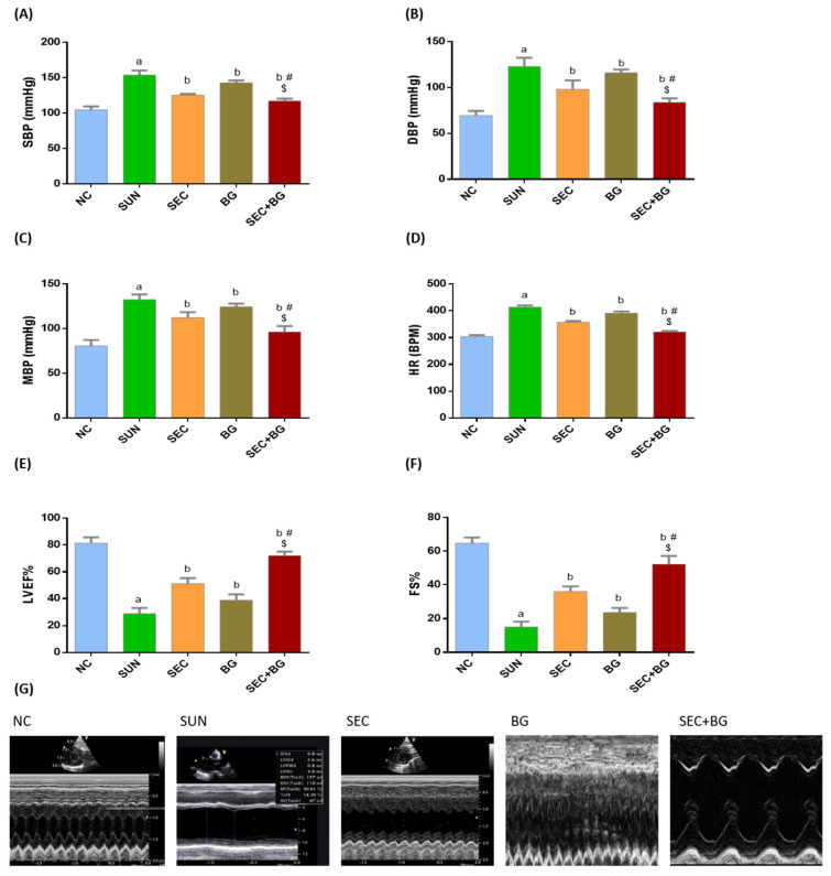
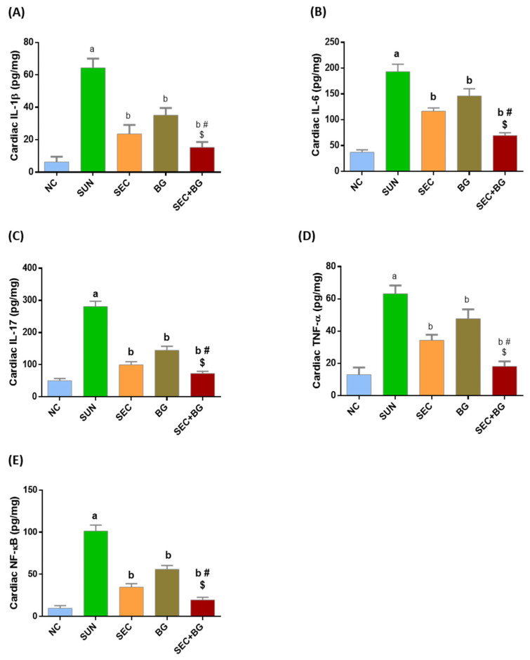
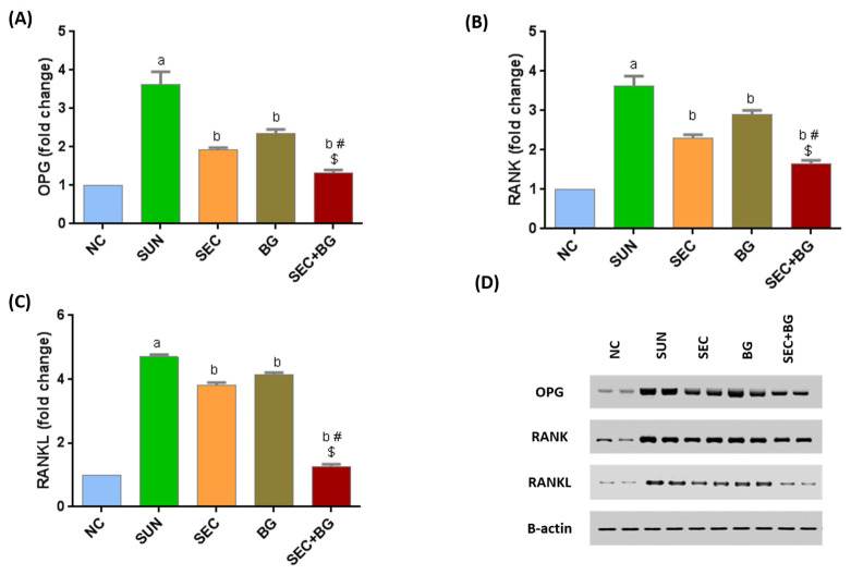
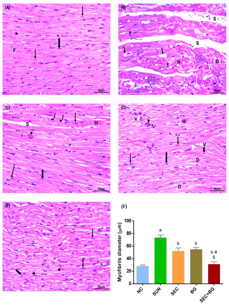
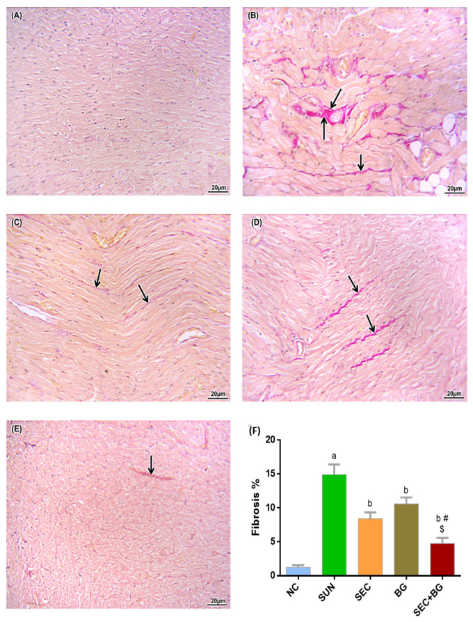
Sunitinib has been associated with several cardiotoxic effects such as cardiac fibrosis. The present study was designed to explore the role of interleukin (IL)-17 in sunitinib-induced myocardial fibrosis (MF) in rats and whether its neutralization and/or administration of black garlic (BG), a form of fermented raw garlic (Allium sativum L.), could extenuate this adverse effect. Male Wistar albino rats received sunitinib (25 mg/kg three times a week, orally) and were co-treated with secukinumab (3 mg/kg, subcutaneously, three times total) and/or BG (300 mg/kg/day, orally) for four weeks. Administration of sunitinib induced significant increase in cardiac index, cardiac inflammatory markers, and cardiac dysfunction that were ameliorated by both secukinumab and BG, and to a preferable extent, with the combined treatment. Histological examination revealed disruption in the myocardial architecture and interstitial fibrosis in cardiac sections of the sunitinib group, which were reversed by both secukinumab and BG treatments. Both drugs and their co-administration restored normal cardiac functions, downregulated cardiac inflammatory cytokines, mainly IL-17 and NF-κB, along with increasing the MMP1/TIMP1 ratio. Additionally, they attenuated sunitinib-induced upregulation of the OPG/RANK/RANKL axis. These findings highlight another new mechanism through which sunitinib can induce interstitial MF. The current results propose that neutralizing IL-17 by secukinumab and/or supplementation with BG can be a promising therapeutic approach for ameliorating sunitinib-induced MF.

Keywords: IL-17; OPG/RANK/RANKL; black garlic; interstitial myocardial fibrosis; secukinumab; sunitinib.


Conflict of interest statement

The authors declare no conflict of interest.

Figures

Figure 1
Figure 1
Effect of treatment with secukinumab and/or black garlic supplement on cardiac hemodynamic parameters and left ventricular function of SUN-administered rats. (A) SBP: systolic blood pressure. (B) DBP: diastolic blood pressure. (C) MBP: mean blood pressure. (D) HR: heart rate. (E) LVEF%: left ventricular ejection fraction. (F) FS%: fractional shortening. (G) Representative images of M-mode tracings through the left ventricles of the rats. Bars and error bars represent mean ±SD. (n = 6/group). NC: normal control; SUN: sunitinib; SEC: secukinumab; BG: black garlic. a p < 0.001 vs. NC, b p < 0.05 vs. SUN, # p < 0.01 vs. BG, $ p < 0.01 vs. SEC.
Figure 2
Figure 2
Effect of treatment with secukinumab and/or black garlic supplement on cardiac inflammatory markers and cytokines of SUN-administered rats. (A) IL-1β: interleukin-1β. (B) IL-6. (C) IL-17. (D) TNF-α: tumor necrosis factor-α. (E) NF-κB: nuclear factor kappa B. NC: normal control; SUN: sunitinib; SEC: secukinumab; BG: black garlic. Bars and error bars represent mean ± SD. (n = 6/group). a p < 0.001 vs. NC, b p < 0.001 vs. SUN, # p < 0.01 vs. BG, $ p < 0.001 vs. SEC.
Figure 3
Figure 3
Effect of treatment with secukinumab and/or black garlic supplement on OPG/RANK/RANKL axis of SUN-administered rats. Quantitative analysis of: (A) Osteoprotegerin (OPG) expression, (B) receptor activator of nuclear factor-κB (RANK) expression, (C) RANK ligand (RANKL) expression; relative to β-actin, and (D) representative Western blot images. Bars and error bars represent mean ± SD. (n = 6/group). NC: normal control; SUN: sunitinib; SEC: secukinumab; BG: black garlic. a p < 0.001 vs. NC, b p < 0.001 vs. SUN, # p < 0.001 vs. BG, $ p < 0.001 vs. SEC.
Figure 4
Figure 4
Representative photomicrographs of H&E-stained cardiac sections from experimental groups (H&E staining ×400, scale bar = 50 μm). (A) Normal control group showing normal striated myocardial fibers with acidophilic cytoplasm and vesicular nuclei (arrows) and separated by few connective tissue containing fibroblasts with their flat nuclei (thick arrow). Most cardiac fibers display intercalated discs (arrowhead). Blood vessels are not congested (*). (B) SUN group displayed disordered myocardial structure with apparent inflammatory cellular infiltration (ovale). Myofibers are curly and have acidophilic cytoplasm with hyaline appearance (H). Some cardiac fibers are fragmented and dissolved (D). Most of the nuclei are dark and pyknotic (arrowhead). Wide spaces containing extravasated blood (S) and vacuoles (arrow) were seen between cardiac myofibrils. Blood vessels are congested (*). (C,D) represents SEC and BG groups, respectively, showed apparently better cardiac muscle structure with mild cellular inflammatory infiltrate (arrowhead) and congested blood vessels (*). Some cardiac muscle fibers have bright nuclei (arrow). Other muscles fibers are dissolved (D) and have dark shrunken nuclei (thick arrow) with hyaline outlook (H). Vacuoles (V) are present between the affected muscle fibers. SEC group exhibited the presence of intercalated discs (curved arrow). (E) SEC + BG group showed marked improvement in the cardiac muscle architecture with near to normal cardiac muscle structure. (F) Myofibril diameter (μm). NC: normal control; SUN: sunitinib; SEC: secukinumab; BG: black garlic. Bars with error bar represent mean ± SD. a p < 0.001 vs. NC, b p < 0.001 vs. SUN, # p < 0.001 vs. BG, $ p < 0.001 vs. SEC.
Figure 5
Figure 5
Representative photomicrographs of Van Gieson-stained sections of the heart (Van Gieson ×200, scale bar = 20 μm). (A) NC group showing a normal cardiac histological structure. (B) SUN group showing extensive collagen fibers indicated by the arrows. (C) SEC group showing few collagen fibers (arrows). (D) BG group showing moderate amount of collagen deposition (arrows). (E) SEC + BG group revealing remarkable reduction in collagen deposition with nearly normal structure (arrow). (F) Fibrosis%. NC: normal control; SUN: sunitinib; SEC: secukinumab; BG: black garlic; Red color indicates fibrosis. Bars with error bar represent mean ± SD. a p < 0.001 vs. NC, b p < 0.001 vs. SUN, # p < 0.001 vs. BG, $ p < 0.001 vs. SEC.

Similar articles

Cited by

References

    1. Gorini S., De Angelis A., Berrino L., Malara N., Rosano G., Ferraro E. Chemotherapeutic Drugs and Mitochondrial Dysfunction: Focus on Doxorubicin, Trastuzumab, and Sunitinib. Oxid. Med. Cell. Longev. 2018;2018:7582730. doi: 10.1155/2018/7582730. - DOI - PMC - PubMed
    1. Yang Y., Bu P. Progress on the cardiotoxicity of sunitinib: Prognostic significance, mechanism and protective therapies. Chem. Biol. Interact. 2016;257:125–131. doi: 10.1016/j.cbi.2016.08.006. - DOI - PubMed
    1. Richards C.J., Je Y., Schutz F., Heng D., Dallabrida S., Moslehi J., Choueiri T.K. Incidence and Risk of Congestive Heart Failure in Patients with Renal and Nonrenal Cell Carcinoma Treated with Sunitinib. J. Clin. Oncol. 2011;29:3450–3456. doi: 10.1200/JCO.2010.34.4309. - DOI - PubMed
    1. Formiga M.N., Fanelli M.F. Aortic dissection during antiangiogenic therapy with sunitinib. A case report. Sao Paulo Med. J. 2015;133:275–277. doi: 10.1590/1516-3180.2013.7380002. - DOI - PMC - PubMed
    1. Sourdon J., Facchin C., Certain A., Viel T., Robin B., Lager F., Marchiol C., Balvay D., Yoganathan T., Favier J., et al. Sunitinib-induced cardiac hypertrophy and the endothelin axis. Theranostics. 2021;11:3830–3838. doi: 10.7150/thno.49837. - DOI - PMC - PubMed