

Orf Infection on the Scalp of a Taiwanese Woman: A Case Report and Literature Review
- PMID: 36836716
- PMCID: PMC9966865
- DOI: 10.3390/life13020358
Orf Infection on the Scalp of a Taiwanese Woman: A Case Report and Literature Review
Abstract
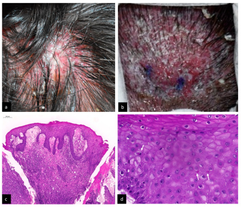
Background: Orf, or ecthyma contagiosum, is a zoonosis caused by Parapoxvirus that infects sheep and goats. Human transmission typically occurs in persons in contact with the infected animals or contaminated fomites and environment. In humans, it generally occurs as solitary or multiple skin lesions on the hands or fingers. Involvement of the head region has rarely been reported.
Case presentation: We report an unusual case with multiple orf lesions on the scalp of a middle-aged woman, along with a review of previously reported Orf cases on the head region.
Conclusions: Although Orf infection rarely happens on the head region, it should be considered in the differential diagnosis of cases with relevant animal exposure.
Keywords: Orf; Orf virus; Parapoxvirus; ecthyma contagiosum; zoonosis.
Conflict of interest statement
The authors declare no conflict of interest.
Figures
Similar articles
-
Solitary facial lesion of orf: An unusual presentation.North Clin Istanb. 2021 Nov 18;8(6):626-628. doi: 10.14744/nci.2020.59254. eCollection 2021. North Clin Istanb. 2021. PMID: 35284790 Free PMC article.
-
A case of orf (ecthyma contagiosum) with multiple lesions.J Pak Med Assoc. 2013 Jun;63(6):786-7. J Pak Med Assoc. 2013. PMID: 23901689
-
Solitary Subungual Orf.J Hand Surg Am. 2022 Feb;47(2):194.e1-194.e3. doi: 10.1016/j.jhsa.2020.12.010. Epub 2021 Mar 1. J Hand Surg Am. 2022. PMID: 33663886
-
A Review on Human Orf: A Neglected Viral Zoonosis.Res Rep Trop Med. 2021 Jul 8;12:153-172. doi: 10.2147/RRTM.S306446. eCollection 2021. Res Rep Trop Med. 2021. PMID: 34267574 Free PMC article. Review.
-
Orf Virus Infection in Humans: A Review With a Focus on Advances in Diagnosis and Treatment.J Drugs Dermatol. 2017 Jul 1;16(7):684-689. J Drugs Dermatol. 2017. PMID: 28697220 Review.
Cited by
-
The whole genome analysis of the wild-type and attenuated orf virus reveals that ORF022 facilitates viral replication.BMC Genomics. 2025 May 15;26(1):488. doi: 10.1186/s12864-025-11663-1. BMC Genomics. 2025. PMID: 40375129 Free PMC article.
References
Publication types
LinkOut - more resources
Full Text Sources
