. 2023 Jan 29;13(2):367.
doi: 10.3390/life13020367.

Arachidonic Acid Added during the Differentiation Phase of 3T3-L1 Cells Exerts Anti-Adipogenic Effect by Reducing the Effects of Pro-Adipogenic Prostaglandins

Affiliations

Arachidonic Acid Added during the Differentiation Phase of 3T3-L1 Cells Exerts Anti-Adipogenic Effect by Reducing the Effects of Pro-Adipogenic Prostaglandins

Michael N N Nartey et al. Life (Basel). .

Abstract

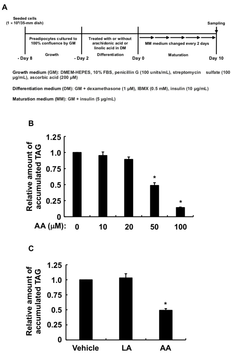
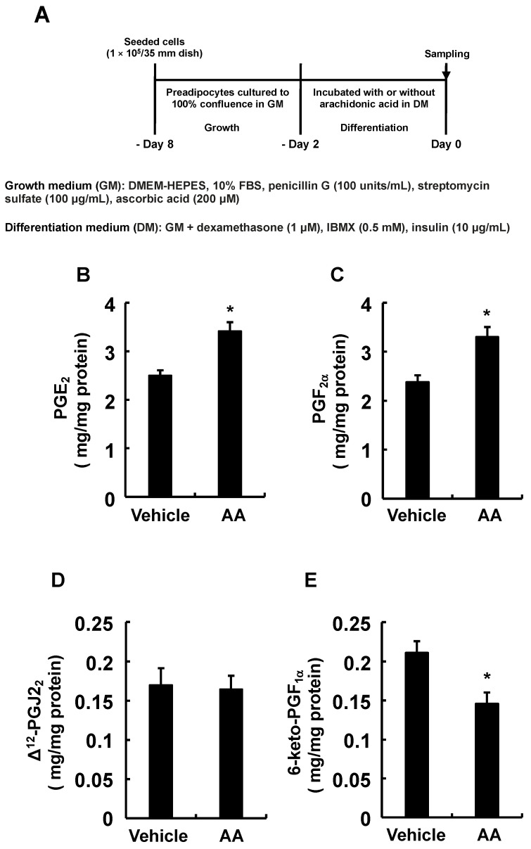
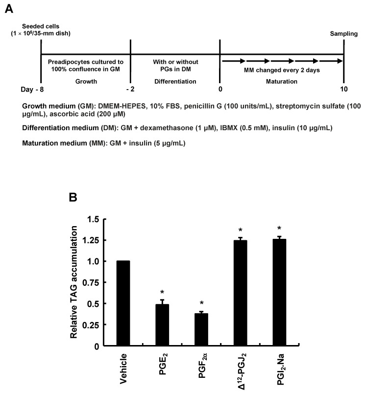
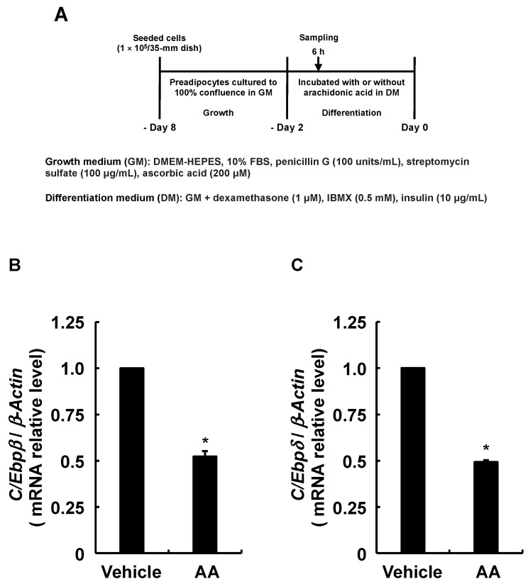
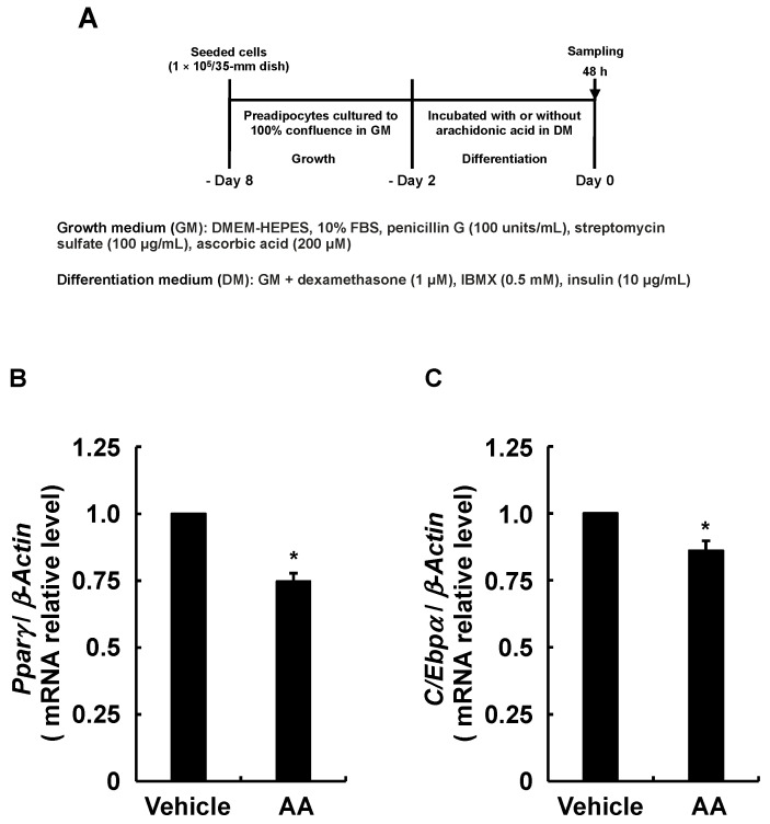
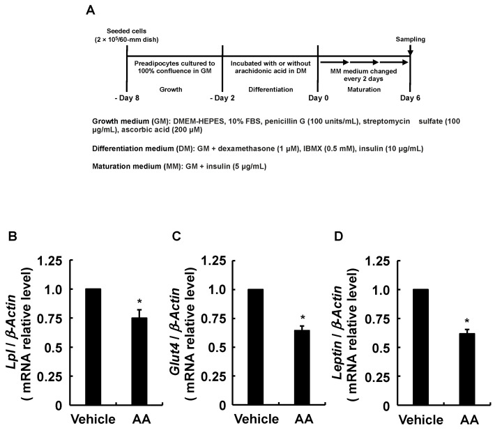
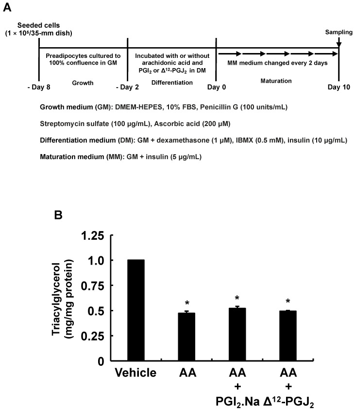
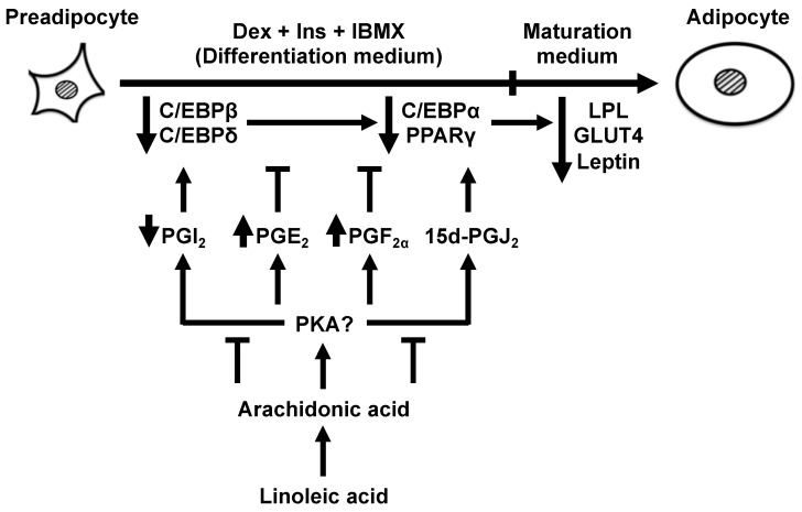
A linoleic acid (LA) metabolite arachidonic acid (AA) added to 3T3-L1 cells is reported to suppress adipogenesis. The purpose of the present study aimed to clarify the effects of AA added during the differentiation phase, including adipogenesis, the types of prostaglandins (PG)s produced, and the crosstalk between AA and the PGs produced. Adipogenesis was inhibited by AA added, while LA did not. When AA was added, increased PGE2 and PGF production, unchanged Δ12-PGJ2 production, and reduced PGI2 production were observed. Since the decreased PGI2 production was reflected in decreased CCAAT/enhancer-binding protein-β (C/EBPβ) and C/EBPδ expression, we expected that the coexistence of PGI2 with AA would suppress the anti-adipogenic effects of AA. However, the coexistence of PGI2 with AA did not attenuate the anti-adipogenic effects of AA. In addition, the results were similar when Δ12-PGJ2 coexisted with AA. Taken together, these results indicated that the metabolism of ingested LA to AA is necessary to inhibit adipogenesis and that exposure of AA to adipocytes during only the differentiation phase is sufficient. As further mechanisms for suppressing adipogenesis, AA was found not only to increase PGE2 and PGF and decrease PGI2 production but also to abrogate the pro-adipogenic effects of PGI2 and Δ12-PGJ2.

Keywords: 6-PUFAs; adipogenesis; arachidonic acid; prostaglandin; the differentiation phase.


Conflict of interest statement

The authors declare no conflict of interest.

Figures

Figure 1
Figure 1
Effects of AA or LA added during the differentiation phase of 3T3-L1 cells on intracellular TAG levels after maturation. (A) Experimental procedure. We seeded 3T3-L1 cells (1 × 10⁵/dish) in 35 mm dishes containing 2 mL of GM and incubated them until they reached 100% confluence. Confluent cells were cultured for 48 h in 2 mL of DM-containing vehicle, indicated concentrations of AA, or LA (50 µM), followed by 2 mL of fresh MM every 2 days for 10 days. Intracellular TAG levels were analyzed in terminally differentiated mature adipocytes collected on day 10. (B,C) Intracellular TAG levels in cultured adipocytes. Data represent means ± SEM of n = 3 experiments for (B,C). * p < 0.05 vs. control (vehicle in DM) (Dunnett tests). GM, growth medium; DM, differentiation medium; MM, maturation medium; AA, arachidonic acid; LA, linoleic acid; TAG, triacylglycerol; SEM, standard error of the mean.
Figure 2
Figure 2
Effect of AA added during the differentiation phase of 3T3-L1 cells on endogenous PG biosynthesis. (A) Experimental procedure. We seeded 3T3-L1 cells (1 × 10⁵/dish) in 35 mm dishes containing 2 mL of GM and cultured them until they reached 100% confluence. Confluent cells were cultured for 48 h in 2 mL of DM-containing vehicle or AA (50 µM). The culture media was collected to determine PGs. (B) PGE2, (C) PGF, (D) Δ12-PGJ2, (E) 6-keto-PGF assessed by ELISA using specific antibodies. Data are shown as means ± SEM of n = 3 experiments for (BE). * p < 0.05 vs. control (vehicle in DM) (Student t-tests). GM, growth medium; DM, differentiation medium; AA, arachidonic acid; PG, prostaglandin; PGE2, prostaglandin E2; PGF, prostaglandin F; Δ12-PGJ2, Δ12-prostaglandin J2; 6-keto-PGF1α, 6-keto-prostaglandin F; SEM, standard error of the mean.
Figure 3
Figure 3
Effects of PGs added during the differentiation phase of 3T3-L1 cells on intracellular TAG levels after maturation. (A) Experimental procedure. We seeded 3T3-L1 cells (1 × 10⁵/dish) in 35 mm dishes containing 2 mL of GM and incubated them until they reached 100% confluence. Confluent cells were cultured for 48 h in 2 mL of DM-containing vehicle, PGE2 (1 µM), PGF (1 µM), Δ12-PGJ2 (1 µM), or PGI2. Na (100 nM), followed by 2 mL of fresh MM every 2 days for 10 days. Intracellular TAG levels were analyzed in terminally differentiated mature adipocytes collected on day 10. (B) Intracellular TAG levels in cultured adipocytes. Data represent means ± SEM of n = 3 experiments for (B). * p < 0.05 vs. control (vehicle in DM) (Dunnett tests). GM, growth medium; DM, differentiation medium; MM, maturation medium; PG, prostaglandin; PGE2, prostaglandin E2; PGF, prostaglandin F; Δ12-PGJ2, Δ12-prostaglandin J2; PGI2.Na, prostaglandin I2.Na; TAG, triacylglycerol; SEM, standard error of the mean.
Figure 4
Figure 4
Effects of AA added during the differentiation phase of 3T3-L1 cells on expression of C/Ebpβ and C/Ebpδ. (A) Experimental procedure. We seeded 3T3-L1 cells (2 × 10⁵/dish) in 60 mm dishes containing 4 mL of GM and incubated them until they reached confluence. Thereafter, cells were incubated with DM containing vehicle or AA (50 µM) during the differentiation phase for 6 h. Expression of mRNA for (B) C/Ebpβ and (C) C/Ebpδ determined by RT-qPCR. Data are shown as means ± SEM of n = 3 experiments for (B,C). * p < 0.05 vs. control (vehicle in DM) (Student t-tests). GM, growth medium; DM, differentiation medium; AA, arachidonic acid; C/Ebpβ, CCAAT/enhancer-binding protein-β; C/Ebpδ, CCAAT/enhancer-binding protein-δ; SEM, standard error of the mean.
Figure 5
Figure 5
Effects of AA added during the differentiation phase of 3T3-L1 cells on expression of Pparγ and C/Ebpα. (A) Experimental procedure. We seeded 3T3-L1 cells (2 × 10⁵/dish) in 60 mm dishes in 4 mL of GM and incubated them until they reached confluence. Thereafter, cells were incubated with DM containing vehicle or AA (50 µM) during the differentiation phase for 48 h. Expression of mRNA for (B) Pparγ and (C) C/Ebpα determined by RT-qPCR. Data are shown as means ± SEM of n = 3 experiments for (B,C). * p < 0.05 vs. control (vehicle in DM) (Student t-tests). GM, growth medium; DM, differentiation medium; AA, arachidonic acid; Pparγ, proliferator-activated receptor-γ; C/Ebpα, CCAAT/enhancer-binding protein-α; SEM, standard error of the mean.
Figure 6
Figure 6
Effects of AA added during the differentiation phase of 3T3-L1 cells on expression of Lpl, Glut4, and Leptin. (A) Experimental procedure. We seeded 3T3-L1 cells (2 × 10⁵/dish) in 60 mm dishes containing 4 mL of GM and incubated them until they reached confluence. Thereafter, cells were incubated with DM-containing vehicle or AA (50 µM) during the differentiation phase, followed by 6 days in fresh MM that was replaced every 2 days. Expression of mRNA for (B) Lpl, (C) Glut4, and (D) Leptin determined by RT-qPCR. Data are shown as means ± SEM of n = 3 experiments for (BD). * p < 0.05 vs. control (vehicle in DM) (Student t-tests). GM, growth medium; DM, differentiation medium; MM, maturation medium; AA, arachidonic acid; Lpl, lipoprotein lipase; Glut4, glucose transporter 4; SEM, standard error of the mean.
Figure 7
Figure 7
Effects of PGI2 or Δ12-PGJ2 on 3T3-L1 cells incubated with AA during the differentiation phase of 3T3-L1 cells. (A) Experimental procedure. We seeded and incubated 3T3-L1 cells (1 × 10⁵/dish) in 35 mm dishes in 2 mL of GM until they reached 100% confluence. The cells were then incubated for 48 h with 2 mL of DM containing vehicle, AA (50 µM), and PGI2.Na (100 nM) or Δ12-PGJ2 (1 µM). The differentiation medium was replaced with 2 mL of fresh MM every 2 days thereafter. Intracellular TAG levels were analyzed in terminally differentiated mature adipocytes on day 10. (B) Intracellular TAG levels in cultured adipocytes. Data are shown as means ± SEM of n = 3 experiments for (B). * p < 0.05 vs. control (vehicle in DM) (Tukey-Kramer tests). GM, growth medium; DM, differentiation medium; MM, maturation medium; AA, arachidonic acid; PGI2.Na, prostaglandin I2.Na; Δ12-PGJ2, Δ12-prostaglandin J2; TAG, triacylglycerol; SEM, standard error of the mean.
Figure 8
Figure 8
Schematic representation of arachidonic acid function during the differentiation phase of 3T3-L1 cells and subsequent effects on adipogenesis induced by 3-isobutyl-1-methylxanthine, dexamethasone, and insulin. Increased PGE2 and PGF production, whereas the reduced PGI2 production together with the suppression of PGI2 and Δ12-PGJ2 actions by arachidonic acid during the differentiation phase inhibited MDI-induced adipogenesis during the maturation phase. C/EBPα, CCAAT/enhancer-binding protein-α; C/EBPβ, CCAAT/enhancer-binding protein-β; C/EBPδ, CCAAT/enhancer-binding protein-δ; DEX, dexamethasone; Ins, insulin; IBMX, 3-isobutyl-1-methylxanthine; PPARγ, peroxisome proliferator-activated receptor-γ; LPL, lipoprotein lipase; GLUT4, glucose transporter 4; PG, prostaglandin.

Similar articles

Cited by

References

    1. Bennett C.N., Ross S.E., Longo K.A., Bajnok L., Hemati N., Johnson K.W., Harrison S.D., MacDougald O.A. Regulation of Wnt signaling during adipogenesis. J. Biol. Chem. 2002;277:30998–31004. doi: 10.1074/jbc.M204527200. - DOI - PubMed
    1. Kopelman P.G. Obesity as a medical problem. Nature. 2000;404:635–643. doi: 10.1038/35007508. - DOI - PubMed
    1. Green H., Kehinde O. Sublines of mouse 3T3 cells that accumulate lipid. Cell. 1974;1:113–116. doi: 10.1016/0092-8674(74)90126-3. - DOI
    1. Green H., Kehinde O. An established preadipose cell line and its differentiation in culture. II. Factors affecting the adipose conversion. Cell. 1975;5:19–27. doi: 10.1016/0092-8674(75)90087-2. - DOI - PubMed
    1. Cao Z., Umek R.M., McKnight S.L. Regulated expression of three C/EBP isoforms during adipose conversion of 3T3-L1 cells. Genes Dev. 1991;5:1538–1552. doi: 10.1101/gad.5.9.1538. - DOI - PubMed