

The Many Roles of Dermoscopy in Melanoma Detection
- PMID: 36836834
- PMCID: PMC9964307
- DOI: 10.3390/life13020477
The Many Roles of Dermoscopy in Melanoma Detection
Abstract
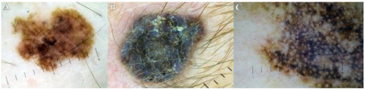
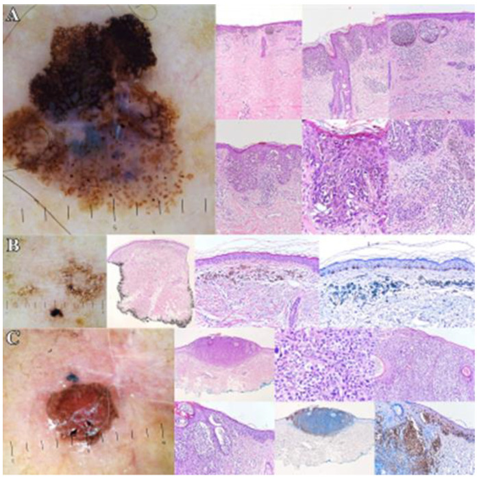
Dermoscopy is a non-invasive method of examination that aids the clinician in many ways, especially in early skin cancer detection. Melanoma is one of the most aggressive forms of skin cancer that can affect individuals of any age, having an increasing incidence worldwide. The gold standard for melanoma diagnosis is histopathological examination, but dermoscopy is also very important for its detection. To highlight the many roles of dermoscopy, we analyzed 200 melanocytic lesions. The main objective of this study was to detect through dermoscopy hints of melanomagenesis in the studied lot. The most suspicious were 10 lesions which proved to be melanomas confirmed through histopathology. The second objective of this study was to establish if dermoscopy can aid in estimating the Breslow index (tumoral thickness) of the melanomas and to compare the results to the histopathological examination. We found that the tumoral thickness may be estimated through dermoscopy, but the histopathological examination is superior. To conclude, the aim of this study was to showcase the versatility and many roles of dermoscopy, besides being one of the most important tools for early melanoma diagnosis.
Keywords: dermoscopy; melanoma; melanomagenesis; nevi; tumoral thickness.
Conflict of interest statement
The authors declare no conflict of interest.
Figures
Similar articles
-
Role of In Vivo Reflectance Confocal Microscopy in the Analysis of Melanocytic Lesions.Acta Dermatovenerol Croat. 2018 Apr;26(1):64-67. Acta Dermatovenerol Croat. 2018. PMID: 29782304 Review.
-
Long-Term Sequential Digital Dermoscopy of Low-Risk Patients May Not Improve Early Diagnosis of Melanoma Compared to Periodical Handheld Dermoscopy.Cancers (Basel). 2023 Feb 10;15(4):1129. doi: 10.3390/cancers15041129. Cancers (Basel). 2023. PMID: 36831472 Free PMC article.
-
Detection of primary melanoma in individuals at extreme high risk: a prospective 5-year follow-up study.JAMA Dermatol. 2014 Aug;150(8):819-27. doi: 10.1001/jamadermatol.2014.514. JAMA Dermatol. 2014. PMID: 24964862
-
Association of Patient Risk Factors and Frequency of Nevus-Associated Cutaneous Melanomas.JAMA Dermatol. 2016 Mar;152(3):291-8. doi: 10.1001/jamadermatol.2015.3775. JAMA Dermatol. 2016. PMID: 26536613
-
Dermoscopy for the pediatric dermatologist part III: dermoscopy of melanocytic lesions.Pediatr Dermatol. 2013 May-Jun;30(3):281-93. doi: 10.1111/pde.12041. Epub 2012 Dec 18. Pediatr Dermatol. 2013. PMID: 23252411 Review.
Cited by
-
Advancing dermoscopy through a synthetic hair benchmark dataset and deep learning-based hair removal.J Biomed Opt. 2024 Nov;29(11):116003. doi: 10.1117/1.JBO.29.11.116003. Epub 2024 Nov 19. J Biomed Opt. 2024. PMID: 39564076 Free PMC article.
-
The Accuracy of Skin Cancer Detection Rates with the Implementation of Dermoscopy Among Dermatology Clinicians: A Scoping Review.J Clin Aesthet Dermatol. 2024 Sep-Oct;17(9-10 Suppl 1):S18-S27. J Clin Aesthet Dermatol. 2024. PMID: 39386002 Free PMC article.
-
Fluorescence in situ hybridization in melanoma diagnosis: Pros and cons.Exp Ther Med. 2025 Apr 14;29(6):118. doi: 10.3892/etm.2025.12868. eCollection 2025 Jun. Exp Ther Med. 2025. PMID: 40297620 Free PMC article.
References
-
- Rosendahl C., Marozava A. A Handbook for Hunters of Skin Cancer and Melanoma. Scion Publishing Ltd.; Banbury, UK: 2019. Dermatoscopy and Skin Cancer.
-
- Sainz-Gaspar L., Sánchez-Bernal J., Noguera-Morel L., Hernández-Martín A., Colmenero I., Torrelo A. Spitz Nevus and Other Spitzoid Tumors in Children—Part 1: Clinical, Histopathologic, and Immunohistochemical Features. Actas Dermosifiliogr. (Engl. Ed.) 2020;111:7–19. doi: 10.1016/j.ad.2019.02.011. (In English, Spanish) - DOI - PubMed
-
- Ward W.H., Lambreton F., Goel N., Jian Q.Y., Farma J.M. Clinical Presentation and Staging of Melanoma. In: Ward W.H., Farma J.M., editors. Cutaneous Melanoma: Etiology and Therapy [Internet] Codon Publications; Brisbane, Australia: 2017. [(accessed on 12 December 2022)]. Chapter 6. Available online: https://www.ncbi.nlm.nih.gov/books/NBK481857/ - DOI - PubMed
LinkOut - more resources
Full Text Sources
