Review
. 2023 Feb 15;13(2):539.
doi: 10.3390/life13020539.

The Mechanism of Hyperglycemia-Induced Renal Cell Injury in Diabetic Nephropathy Disease: An Update

Affiliations
Review

The Mechanism of Hyperglycemia-Induced Renal Cell Injury in Diabetic Nephropathy Disease: An Update

Tong Wu et al. Life (Basel). .

Abstract

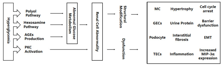
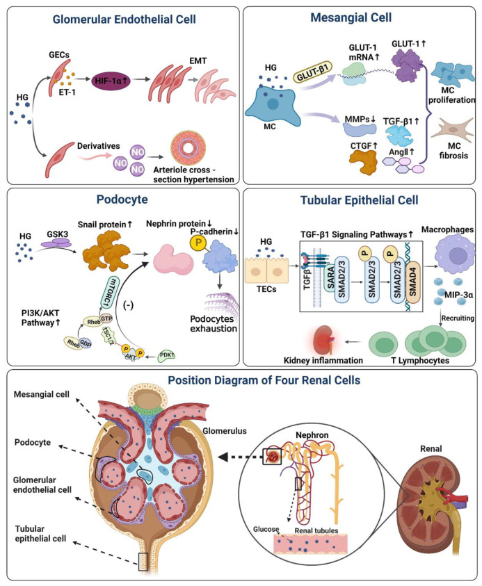
Diabetic Nephropathy (DN) is a serious complication of type I and II diabetes. It develops from the initial microproteinuria to end-stage renal failure. The main initiator for DN is chronic hyperglycemia. Hyperglycemia (HG) can stimulate the resident and non-resident renal cells to produce humoral mediators and cytokines that can lead to functional and phenotypic changes in renal cells and tissues, interference with cell growth, interacting proteins, advanced glycation end products (AGEs), etc., ultimately resulting in glomerular and tubular damage and the onset of kidney disease. Therefore, poor blood glucose control is a particularly important risk factor for the development of DN. In this paper, the types and mechanisms of DN cell damage are classified and summarized by reviewing the related literature concerning the effect of hyperglycemia on the development of DN. At the cellular level, we summarize the mechanisms and effects of renal damage by hyperglycemia. This is expected to provide therapeutic ideas and inspiration for further studies on the treatment of patients with DN.

Keywords: diabetic nephropathy; hyperglycemia; mechanism; renal cell injury.


Conflict of interest statement

The authors declare no conflict of interest.

Figures

Figure 1
Figure 1
Hyperglycemia interferes with the activation of the polyol pathway, hexosamine pathway, AGEs production, and PKC, leading to abnormal glucose metabolism. Abnormal glucose metabolism accelerates renal structural modification and dysfunction, as well as kidney damage to MCs, GECs, adipocytes, and TECs. AGEs, advanced glycation end products; PKC, protein kinase C; MC, mesangial cell; GECs, glomerular endothelial cells; TECs, tubular epithelial cells; EMT, endothelial-interstitial transformation; MIP-3α, macrophage inflammatory protein-3α.
Figure 2
Figure 2
The effect and mechanism of high glucose on four renal cells in DN. Mesangial Cell Figure: Under the stimulation of a high glucose environment, it will induce MC to produce a large amount of GLUT-1 protein, while high sugar will affect the activity of MMPs, and induce the production of TGF-β1, Ang II, and CTGF. Finally, it will aggravate MC proliferation, sclerosis, and accelerate renal lesions. Glomerular Endothelial Cells figure: Hyperglycemia induces ET-1 production in GECs, and ET-1 mediates high glucose-induced EMT in GECs under the regulation of HIF-1α. glucose-mediated NO loss can directly cause arteriole cross-section hypertension and aggravate renal microvascular injury. Podocyte Figure: Reduced GSK3 activity in damaged podocytes leads to increased expression of Snail in podocytes, and upregulated Snail suppresses protein expression of P-calmodulin and nephrin, finally causing podocyte depletion. Activation of PI3K/AKT signaling pathway in podocytes by high glucose likewise inhibits protein expression of podocalyxin and nephrin, which on also induces podocyte phenotypic transformation. Tubular Epithelial Cells Figure: Exposure of TECs to HG levels increased macrophage inflammatory protein-3α (MIP-3α) expression through a TGF-β1-dependent pathway, recruiting memory T lymphocytes and inducing renal inflammation. HG, hyperglycemia; GLUT-1, glucose transporter-1; MMPs, matrix metalloproteinases; TGF-β1, transforming growth factor; CTGF, connective tissue growth factor; AngII, circulating angiotensin II; HIF-1α, hypoxia-inducible factor-1α; ET-1, endothelin-1; EMT, endothelial-interstitial transformation; GSK3, glycogen synthase kinase-3; PDK1, 3-phosphoinositide-dependent protein kinase-1; AKT, protein kinases B; mTOR, mammalian target of rapamycin; TSC1/2, tuberous sclerosis1/2; MIP-3α, macrophage inflammatory protein-3α; (−), negative Feedback; ↑, up-regulation; ↓, down-regulation.

References

    1. Thomas S., Karalliedde J. Diabetic nephropathy. Medicine. 2015;43:20–25. doi: 10.1016/j.mpmed.2014.10.007. - DOI
    1. Rask-Madsen C., King G.L. Vascular complications of diabetes: Mechanisms of injury and protective factors. Cell Metab. 2013;17:20–33. doi: 10.1016/j.cmet.2012.11.012. - DOI - PMC - PubMed
    1. Schrijvers B.F., De Vriese A.S., Flyvbjerg A. From hyperglycemia to diabetic kidney disease: The role of metabolic, hemodynamic, intracellular factors and growth factors/cytokines. Endocr. Rev. 2004;25:971–1010. doi: 10.1210/er.2003-0018. - DOI - PubMed
    1. Tang G., Li S., Zhang C., Chen H., Wang N., Feng Y. Clinical efficacies, underlying mechanisms and molecular targets of Chinese medicines for diabetic nephropathy treatment and management. Acta Pharm. Sin. B. 2021;11:2749–2767. doi: 10.1016/j.apsb.2020.12.020. - DOI - PMC - PubMed
    1. Tervaert T.W., Mooyaart A.L., Amann K., Cohen A.H., Cook H.T., Drachenberg C.B., Ferrario F., Fogo A.B., Haas M., de Heer E., et al. Pathologic classification of diabetic nephropathy. J. Am. Soc. Nephrol. 2010;21:556–563. doi: 10.1681/ASN.2010010010. - DOI - PubMed

LinkOut - more resources