

Femoral Anteversion Measured by the Surgical Transepicondylar Axis Is Correlated with the Tibial Tubercle-Roman Arch Distance in Patients with Lateral Patellar Dislocation
- PMID: 36837583
- PMCID: PMC9959396
- DOI: 10.3390/medicina59020382
Femoral Anteversion Measured by the Surgical Transepicondylar Axis Is Correlated with the Tibial Tubercle-Roman Arch Distance in Patients with Lateral Patellar Dislocation
Abstract
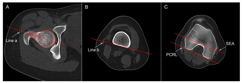
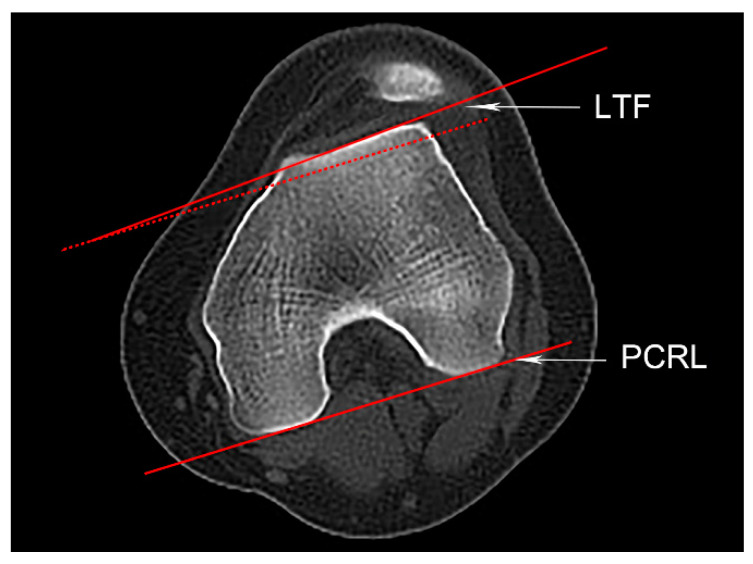
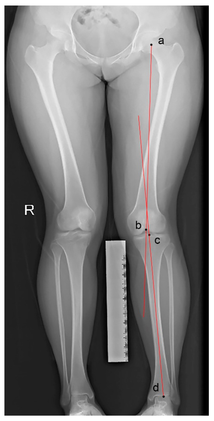
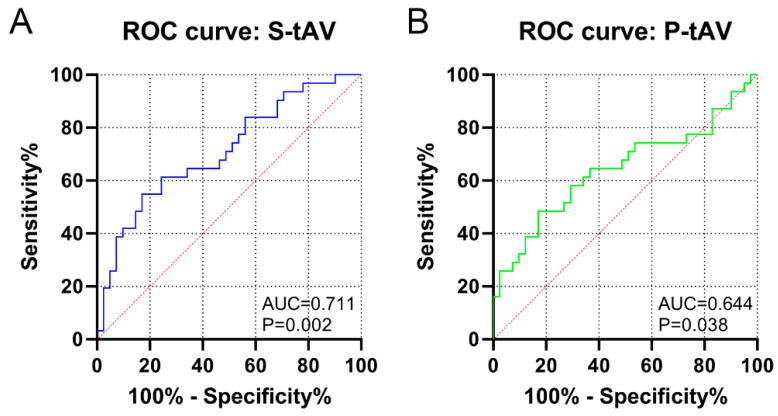
Background and Objectives: Various predisposing factors for lateral patellar dislocation (LPD) have been identified, but the relation between femoral rotational deformity and the tibial tubercle-Roman arch (TT-RA) distance remains elusive. Materials and Methods: We conducted this study including 72 consecutive patients with unilateral LPD. Femoral anteversion was measured by the surgical transepicondylar axis (S-tAV), and the posterior condylar reference line (P-tAV), TT-RA distance, trochlear dysplasia, knee joint rotation, patellar height, and hip-knee-ankle angle were measured by CT images or by radiographs. The correlations among these parameters were analyzed, and the parameters were compared between patients with and without a pathological TT-RA distance. Binary regression analysis was performed, and receiver operating characteristic curves were obtained. Results: The TT-RA distance was correlated with S-tAV (r = 0.360, p = 0.002), but the correlation between P-tAV and the TT-RA distance was not significant. S-tAV had an AUC of 0.711 for predicting a pathological TT-RA, with a value of >18.6° indicating 54.8% sensitivity and 82.9% specificity. S-tAV revealed an OR of 1.13 (95% CI [1.04, 1.22], p = 0.003) with regard to the pathological TT-RA distance by an adjusted regression model. Conclusions: S-tAV was significantly correlated with the TT-RA distance, with a correlation coefficient of 0.360, and was identified as an independent risk factor for a pathological TT-RA distance. However, the TT-RA distance was found to be independent of P-tAV.
Keywords: TT-RA distance; femoral anteversion; patellar dislocation; surgical transepicondylar axis; tibial tubercle osteotomy.
Conflict of interest statement
The authors declare no conflict of interest.
Figures


Similar articles
-
Femoral anteversion angle is more advantageous than TT-TG distance in evaluating patellar dislocation: A retrospective cohort study.Knee Surg Sports Traumatol Arthrosc. 2025 May;33(5):1721-1727. doi: 10.1002/ksa.12475. Epub 2024 Sep 18. Knee Surg Sports Traumatol Arthrosc. 2025. PMID: 39290196
-
Tibial tubercle-Roman arch (TT-RA) distance is superior to tibial tubercle-trochlear groove (TT-TG) distance when evaluating coronal malalignment in patients with knee osteoarthritis.Eur Radiol. 2022 Dec;32(12):8404-8413. doi: 10.1007/s00330-022-08924-y. Epub 2022 Jun 22. Eur Radiol. 2022. PMID: 35729426
-
CT and MRI measurements of tibial tubercle lateralization in patients with patellar dislocation were not equivalent but could be interchangeable.Knee Surg Sports Traumatol Arthrosc. 2023 Jan;31(1):349-357. doi: 10.1007/s00167-022-07119-8. Epub 2022 Sep 11. Knee Surg Sports Traumatol Arthrosc. 2023. PMID: 36088618
-
Current evidence advocates use of a new pathologic tibial tubercle-posterior cruciate ligament distance threshold in patients with patellar instability.Knee Surg Sports Traumatol Arthrosc. 2018 Sep;26(9):2733-2742. doi: 10.1007/s00167-017-4716-2. Epub 2017 Sep 16. Knee Surg Sports Traumatol Arthrosc. 2018. PMID: 28918500
-
Tibial Tubercle Osteotomy May Not Provide Additional Benefit in Treating Patellar Dislocation With Increased Tibial Tuberosity-Trochlear Groove Distance: A Systematic Review.Arthroscopy. 2021 May;37(5):1670-1679.e1. doi: 10.1016/j.arthro.2020.12.210. Epub 2020 Dec 24. Arthroscopy. 2021. PMID: 33359817
References
-
- Song Y.F., Wang H.J., Yan X., Yuan F.Z., Xu B.B., Chen Y.R., Ye J., Fan B.S., Yu J.K. Tibial Tubercle Osteotomy May not Provide Additional Benefit in Treating Patellar Dislocation with Increased Tibial Tuberosity-Trochlear Groove Distance: A Systematic Review. Arthroscopy. 2021;37:1670–1679.e1671. doi: 10.1016/j.arthro.2020.12.210. - DOI - PubMed
MeSH terms
LinkOut - more resources
Full Text Sources
