. 2023 Jan 24;13(2):151.
doi: 10.3390/membranes13020151.

Odor Discrimination by Lipid Membranes

Affiliations

Odor Discrimination by Lipid Membranes

Troy W Lowry et al. Membranes (Basel). .

Abstract

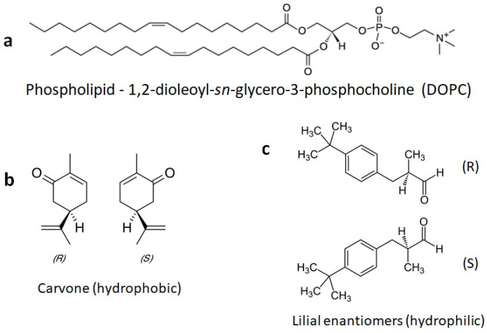
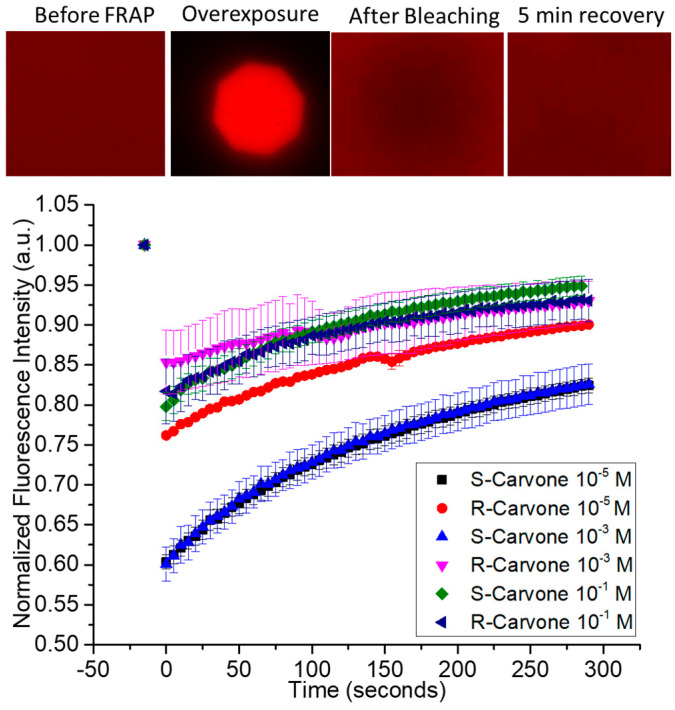
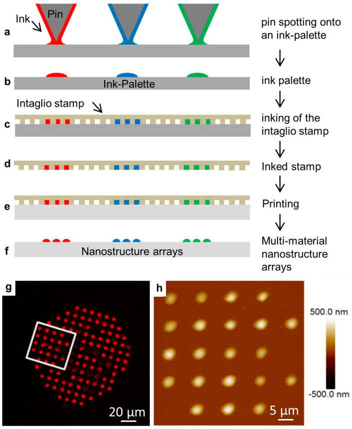
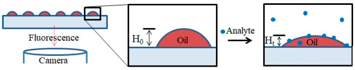
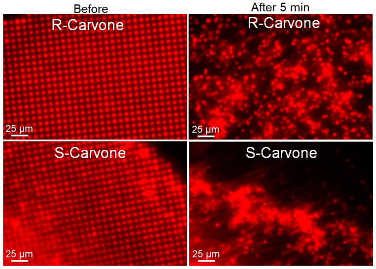
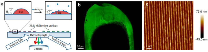
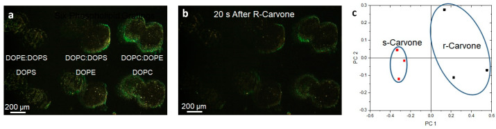
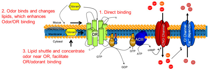
Odor detection and discrimination in mammals is known to be initiated by membrane-bound G-protein-coupled receptors (GPCRs). The role that the lipid membrane may play in odor discrimination, however, is less well understood. Here, we used model membrane systems to test the hypothesis that phospholipid bilayer membranes may be capable of odor discrimination. The effect of S-carvone, R-carvone, and racemic lilial on the model membrane systems was investigated. The odorants were found to affect the fluidity of supported lipid bilayers as measured by fluorescence recovery after photobleaching (FRAP). The effect of odorants on surface-supported lipid multilayer microarrays of different dimensions was also investigated. The lipid multilayer micro- and nanostructure was highly sensitive to exposure to these odorants. Fluorescently-labeled lipid multilayer droplets of 5-micron diameter were more responsive to these odorants than ethanol controls. Arrays of lipid multilayer diffraction gratings distinguished S-carvone from R-carvone in an artificial nose assay. Our results suggest that lipid bilayer membranes may play a role in odorant discrimination and molecular recognition in general.

Keywords: biosensor; droplet; enantioselectivity; lipid; lithography; microarray; nanointaglio; nose; odorant.


Conflict of interest statement

The authors declare no conflict of interest.

Figures

Figure 1
Figure 1
Chemical structures of a phospholipid and the odorants used here. (a) The phospholipid DOPC is commonly used to form fluid model membranes in water at room temperature. (b) The odorants S-carvone and R-carvone. (c) The odorant lilial, which was used here as a racemic mixture.
Figure 2
Figure 2
Fluorescence recovery after photobleaching of supported lipid bilayers upon exposure to enantiomeric odorants S-carvone and R-carvone. Top—examples of data from the FRAP experiment. A fluorescently labeled supported lipid bilayer was exposed to high-intensity light through an aperture to bleach a circular area within the spot. The fluorescence intensity of the bleached spot was measured as lipids diffused into it from the unbleached region. Represented are FRAP data from the two enantiomers, measured at 3 different concentrations.
Figure 3
Figure 3
The nanointaglio printing process. (af) Schematic illustration of the nanointaglio process. (a,b) An ink palette is prepared by spotting inks onto surface. (c,d) A microstructured polymer stamp is inked by placing the stamp in contact with the palette. (e) The inked stamp is used to print from the recesses of the stamp. Initial prints are discarded as excess ink is removed from the surface of the stamp until intaglio printing occurs. (f) The printed droplet arrays. (g) Fluorescence micrograph of a printed droplet array. (h) Atomic force micrograph of the area outlined in (g).
Figure 4
Figure 4
Schematic illustration of a lipid multilayer fluorescence assay. Fluorescently-labeled arrays of lipid droplets on a surface can be observed with a fluorescence microscope. Upon exposure to an analyte, the change in intensity or structure of the lipid arrays is monitored.
Figure 5
Figure 5
Lipid multilayer fluorescence assay upon exposure to R-carvone and S-carvone. Both enantiomers were found to significantly disrupt the lipid multilayer structures. Lipid dots exposed to 1 mM R-carvone demonstrated spreading and dissolution, while lipid multilayers exposed to 1 mM S-carvone tended to demonstrate more tension and remodeling effects.
Figure 6
Figure 6
Lipid multilayer gratings. (a) Schematic showing the assay of exposing lipid multilayer gratings to an analyte while monitoring the diffraction from the arrays. (b) An example of an optical image of a DOPC-based lipid multilayer grating taken according to the setup shown in (a). (c) AFM image of a DOPC lipid multilayer grating.
Figure 7
Figure 7
Lipid multilayer gratings respond to carvone vapors with enantiomer dependence. (a) Lipid multilayer gratings of DOPC, DOPE, DOPS, 1:1 DOPC:DOPE, 1:1 DOPC:DOPS, and 1:1 DOPE:DOPS were fabricated on a polystyrene surface. The sample was illuminated by white light from an angle and an image was taken of the light diffracted from the gratings as illustrated in Figure 5. (b) The gratings were then exposed to carvone vapors at saturation. (c) Utilizing three different experiments for each enantiomer, clustering was observed using multivariable analysis using 20 to 40 s time point data to construct the response of the gratings in PCA space.
Figure 8
Figure 8
Possibilities for how the lipid bilayer membrane could assist in the molecular binding of odorants. Schematic depicting the odorant receptor (OR) G-protein-coupled receptor cascade and the three possibilities for molecular triggering of the cascade. 1. Direct binding of the odorant to the OR. 2. Lipid reorganization from odorants could provide access for the odorant to arrive in the binding site of the OR. 3. Lipids shuttle and concentrate odorants near the OR to facilitate OR/odorant binding. Golf = G-protein olfaction, ACIII = Adenyl cyclase 3, CNG = cyclic nucleotide.

References

    1. Buck L., Axel R. A novel multigene family may encode odorant receptors: A molecular basis for odor recognition. Cell. 1991;65:175–187. doi: 10.1016/0092-8674(91)90418-X. - DOI - PubMed
    1. Firestein S., Darrow B., Shepherd G.M. Activation of the sensory current in salamander olfactory receptor neurons depends on a G protein-mediated cAMP second messenger system. Neuron. 1991;6:825–835. doi: 10.1016/0896-6273(91)90178-3. - DOI - PubMed
    1. Jones D.T., Reed R.R. Golf: An olfactory neuron specific-G protein involved in odorant signal transduction. Science. 1989;244:790–795. doi: 10.1126/science.2499043. - DOI - PubMed
    1. Jones D.T., Masters S.B., Bourne H.R., Reed R.R. Biochemical characterization of three stimulatory GTP-binding proteins. The large and small forms of Gs and the olfactory-specific G-protein, Golf. J. Biol. Chem. 1990;265:2671–2676. doi: 10.1016/S0021-9258(19)39853-9. - DOI - PubMed
    1. Bruch R.C. Signal transducing GTP-binding proteins in olfaction. Comp. Biochem. Physiol. A-Physiol. 1990;95:27–29. doi: 10.1016/0300-9629(90)90005-D. - DOI - PubMed

LinkOut - more resources