Case Reports
. 2023 Jan 31;13(2):209.
doi: 10.3390/metabo13020209.

Mucopolysaccharidosis Type 1 among Children-Neuroradiological Perspective Based on Single Centre Experience and Literature Review

Affiliations
Case Reports

Mucopolysaccharidosis Type 1 among Children-Neuroradiological Perspective Based on Single Centre Experience and Literature Review

Magdalena Machnikowska-Sokołowska et al. Metabolites. .

Abstract

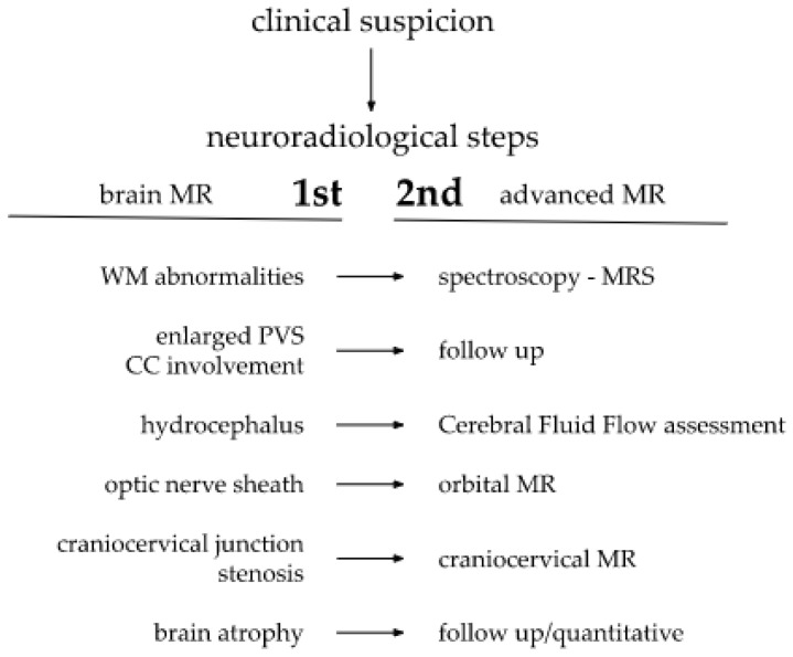
Mucopolysaccharidosis 1 (MPS 1) is a group of rare lysosomal genetic disorders resulting from the accumulation of undegraded glycosaminoglycans (GAGs) leading to multiorgan damage. Neurological symptoms vary from mild to severe. Neuroimaging-mainly magnetic resonance (MRI)-plays a crucial role in disease diagnosis and monitoring. Early diagnosis is of the utmost importance due to the necessity of an early therapy implementation. New imaging tools like MR spectroscopy (MRS), semiquantitative MRI analysis and applying scoring systems help substantially in MPS 1 surveillance. The presented analysis of neuroimaging manifestations is based on 5 children with MPS 1 and a literature review. The vigilance of the radiologist based on knowledge of neuroradiological patterns is highlighted.

Keywords: GAGs; Hurler disease; MPS 1; glycosaminoglycans; magnetic resonance imaging; mucopolysaccharidosis 1; neuroradiology.


Conflict of interest statement

The authors declare no conflict of interest.

Figures

Figure 1
Figure 1
Brain and cervical spine MRI. Patient 1. (younger sister) (a) axial T2; (b) sagittal T2 FLAIR, (c) sagittal T1; (d) sagittal spinal T2. Findings: delayed myelination, slight skiboot sella turcica shape; suspicion of dens hypoplasia—for observation (age 4 mo).
Figure 2
Figure 2
Brain MRI. Patient 2. (a,c,e) axial T2; (b,d) axial T2 FLAIR; (f) sagittal T1. Findings: (a,c,d) enlarged PVS with characteristic location in corpus callosum; (ad) abnormal white matter signals; (e) posterior fossa horns; (f) J-shaped sella turcica, dysplastic dens.
Figure 3
Figure 3
Brain MRI. Patient 3. (a,e,d) T2 axial; (f) T2 coronal; (b,c) T2 sagittal; (b) T1 contrast-enhanced; (c) T2 FLAIR. Findings: (a) delayed myelination; (b,c) J-shaped sella turcica, dysplastic dens; (d) temporal bone air cells effusion, posterior fossa horns; (e,f) enlarged optic nerve sheath.
Figure 4
Figure 4
Brain MRI. Patient 4, 1st exam. (ad) axial T1 & T2; (eh) sagittal T1, T2, Flair T2, T1. Findings: (a,c,g) enlarged periventricular spaces with characteristic location in corpus callosum; (a,c,g) abnormal white matter signals; (b) posterior fossa horns, mastoid cells fluid; (d) optic nerve sheath enlargement, ethmoid sinus fluid; (e,f,h) J-shaped sella turcica, dysplastic dens; (e,g) skull deformation.
Figure 5
Figure 5
Brain and cervical spine MRI. Patient 4, follow up: (a,b,h) axial T2; (c,d,g); sagittal brain (c) T1; (d,g) T2; (e,f) sagittal spine (e) T2, (f) T1. Findings: (ad) enlarged PVS with characteristic location in corpus callosum; (a,b,d,g) abnormal white matter signals;(e,f) vertebral spaces and bodies deformity, J-shaped sella turcica, dysplastic dens, (h) mastoid cells fluid, discrete posterior fossa horns, (ad,g) skull deformity and enlarged frontal subarachnoid spaces.
Figure 6
Figure 6
Brain and spine MRI. Patient 5. (a,b,f) axial T2; (c,d) sagittal brain; (c) T2 FLAIR; (d,g) T1; (e) sagittal spine; (e) T2; Findings: (a) enlarged PVS with characteristic location in corpus callosum; (b) optic nerve sheath enlargement; (c,d) flattened anterior border of sella turcica, dysplastic dens; (f) mastoid cells fluid; (e) vertebral and intervertebral discs deformation, cervical spine kyphosis and stenosis.
Scheme 1
Scheme 1
Proposed steps of MR neuroimaging.

References

    1. Barkovich A.J., Raybaud C. Pediatric Neuroimaging. Wolters Kluwer; Philadelphia, PA, USA: 2018.
    1. Scott H.S., Ashton L.J., Eyre H.J., Baker E., Brooks D.A., Callen D.F., Sutherland G.R., Morris C.P., Hopwood J.J. Chromosomal localization of the human alpha-L-iduronidase gene (IDUA) to 4p16.3. Am. J. Hum. Genet. 1990;47:802–807. - PMC - PubMed
    1. Kubaski F., Poswar F.D.O., Michelin-Tirelli K., Burin M.G., Rojas-Málaga D., Brusius-Facchin A.C., Leistner-Segal S., Giugliani R. Diagnosis of Mucopolysaccharidoses. Diagnostics. 2020;10:172. doi: 10.3390/diagnostics10030172. - DOI - PMC - PubMed
    1. Jurecka A., Ługowska A., Golda A., Czartoryska B., Tylki-Szymańska A. Prevalence rates of mucopolysaccharidoses in Poland. J. Appl. Genet. 2014;56:205–210. doi: 10.1007/s13353-014-0262-5. - DOI - PMC - PubMed
    1. Khan S.A., Peracha H., Ballhausen D., Wiesbauer A., Rohrbach M., Gautschi M., Mason R.W., Giugliani R., Suzuki Y., Orii K.E., et al. Epidemiology of mucopolysaccharidoses. Mol. Genet. Metab. 2017;121:227–240. doi: 10.1016/j.ymgme.2017.05.016. - DOI - PMC - PubMed

Publication types

LinkOut - more resources