. 2023 Feb 14;13(2):275.
doi: 10.3390/metabo13020275.

Metabolomics-Based Profiling via a Chemometric Approach to Investigate the Antidiabetic Property of Different Parts and Origins of Pistacia lentiscus L

Affiliations

Metabolomics-Based Profiling via a Chemometric Approach to Investigate the Antidiabetic Property of Different Parts and Origins of Pistacia lentiscus L

Chabha Sehaki et al. Metabolites. .

Abstract

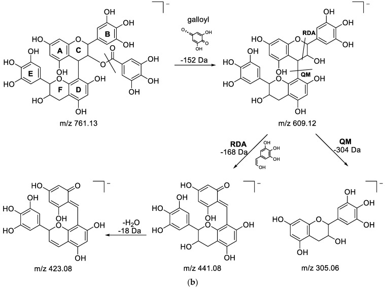
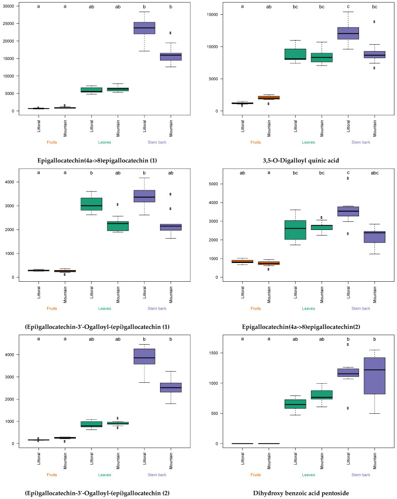
Pistacia lentiscus L. is a medicinal plant that grows spontaneously throughout the Mediterranean basin and is traditionally used to treat diseases, including diabetes. The aim of this work consists of the evaluation of the α-glucosidase inhibitory effect (i.e., antidiabetic activity in vitro) of different extracts from the leaves, stem barks and fruits of P. lentiscus harvested on mountains and the littoral of Tizi-Ouzou in Algeria. Metabolomic profiling combined with a chemometric approach highlighted the variation of the antidiabetic properties of P. lentiscus according to the plant's part and origin. A multiblock OPLS analysis showed that the metabolites most involved in α-glucosidase inhibition activity were mainly found in the stem bark extracts. The highest inhibitory activity was found for the stem bark extracts, with averaged inhibition percentage values of 84.7% and 69.9% for the harvested samples from the littoral and mountain, respectively. On the other hand, the fruit extracts showed a lower effect (13.6%) at both locations. The UHPLC-ESI-HRMS characterization of the metabolites most likely responsible for the α-glucosidase-inhibitory activity allowed the identification of six compounds: epigallocatechin(4a>8)epigallocatechin (two isomers), (epi)gallocatechin-3'-O-galloyl-(epi)gallocatechin (two isomers), 3,5-O-digalloylquinic acid and dihydroxy benzoic acid pentoside.

Keywords: P. lentiscus; UHPLC-ESI-HRMS; antidiabetic; metabolomic approach; phytochemical profiling; α-glucosidase.


Conflict of interest statement

The authors declare no conflict of interest. The funders had no role in the design of the study; in the collection, analyses, or interpretation of data; in the writing of the manuscript; or in the decision to publish the results.

Figures

Figure 1
Figure 1
Boxplot showing α-glucosidase-inhibitory effect of lentisk leaves, stems barks and fruits from the mountain and littoral. (The means and medians are presented as black dots in the center of each box plot and by bars, respectively. The significance of the α-glucosidase inhibition variation is represented by letters (groups, a, b and ab) according to pairwise comparison through the Kruskal–Wallis test adjusted with Bonferroni correction).
Figure 2
Figure 2
Multiblock OPLS analysis performed on the UHPLC-ESI-HRMS and α-glucosidase inhibition data: (A) score plot—lentisk samples: ▪ Littoral/Δ Mountain; (B) loading plot—metabolites detected: ▪ ions with negative ionization/Δ ions with positive ionization: metabolites; M1: epigallocatechin(4a->8)epigallocatechin (1); M2: 3,5-O-digalloylquinic acid; M3: (epi)gallocatechin-3′-O-galloyl-(epi)gallocatechin (1); M4: epigallocatechin(4a->8)epigallocatechin (2); M5: (epi)gallocatechin-3′-O-galloyl-(epi)gallocatechin (2); M6:dihydroxy benzoic acid pentoside.
Figure 3
Figure 3
Proposed fragmentation pathways for epigallocatechin(4a->8)epigallocatechin.
Figure 4
Figure 4
(a) Fragmentation pathways of (epi)gallocatechin-3′-O-galloyl-(epi)gallocatechin showing the formation of the fragmentations at m/z 609.12,591.11 and 465.08. (b) Fragmentation pathways of (epi)gallocatechin-3′-O-galloyl-(epi)gallocatechin showing the formation of the fragment ions at m/z 423.08 and 305.06.
Figure 4
Figure 4
(a) Fragmentation pathways of (epi)gallocatechin-3′-O-galloyl-(epi)gallocatechin showing the formation of the fragmentations at m/z 609.12,591.11 and 465.08. (b) Fragmentation pathways of (epi)gallocatechin-3′-O-galloyl-(epi)gallocatechin showing the formation of the fragment ions at m/z 423.08 and 305.06.
Figure 5
Figure 5
Fragmentation pathway of 3,5-O-digalloylquinic acid.
Figure 6
Figure 6
Fragmentation pathways of dihydroxy benzoic acid pentoside.
Figure 7
Figure 7
Box plots of the metabolite contents in different organs—leaves, stem barks and fruits—from the mountain and littoral. (The significance of the metabolite intensity variation is presented by letters (groups, a, b, c, ab, bc and abc) according to pairwise comparison through the Kruskal-Wallis test adjusted with Bonferroni correction).

References

    1. Benalla W., Bellahcen S., Bnouham M. Antidiabetic medicinal plants as a source of alpha glucosidase inhibitors. Curr. Diabetes Rev. 2010;6:247–254. doi: 10.2174/157339910791658826. - DOI - PubMed
    1. Tundis R., Loizzo M.R., Menichini F. Natural products as α-amylase and α-glucosidase inhibitors and their hypoglycaemic potential in the treatment of diabetes: An update. Mini Rev. Med. Chem. 2010;10:315–331. doi: 10.2174/138955710791331007. - DOI - PubMed
    1. Rees D.A., Alcolado J.C. Animal models of diabetes mellitus. Diabet. Med. 2005;22:359–370. doi: 10.1111/j.1464-5491.2005.01499.x. - DOI - PubMed
    1. Sudesna C., Khunti K., Davies M.J. Type 2 diabetes. Lancet. 2017;389:2239–2251. - PubMed
    1. Vinholes J., Vizzotto M. Synergisms in alpha-glucosidase inhibition and antioxidant activity of camellia sinensis l. kuntze and Eugeniauniflora L. ethanolic extracts. Pharmacogn. Res. 2017;9:101. doi: 10.4103/0974-8490.197797. - DOI - PMC - PubMed

LinkOut - more resources