Review
. 2023 Feb 18;14(2):472.
doi: 10.3390/mi14020472.

Strategies for the Voltammetric Detection of Loop-Mediated Isothermal Amplification

Affiliations
Review

Strategies for the Voltammetric Detection of Loop-Mediated Isothermal Amplification

Jesse M Marangoni et al. Micromachines (Basel). .

Abstract

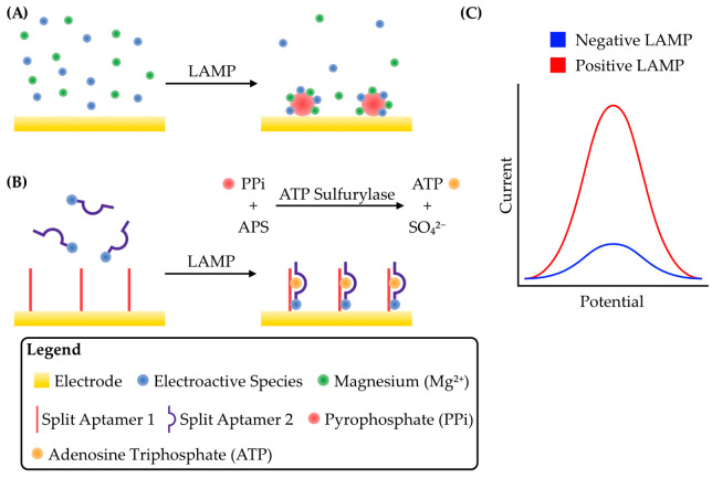
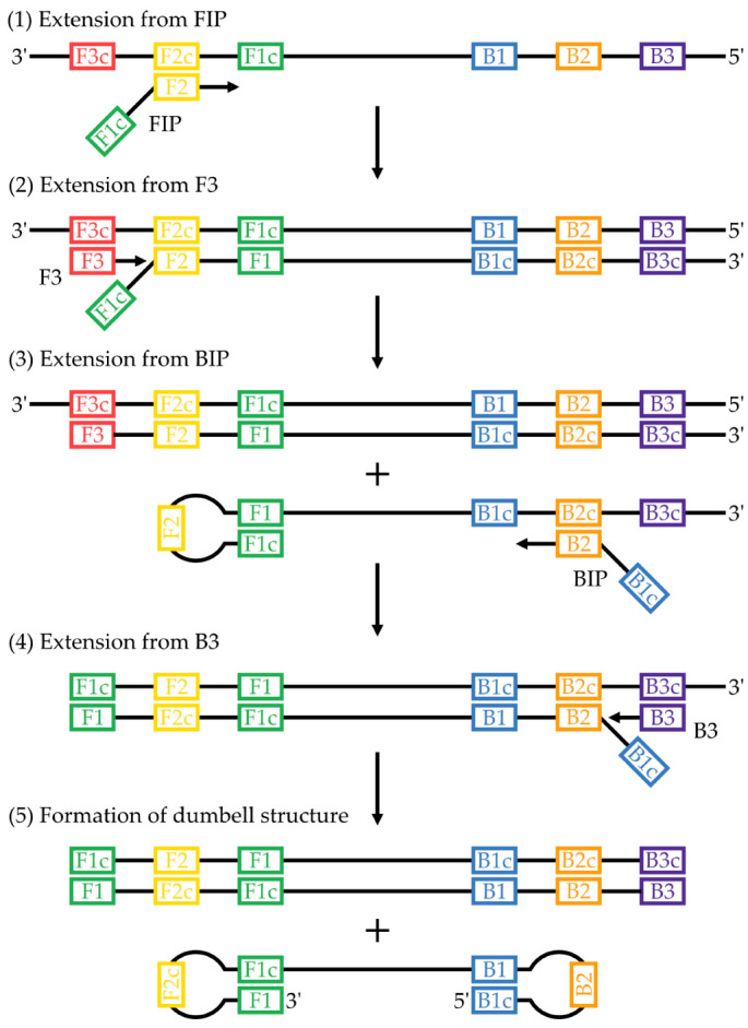
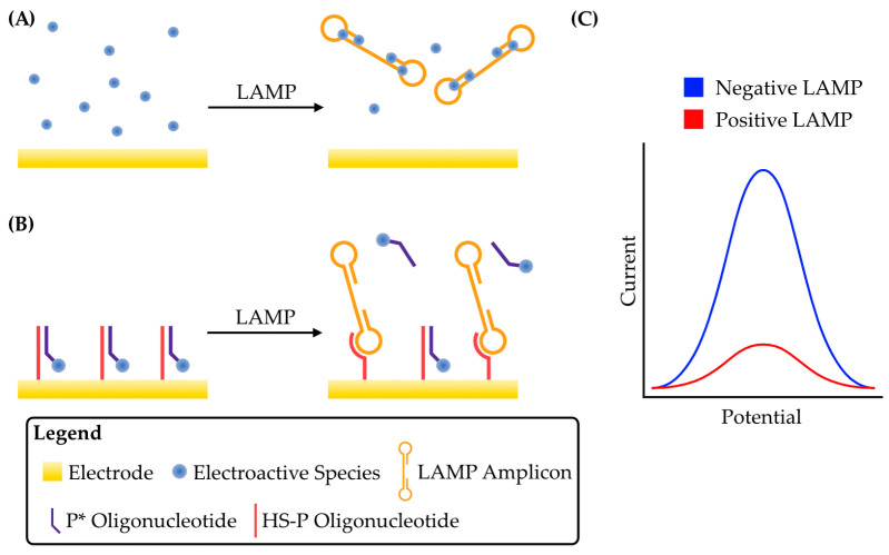
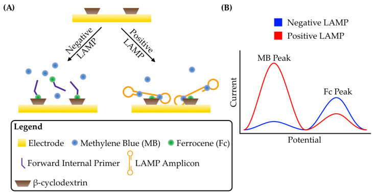
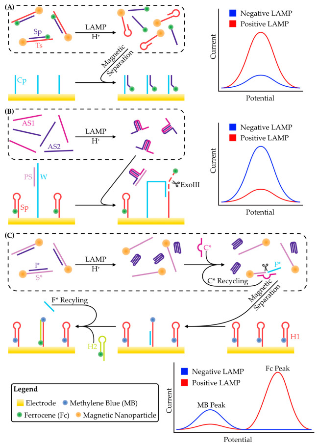
Loop-mediated isothermal amplification (LAMP) is rapidly developing into an important tool for the point-of-use detection of pathogens for both clinical and environmental samples, largely due to its sensitivity, rapidity, and adaptability to portable devices. Many methods are used to monitor LAMP, but not all are amenable to point-of-use applications. Common methods such as fluorescence often require bulky equipment, whereas colorimetric and turbidimetric methods can lack sensitivity. Electrochemical biosensors are becoming increasingly important for these applications due to their potential for low cost, high sensitivity, and capacity for miniaturization into integrated devices. This review provides an overview of the use of voltammetric sensors for monitoring LAMP, with a specific focus on how electroactive species are used to interface between the biochemical products of the LAMP reaction and the voltammetric sensor. Various strategies for the voltammetric detection of DNA amplicons as well as pyrophosphate and protons released during LAMP are presented, ranging from direct DNA binding by electroactive species to the creative use of pyrophosphate-detecting aptamers and pH-sensitive oligonucleotide structures. Hurdles for adapting these devices to point-of-use applications are also discussed.

Keywords: electrochemical biosensors; loop-mediated isothermal amplification; voltammetry.


Conflict of interest statement

The authors declare no conflict of interest.

Figures

Figure 4
Figure 4
Schematic strategies for the detection of pyrophosphate produced through DNA replication during LAMP. (A) Before LAMP, the electroactive species RuHex diffuses freely. Mg2+, a cofactor for DNA polymerases, is also present. As LAMP proceeds, RuHex coprecipitates onto the electrode surface with Mg2+ and the pyrophosphate produced from DNA polymerization, concentrating RuHex on the electrode and greatly increasing the peak faradaic current. Adapted from [72]. (B) An ATP-binding aptamer is split in two, with one half (Split Aptamer 1) conjugated to the electrode while the other (Split Aptamer 2), conjugated to an electroactive species, is free in solution. The enzyme ATP sulfurylase catalyzes a reaction between the pyrophosphate (PPi) produced during LAMP and adenosine phosphosulfate (APS) to produce ATP and sulfate (SO42−). The ATP brings the split aptamer together, concentrating the electroactive species on the electrode and producing a large peak current. Adapted from [75]. (C) Schematic of a typical voltammogram for these detection strategies. Both strategies result in a concentration of an electroactive species at the electrode surface, either by coprecipitation in A or by aptamer formation in B. The net result in both cases is an increase in peak faradaic current during voltammetry.
Figure 1
Figure 1
Mechanism of LAMP up to the formation of the dumbbell structure. (1) The F2 region of primer FIP hybridizes to the F2 region of the template, and replication is initiated. This introduces a region of self-complementarity in the newly synthesized strand. (2) Extension from primer F3 displaces the previously synthesized strand. (3) B2 of BIP hybridizes with B2c of the displaced strand and replication occurs. (4) Extension from B3 displaces the previously synthesized strand. (4) The self-complementary regions form a self-priming dumbbell structure that feeds into the cycling and elongation phases of LAMP. Figure is adapted from [22,23], which also provide a detailed explanation of the entire LAMP mechanism.
Figure 2
Figure 2
Schematic strategies for the detection of LAMP amplicons by sequestration of an electroactive species from the electrode surface. (A) Before LAMP, an electroactive species such as methylene blue or Hoechst 33258 is free to diffuse to the electrode surface, producing a relatively large faradaic current. As LAMP proceeds and amplicons are produced, the electroactive species binds to DNA, reducing its effective concentration and leading to a reduction in peak current. (B) An electrode functionalized with an oligonucleotide probe, HS-P. Before LAMP, a complementary oligonucleotide, P*, conjugated to an electroactive species is free to bind HS-P. This concentrates the electroactive species at the electrode surface and gives a large peak faradaic current. As LAMP amplicons are produced, they compete with P* for binding to HS-P and displace them from the surface, reducing the peak current. Adapted from [40]. (C) Schematic of a typical voltammogram for both detection strategies. In both cases illustrated in A and B, the net effect is sequestration of the electroactive species from the electrode, and therefore the peak current generated during voltammetry is reduced.
Figure 3
Figure 3
Schematic strategy for the detection of LAMP amplicons by concentrating electroactive species near the electrode surface. (A) An electrode is functionalized with β-cyclodextrin. The forward internal primer (FIP) of LAMP is conjugated to ferrocene such that it can be incorporated into the LAMP amplicons. Ferrocene can also bind to β-cyclodextrin. In a negative reaction, Fc-FIP will bind to β-cyclodextrin, producing a relatively large peak faradaic current. Methylene blue in solution will also produce a faradaic current at a different potential. In a positive LAMP reaction, Fc-FIP tethers entire amplicons to the electrode surface. The dsDNA is bound by methylene blue and produces a large ratiometric current between ferrocene and methylene blue. Adapted from [59]. (B) Schematic of a typical voltammogram for this detection strategy. Two peaks are observed at different potentials due to the presence of two electroactive species. While ferrocene is brought close to the electrode through interactions with β-cyclodextrin in both positive and negative LAMP reactions, the incorporation of free Fc-labelled primers into LAMP amplicons reduces their diffusivity and therefore the peak faradaic current decreases. For methylene blue, intercalation into LAMP amplicons effectively concentrates it at the electrode surface and results in an increased peak faradaic current.
Figure 5
Figure 5
Schematic strategies for the detection of protons produced through DNA replication during LAMP. (A) An oligonucleotide, Ts, is conjugated to a magnetic nanoparticle. Another oligonucleotide, Sp, is conjugated to ferrocene and hybridized to Sp. This duplex is pH sensitive, and protons produced during LAMP cause it to denature. Ts is removed by magnetic separation and the purified Sp is hybridized to another oligonucleotide, Cp, that is functionalized to the electrode. This concentrates the Sp-bound ferrocene at the electrode surface and produces a large peak faradaic current, as shown in the schematic voltammogram. Adapted from [78]. (B) The electrode is functionalized with two oligonucleotides: W and Sp. Sp is conjugated to ferrocene and forms a hairpin structure, bringing ferrocene in close contact with the electrode. W is blocked by another oligonucleotide, Ps. Protons produced during LAMP cause two oligonucleotides (AS1 and AS2) to form an i-motif. This i-motif has a region complementarity with Ps and causes its release from W via competitive binding. W can then interact with Sp, breaking the latter’s hairpin structure and making it susceptible to exonuclease III digestion. This releases ferrocene from the electrode and results in a decrease in peak current, as shown in the schematic voltammogram. W is then free to interact with more Sp, releasing more ferrocene, and amplifying the signal through a walking mechanism. Adapted from [80]. (C) An oligonucleotide, S*, is conjugated to a magnetic nanoparticle. Hybridized to S* is another oligonucleotide, I*. Acidification of the solution following LAMP causes I* to form an intramolecular i-motif which favours its release from S*. This allows another oligonucleotide, C*, to hybridize to S*. Together, this duplex forms an autocatalytic deoxyribozyme that releases a short segment of S*, called F*. F* is purified by magnetic separation of S*. An electrode has been functionalized with a hairpin forming oligonucleotide, H1, that is conjugated to methylene blue. The purified F* is applied to the electrode where it can hybridize with H1, breaking the hairpin and displacing methylene blue from the electrode surface. This reduces the peak faradaic current as shown in the schematic voltammogram. Breaking the H1 hairpin also enables another oligonucleotide, H2, which is conjugated to ferrocene, to displace F* and hybridize with H1. This brings ferrocene in close contact with the electrode, increasing its peak faradaic current. The released F* is then free to break more H1 hairpins, amplifying the signal. Adapted from [81].

Similar articles

Cited by

References

    1. Liu Y., Zhan L., Qin Z., Sackrison J., Bischof J.C. Ultrasensitive and Highly Specific Lateral Flow Assays for Point-of-Care Diagnosis. ACS Nano. 2021;15:3593–3611. doi: 10.1021/acsnano.0c10035. - DOI - PubMed
    1. Espy M.J., Uhl J.R., Sloan L.M., Buckwalter S.P., Jones M.F., Vetter E.A., Yao J.D.C., Wengenack N.L., Rosenblatt J.E., Cockerill F.R., et al. Real-Time PCR in Clinical Microbiology: Applications for Routine Laboratory Testing. Clin. Microbiol. Rev. 2006;19:165–256. doi: 10.1128/CMR.19.1.165-256.2006. - DOI - PMC - PubMed
    1. Choi J.R. Development of Point-of-Care Biosensors for COVID-19. Front. Chem. 2020;8:517. doi: 10.3389/fchem.2020.00517. - DOI - PMC - PubMed
    1. Parolo C., de la Escosura-Muñiz A., Merkoçi A. Enhanced Lateral Flow Immunoassay Using Gold Nanoparticles Loaded with Enzymes. Biosens. Bioelectron. 2013;40:412–416. doi: 10.1016/j.bios.2012.06.049. - DOI - PubMed
    1. Gong Y., Zheng Y., Jin B., You M., Wang J., Li X.J., Lin M., Xu F., Li F. A Portable and Universal Upconversion Nanoparticle-Based Lateral Flow Assay Platform for Point-of-Care Testing. Talanta. 2019;201:126–133. doi: 10.1016/j.talanta.2019.03.105. - DOI - PubMed

LinkOut - more resources