. 2023 Feb 4;11(2):401.
doi: 10.3390/microorganisms11020401.

Active Sulfate-Reducing Bacterial Community in the Camel Gut

Affiliations

Active Sulfate-Reducing Bacterial Community in the Camel Gut

Olga V Karnachuk et al. Microorganisms. .

Abstract

The diversity and activity of sulfate-reducing bacteria (SRB) in the camel gut remains largely unexplored. An abundant SRB community has been previously revealed in the feces of Bactrian camels (Camelus bactrianus). This study aims to combine the 16S rRNA gene profiling, sulfate reduction rate (SRR) measurement with a radioactive tracer, and targeted cultivation to shed light on SRB activity in the camel gut. Fresh feces of 55 domestic Bactrian camels grazing freely on semi-arid mountain pastures in the Kosh-Agach district of the Russian Altai area were analyzed. Feces were sampled in early winter at an ambient temperature of -15 °C, which prevented possible contamination. SRR values measured with a radioactive tracer in feces were relatively high and ranged from 0.018 to 0.168 nmol S cm-3 day-1. The 16S rRNA gene profiles revealed the presence of Gram-negative Desulfovibrionaceae and spore-forming Desulfotomaculaceae. Targeted isolation allowed us to obtain four pure culture isolates belonging to Desulfovibrio and Desulforamulus. An active SRB community may affect the iron and copper availability in the camel intestine due to metal ions precipitation in the form of sparingly soluble sulfides. The copper-iron sulfide, chalcopyrite (CuFeS2), was detected by X-ray diffraction in 36 out of 55 analyzed camel feces. In semi-arid areas, gypsum, like other evaporite sulfates, can be used as a solid-phase electron acceptor for sulfate reduction in the camel gastrointestinal tract.

Keywords: Desulforamulus; Desulfovibrio; biogenic chalcopyrite; camels; gut microbiota; sulfate-reduction.


Conflict of interest statement

The authors declare that there are no conflict of interest.

Figures
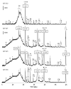
Figure 1
Figure 1
X-ray diffraction patterns of fecal samples KV152, KV147, KV145, and KV136. Letter codes: Ch = chalcopyrite, CuFeS2; Vl = villamaninite, CuS2; Gy = gypsum, CaSO4; Cc = calcite, CaCO3. The diagnostic peaks for muscovite (♦), clinochlore (■), quartz (◊), and albite (+) are indicated. The vertical bar shows the scale of relative counts.
Figure 2
Figure 2
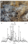
(A) Saline soil with camel feces, sampled for XRD analysis, and (B) X-ray diffraction pattern of the soil sample. Letter codes: Th = thenardite, Na2SO4; Me = melanterite, Fe+2SO4·7H2O. The diagnostic peaks for muscovite (■), clinochlore (●), calcite (□), quartz (◊), and halite (▼) are indicated. The vertical bar shows the scale of relative counts.
Figure 3
Figure 3
Microbial communities of camel feces at the phylum level and sulfate-reducing lineages (Desulfotomaculales and Desulfovibrionales). Relative abundances (% of the total 16S rRNA gene sequences, average of 55 samples) are shown after taxon names.
Figure 4
Figure 4
The sulfate reduction rate (SRR) measured in samples KV104, KV147, and KV 149. The vertical bars show the standard deviation.
Figure 5
Figure 5
16S rRNA gene-based neighbor-joining tree showing the phylogenetic position of strains 1211 and 1198. The percentage of replicate trees in which the associated taxa clustered together in the bootstrap test (1000 replicates) are shown next to the branches. The evolutionary distances were computed using the maximum composite likelihood method and are in the units of the number of base substitutions per site. All ambiguous positions were removed for each sequence pair (pairwise deletion option). There were a total of 1658 positions in the final dataset. Evolutionary analyses were conducted in MEGA11.
Figure 6
Figure 6
16S rRNA gene-based neighbor-joining tree showing the phylogenetic position of strains 1211 and 1198. The percentage of replicate trees in which the associated taxa clustered together in the bootstrap test (1000 replicates) are shown next to the branches. The evolutionary distances were computed using the maximum composite likelihood method and are in the units of the number of base substitutions per site. All ambiguous positions were removed for each sequence pair (pairwise deletion option). There were a total of 1590 positions in the final dataset. Evolutionary analyses were conducted in MEGA11.

References

    1. Gibson G.R., Macfarlane G.T., Cummings J.H. Occurrence of sulphate-reducing bacteria in human faeces and the relationship of dissimilatory sulphate reduction to methanogenesis in the large gut. J. Appl. Bacteriol. 1988;65:103–111. doi: 10.1111/j.1365-2672.1988.tb01498.x. - DOI - PubMed
    1. Deplancke B., Hristova K.R., Oakley H.A., McCracken V.J., Aminov R., Mackie R.I., Gaskins H.R. Molecular Ecological Analysis of the Succession and Diversity of Sulfate-Reducing Bacteria in the Mouse Gastrointestinal Tract. Appl. Environ. Microbiol. 2000;66:2166–2174. doi: 10.1128/AEM.66.5.2166-2174.2000. - DOI - PMC - PubMed
    1. Macfarlane G.T., Cummings J.H., Macfarlane S. Sulphate-reducing bacteria and the human large intestine. In: Barton L.L., Hamilton W.A., editors. Sulphate-Reducing Bacteria. Environmental and Engineered Systems. Cambridge University Press; Cambridge, UK: 2007. pp. 503–523.
    1. Rey F.E., Gonzalez M.D., Cheng J., Wu M., Ahern P.P., Gordon J.I. Metabolic niche of a prominent sulfate-reducing human gut bacterium. Proc. Natl. Acad. Sci. USA. 2013;110:13582–13587. doi: 10.1073/pnas.1312524110. - DOI - PMC - PubMed
    1. Barton L.L., Ritz N.L., Fauque G.D., Lin H.C. Sulfur Cycling and the Intestinal Microbiome. Dig. Dis. Sci. 2017;62:2241–2257. doi: 10.1007/s10620-017-4689-5. - DOI - PubMed

LinkOut - more resources