

Effects of Long-Term Intervention with Losartan, Aspirin and Atorvastatin on Vascular Remodeling in Juvenile Spontaneously Hypertensive Rats
- PMID: 36838830
- PMCID: PMC9965824
- DOI: 10.3390/molecules28041844
Effects of Long-Term Intervention with Losartan, Aspirin and Atorvastatin on Vascular Remodeling in Juvenile Spontaneously Hypertensive Rats
Abstract
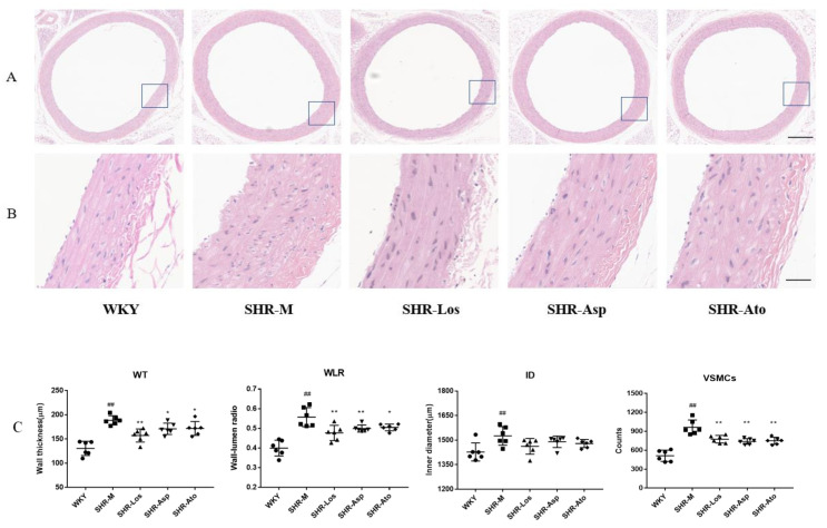
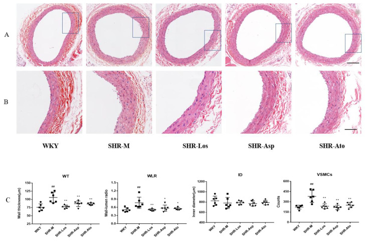
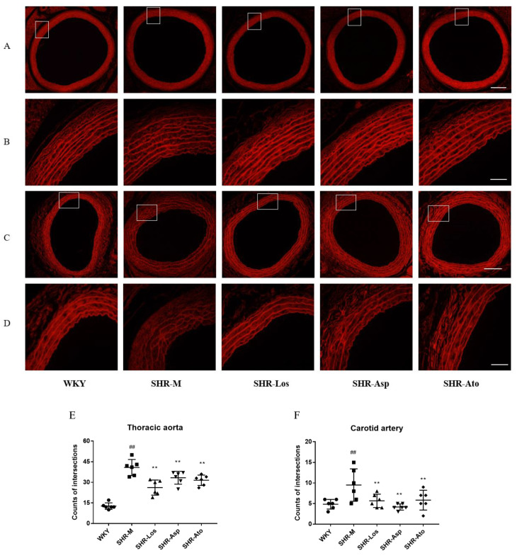
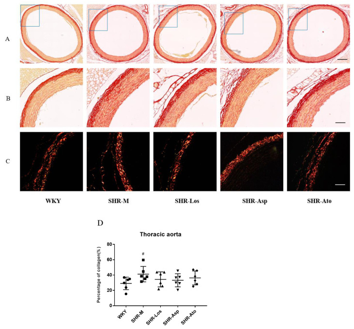
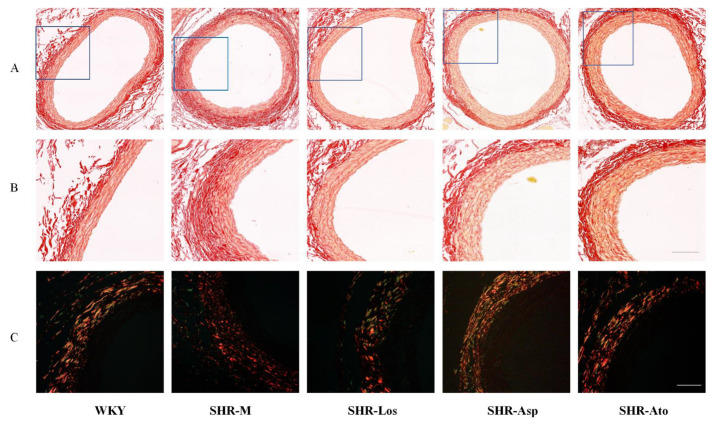
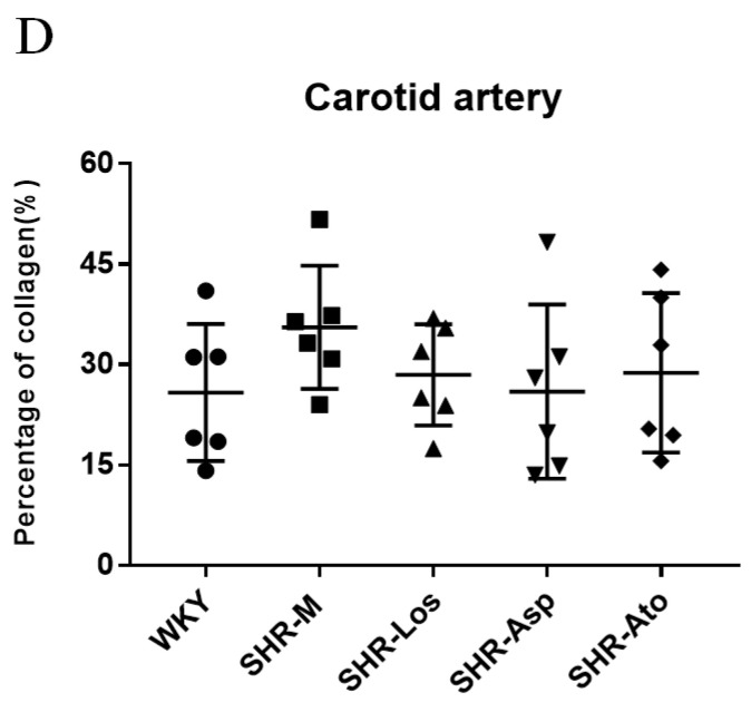
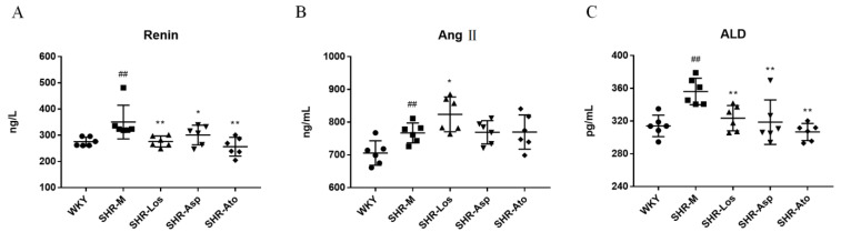
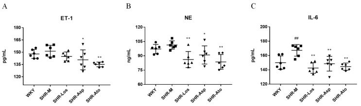
Hypertension in adolescents is associated with adverse cardiac and vascular events. In addition to lowering blood pressure, it is not clear whether pharmacological therapy in early life can improve vascular remodeling. This study aimed to evaluate the effects of long-term administration of losartan, aspirin, and atorvastatin on vascular remodeling in juvenile spontaneously hypertensive rats (SHRs). Losartan, aspirin, and atorvastatin were administered via gavage at doses of 20, 10, and 10 mg/kg/day, respectively, on SHRs aged 6-22 weeks. Paraffin sections of the blood vessels were stained with hematoxylin-eosin (H&E) and Sirius Red to evaluate the changes in the vascular structure and the accumulation of different types of collagen. The plasma levels of renin, angiotensin II (Ang II), aldosterone (ALD), endothelin-1 (ET-1), interleukin-6 (IL-6), and neutrophil elastase (NE) were determined using ELISA kits. After the 16-week treatment with losartan, aspirin, and atorvastatin, the wall thickness of the thoracic aorta and carotid artery decreased. The integrity of the elastic fibers in the tunica media was maintained in an orderly manner, and collagen deposition in the adventitia was retarded. The plasma levels of renin, ALD, ET-1, IL-6, and NE in the SHRs also decreased. These findings suggest that losartan, aspirin, and atorvastatin could improve vascular remodeling beyond their antihypertensive, anti-inflammatory, and lipid-lowering effects. Many aspects of the protection provided by pharmacological therapy are important for the prevention of cardiovascular diseases in adults and older adults.
Keywords: aspirin; atorvastatin; juvenile SHR; losartan; vascular remodeling.
Conflict of interest statement
The authors declare no conflict of interest.
Figures
Similar articles
-
Transient prehypertensive treatment in spontaneously hypertensive rats: a comparison of losartan and amlodipine regarding long-term blood pressure, cardiac and renal protection.Int J Mol Med. 2012 Dec;30(6):1376-86. doi: 10.3892/ijmm.2012.1153. Epub 2012 Oct 9. Int J Mol Med. 2012. PMID: 23064712
-
Effects of Losartan, Atorvastatin, and Aspirin on Blood Pressure and Gut Microbiota in Spontaneously Hypertensive Rats.Molecules. 2023 Jan 6;28(2):612. doi: 10.3390/molecules28020612. Molecules. 2023. PMID: 36677668 Free PMC article.
-
Protective effects of the angiotensin II type 1 (AT1) receptor blockade in low-renin deoxycorticosterone acetate (DOCA)-treated spontaneously hypertensive rats.Clin Sci (Lond). 2004 Mar;106(3):251-9. doi: 10.1042/CS20030299. Clin Sci (Lond). 2004. PMID: 14521506
-
Early treatment with losartan effectively ameliorates hypertension and improves vascular remodeling and function in a prehypertensive rat model.Life Sci. 2017 Mar 15;173:20-27. doi: 10.1016/j.lfs.2017.01.013. Epub 2017 Feb 1. Life Sci. 2017. PMID: 28161159
-
Temporary losartan or captopril in young SHR induces malignant hypertension despite initial normotension.Kidney Int. 2004 Feb;65(2):575-81. doi: 10.1111/j.1523-1755.2004.00410.x. Kidney Int. 2004. PMID: 14717927
Cited by
-
Molecular Mechanisms Underlying Vascular Remodeling in Hypertension.Rev Cardiovasc Med. 2024 Feb 20;25(2):72. doi: 10.31083/j.rcm2502072. eCollection 2024 Feb. Rev Cardiovasc Med. 2024. PMID: 39077331 Free PMC article. Review.
-
Association of early aspirin use with 90-day mortality in patients with sepsis: an PSM analysis of the MIMIC-IV database.Front Pharmacol. 2025 Jan 9;15:1475414. doi: 10.3389/fphar.2024.1475414. eCollection 2024. Front Pharmacol. 2025. PMID: 39850571 Free PMC article.
-
The metabolism of big endothelin-1 axis and lipids affects carotid atherosclerotic plaque stability - the possible opposite effects of treatment with statins and aspirin.Pharmacol Rep. 2025 Jun;77(3):739-750. doi: 10.1007/s43440-025-00714-9. Epub 2025 Mar 10. Pharmacol Rep. 2025. PMID: 40063220 Free PMC article.
References
MeSH terms
Substances
Grants and funding
LinkOut - more resources
Full Text Sources
Medical
Miscellaneous
