

Chitosan-Based Scaffolds for the Treatment of Myocardial Infarction: A Systematic Review
- PMID: 36838907
- PMCID: PMC9962426
- DOI: 10.3390/molecules28041920
Chitosan-Based Scaffolds for the Treatment of Myocardial Infarction: A Systematic Review
Abstract
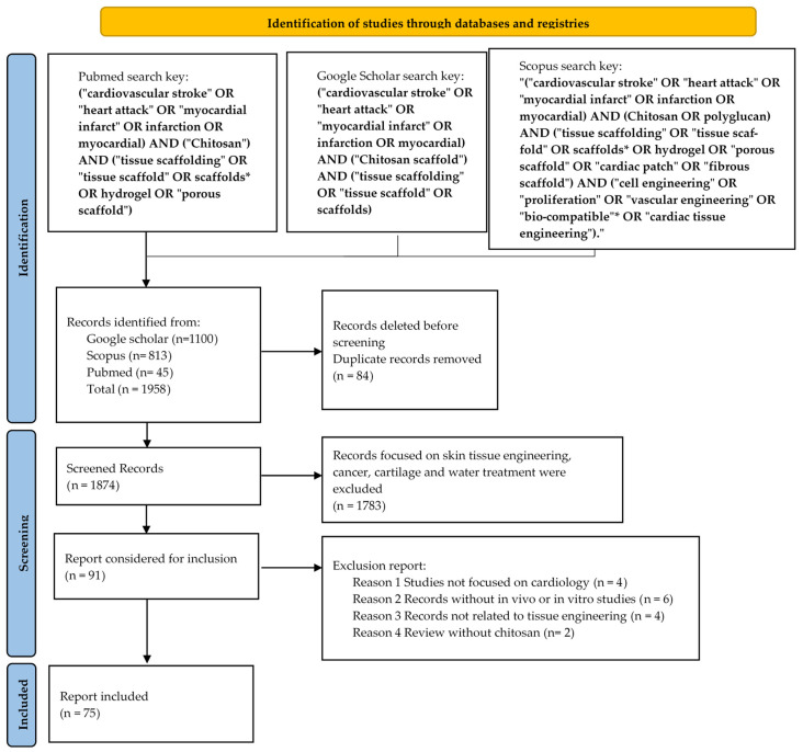
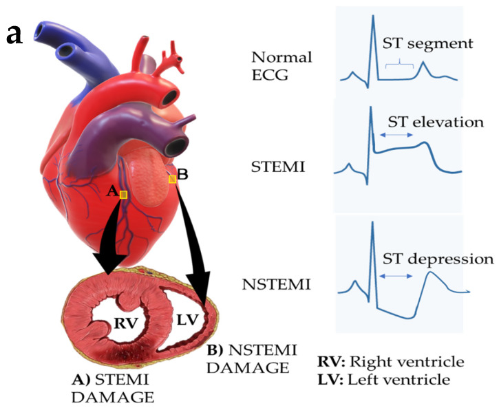
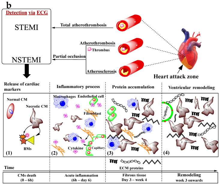
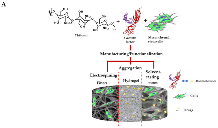
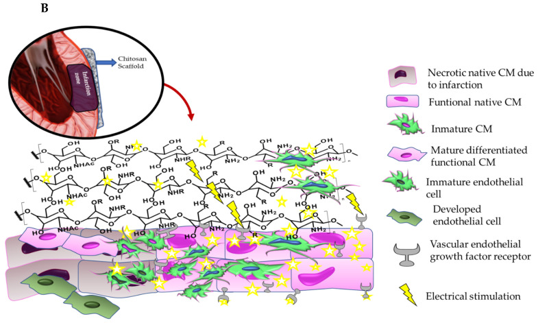
Cardiovascular diseases (CVD), such as myocardial infarction (MI), constitute one of the world's leading causes of annual deaths. This cardiomyopathy generates a tissue scar with poor anatomical properties and cell necrosis that can lead to heart failure. Necrotic tissue repair is required through pharmaceutical or surgical treatments to avoid such loss, which has associated adverse collateral effects. However, to recover the infarcted myocardial tissue, biopolymer-based scaffolds are used as safer alternative treatments with fewer side effects due to their biocompatibility, chemical adaptability and biodegradability. For this reason, a systematic review of the literature from the last five years on the production and application of chitosan scaffolds for the reconstructive engineering of myocardial tissue was carried out. Seventy-five records were included for review using the "preferred reporting items for systematic reviews and meta-analyses" data collection strategy. It was observed that the chitosan scaffolds have a remarkable capacity for restoring the essential functions of the heart through the mimicry of its physiological environment and with a controlled porosity that allows for the exchange of nutrients, the improvement of the electrical conductivity and the stimulation of cell differentiation of the stem cells. In addition, the chitosan scaffolds can significantly improve angiogenesis in the infarcted tissue by stimulating the production of the glycoprotein receptors of the vascular endothelial growth factor (VEGF) family. Therefore, the possible mechanisms of action of the chitosan scaffolds on cardiomyocytes and stem cells were analyzed. For all the advantages observed, it is considered that the treatment of MI with the chitosan scaffolds is promising, showing multiple advantages within the regenerative therapies of CVD.
Keywords: biopolymers; cardiac tissue engineering; chitosan scaffolds; heart attack; natural polysaccharide.
Conflict of interest statement
The authors declare no conflict of interest.
Figures
Similar articles
-
A rapid and systematic review of the clinical effectiveness and cost-effectiveness of topotecan for ovarian cancer.Health Technol Assess. 2001;5(28):1-110. doi: 10.3310/hta5280. Health Technol Assess. 2001. PMID: 11701100
-
Health professionals' experience of teamwork education in acute hospital settings: a systematic review of qualitative literature.JBI Database System Rev Implement Rep. 2016 Apr;14(4):96-137. doi: 10.11124/JBISRIR-2016-1843. JBI Database System Rev Implement Rep. 2016. PMID: 27532314
-
Cost-effectiveness of using prognostic information to select women with breast cancer for adjuvant systemic therapy.Health Technol Assess. 2006 Sep;10(34):iii-iv, ix-xi, 1-204. doi: 10.3310/hta10340. Health Technol Assess. 2006. PMID: 16959170
-
Effects of a gluten-reduced or gluten-free diet for the primary prevention of cardiovascular disease.Cochrane Database Syst Rev. 2022 Feb 24;2(2):CD013556. doi: 10.1002/14651858.CD013556.pub2. Cochrane Database Syst Rev. 2022. PMID: 35199850 Free PMC article.
-
Home treatment for mental health problems: a systematic review.Health Technol Assess. 2001;5(15):1-139. doi: 10.3310/hta5150. Health Technol Assess. 2001. PMID: 11532236
Cited by
-
Application of Pro-angiogenic Biomaterials in Myocardial Infarction.ACS Omega. 2024 Aug 27;9(36):37505-37529. doi: 10.1021/acsomega.4c04682. eCollection 2024 Sep 10. ACS Omega. 2024. PMID: 39281944 Free PMC article. Review.
-
Chitosan-Glycolic Acid Gel Modification of Chloride Ion Transport in Mammalian Skin: An In Vitro Study.Molecules. 2023 Sep 12;28(18):6581. doi: 10.3390/molecules28186581. Molecules. 2023. PMID: 37764357 Free PMC article.
-
Motion Sensing by a Highly Sensitive Nanogold Strain Sensor in a Biomimetic 3D Environment.ACS Appl Mater Interfaces. 2024 Oct 23;16(42):56599-56610. doi: 10.1021/acsami.4c08105. Epub 2024 Sep 10. ACS Appl Mater Interfaces. 2024. PMID: 39253872 Free PMC article.
-
Melatonin/Chitosan Biomaterials for Wound Healing and Beyond: A Multifunctional Therapeutic Approach.Int J Mol Sci. 2025 Jun 20;26(13):5918. doi: 10.3390/ijms26135918. Int J Mol Sci. 2025. PMID: 40649698 Free PMC article. Review.
-
Biological Macromolecule-Based Scaffolds for Urethra Reconstruction.Biomolecules. 2023 Jul 26;13(8):1167. doi: 10.3390/biom13081167. Biomolecules. 2023. PMID: 37627232 Free PMC article. Review.
References
-
- Rathore V. Risk Factors of Acute Myocardial Infarction: A Review. Eurasian J. Med. Investig. 2018;2:1–7. doi: 10.14744/ejmi.2018.76486. - DOI
-
- Virani S.S., Alonso A., Benjamin E.J., Bittencourt M.S., Callaway C.W., Carson A.P., Chamberlain A.M., Chang A.R., Cheng S., Delling F.N., et al. Heart disease and stroke statistics—2020 update: A report from the American Heart Association. Circulation. 2020;141:e139–e596. doi: 10.1161/CIR.0000000000000757. - DOI - PubMed
Publication types
MeSH terms
Substances
LinkOut - more resources
Full Text Sources
Medical
