Review
. 2023 Feb 9;13(4):674.
doi: 10.3390/nano13040674.

Application of Polypyrrole-Based Electrochemical Biosensor for the Early Diagnosis of Colorectal Cancer

Affiliations
Review

Application of Polypyrrole-Based Electrochemical Biosensor for the Early Diagnosis of Colorectal Cancer

Xindan Zhang et al. Nanomaterials (Basel). .

Abstract

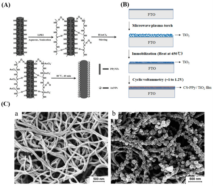
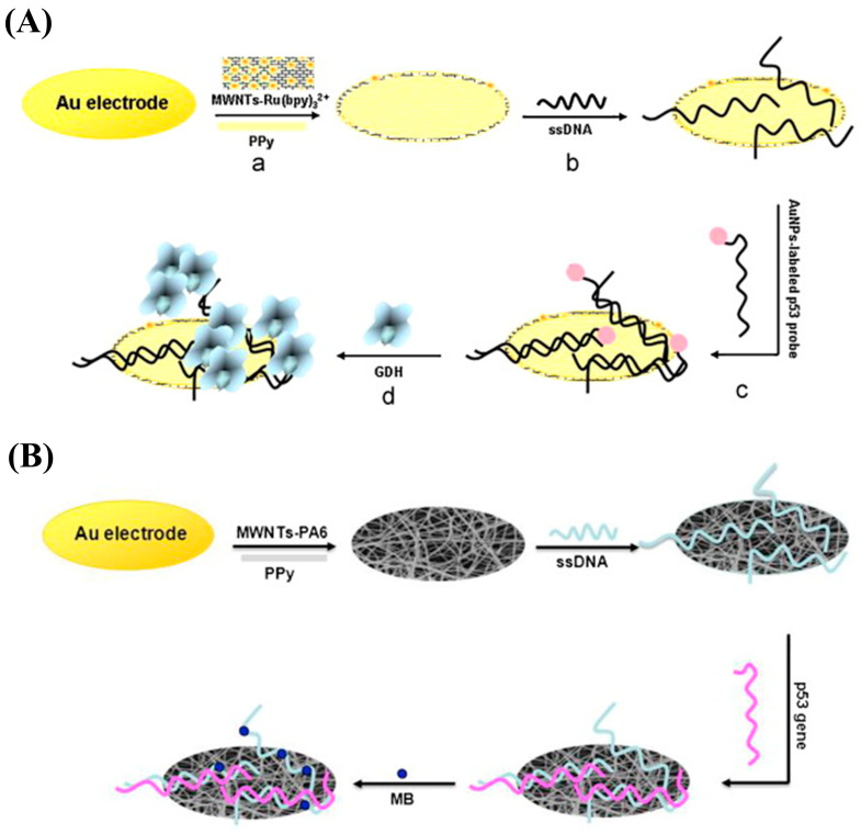
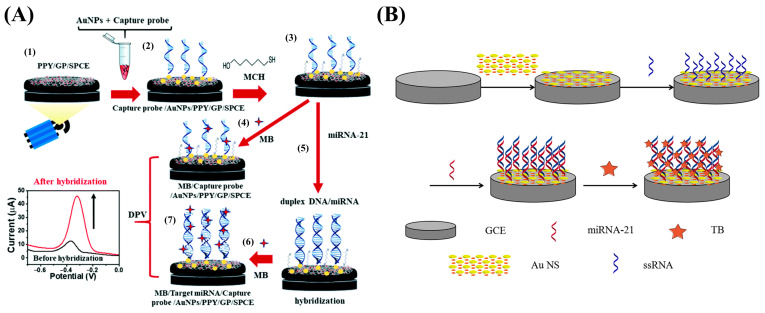
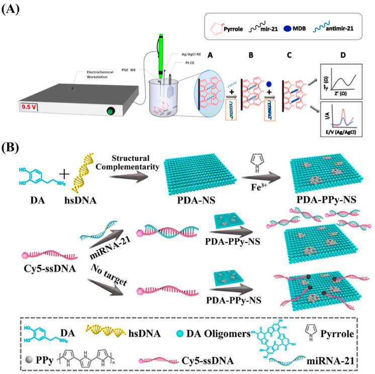
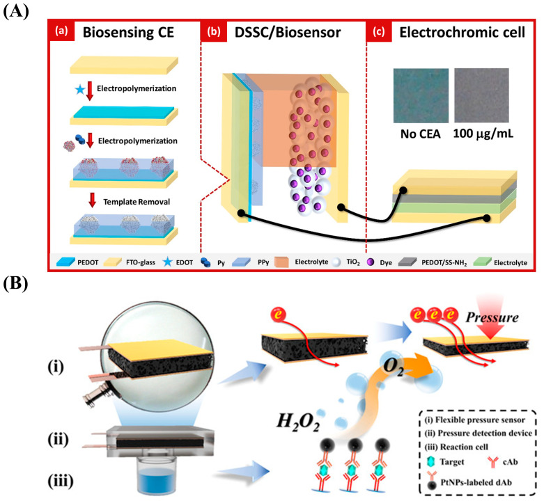
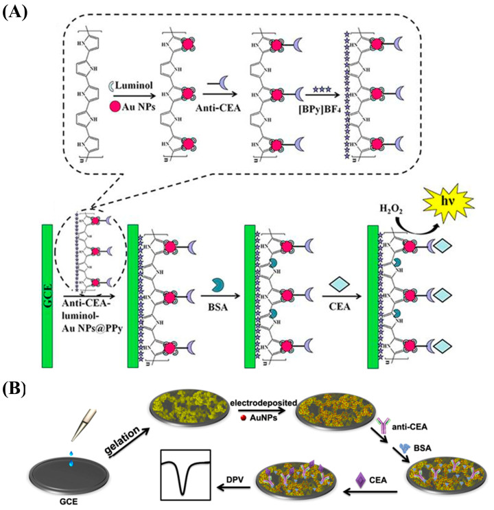
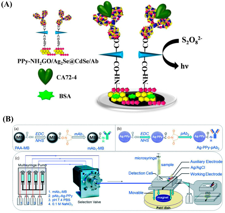
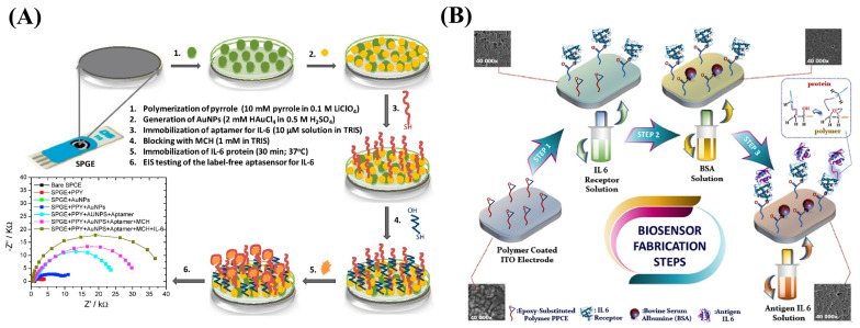
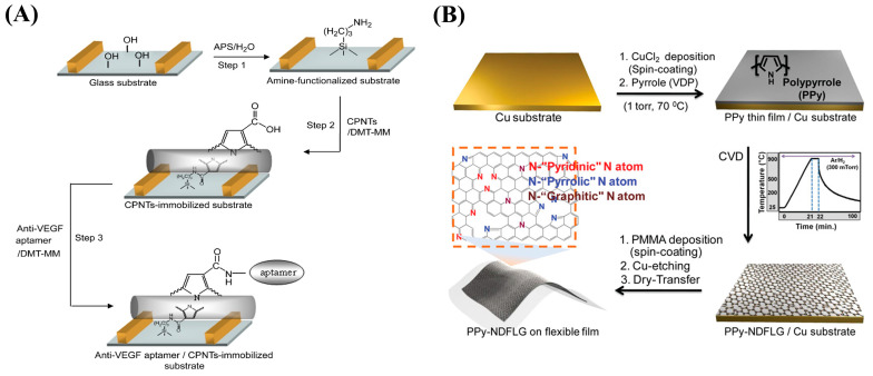
Although colorectal cancer (CRC) is easy to treat surgically and can be combined with postoperative chemotherapy, its five-year survival rate is still not optimistic. Therefore, developing sensitive, efficient, and compliant detection technology is essential to diagnose CRC at an early stage, providing more opportunities for effective treatment and intervention. Currently, the widely used clinical CRC detection methods include endoscopy, stool examination, imaging modalities, and tumor biomarker detection; among them, blood biomarkers, a noninvasive strategy for CRC screening, have shown significant potential for early diagnosis, prediction, prognosis, and staging of cancer. As shown by recent studies, electrochemical biosensors have attracted extensive attention for the detection of blood biomarkers because of their advantages of being cost-effective and having sound sensitivity, good versatility, high selectivity, and a fast response. Among these, nano-conductive polymer materials, especially the conductive polymer polypyrrole (PPy), have been broadly applied to improve sensing performance due to their excellent electrical properties and the flexibility of their surface properties, as well as their easy preparation and functionalization and good biocompatibility. This review mainly discusses the characteristics of PPy-based biosensors, their synthetic methods, and their application for the detection of CRC biomarkers. Finally, the opportunities and challenges related to the use of PPy-based sensors for diagnosing CRC are also discussed.

Keywords: biomarkers; colorectal cancer; early diagnosis; electrochemical biosensor; polypyrrole.


Conflict of interest statement

The authors declare no conflict of interest.

Figures

Figure 1
Figure 1
Electrochemical technologies commonly used in electrochemical detection.
Figure 2
Figure 2
Synthesis methods for PPy.
Figure 3
Figure 3
Synthesized PPy morphologies and their applications.
Figure 4
Figure 4
(A) Schematic diagram of PPy-based enzymatic sensor. Reproduced from [100] with permission from Elsevier. (B) Schematic diagram of the electrochemical platform for polypyrrole–poly(3,4-ethylenedioxythiophene)–gold (PPy-PEDOT-Au). Reproduced from [104] with permission from Elsevier. (C) Schematic diagram of the fabrication process for polypyrrole-reduced graphene oxide/gold nanoparticles (PPy-rGO/AuNPs) for use in biosensors. Reproduced from [105] with permission from Elsevier.
Figure 5
Figure 5
(A) Schematic diagram of the preparation procedure for the polypyrrole nanowire (PPyNW)-based aptasensor. Reproduced from [106] with permission from Elsevier. (B) Schematic diagram of the preparation of molecularly imprinted polypyrrole nanotubes (MIPNs) and the MIPN-based glyphosate platform. Reproduced from [108] with permission from Elsevier. (C) Schematic diagram of the fabrication process for the biosensor based on imprinted polypyrrole film from bacteria. Reproduced from [109] with permission from the American Chemical Society.
Figure 6
Figure 6
(A) Schematic diagram of the synthesis of polypyrrole nanofiber-supporting Au nanoparticles (Au/PPyNFs). Reproduced from [112] with permission from Elsevier. (B) Schematic diagram of the preparation of chitosan–polypyrrole/titanium oxide (CS-PPy/TiO2) nanocomposite films on a fluorine-doped tin oxide-coated glass slide (FTO). Reproduced from [113] with permission from MDPI. (C) Scanning electron microscope images of PPyNWs (a) and copper oxide (CuxO) nanoparticle-modified PPyNWs (b). Reproduced from [114] with permission from Elsevier.
Figure 7
Figure 7
(A) Schematic illustration of the fabrication of a PPy-covered multiwalled carbon nanotube and ruthenium (II) tris-(bipyridine) (MWNT-Ru(bpy)32+-PPy) biosensor for the wild-type p53 sequence (wtp53) assay based on electrochemical luminescence (ECL). Reproduced from [126] with permission from Elsevier. (B) Schematic illustration of the preparation of a multi-walled carbon nanotube–nylon 6–polypyrrole (MWNT-PA6-PPy) biosensor for wtp53 detection based on electrospinning technology. Reproduced from [127] with permission from Elsevier.
Figure 8
Figure 8
(A) Schematic representation of the strategy based on a gold nanoparticle/polypyrrole/graphene (AuNP/PPy/GP) nanocomposite. Reproduced from [139] with permission from the Royal Society of Chemistry. (B) Schematic representation of the approach using a PPy-AuNP superlattice (PPy-AuNS). Reproduced from [140] with permission from Elsevier.
Figure 9
Figure 9
(A) Schematic diagram of the biosensor fabrication process utilizing PPy electrodeposition. Reproduced from [141] with permission from Elsevier. (B) Schematic diagram of the preparation procedure for the hybrid polydopamine/polypyrrole nanosheet (PDA-PPy-NS) biosensor for miRNA-21 determination. Reproduced from [83] with permission from the American Chemical Society.
Figure 10
Figure 10
(A) Schematic diagram of the design of a self-powered and self-signaled biosensing platform based on biosensing, molecular imprinting technology, a dye-sensitized solar cell (DSSC), and electrochromic technology. Reproduced from [158] with permission from Elsevier. (B) Schematic diagram of a flexible pressure sensor based on the elastic three-dimensional (3D) structure of PPy foam. Reproduced from [143] with permission from the American Chemical Society.
Figure 11
Figure 11
(A) Schematic diagram of the preparation of an anti-CEA–luminol–AuNP@PPy-based immunosensor for CEA detection. Reproduced from [144] with permission from Nature Publishing Group. (B) Schematic diagram of the electrochemical method for CEA determination based on 3D continuous conducting network nanocomposites composed of PPy hydrogel loaded with AuNPs. Reproduced from [147] with permission from Nature Publishing Group.
Figure 12
Figure 12
(A) Schematic diagram of the polypyrrole-intercalated aminated graphene/Ag2Se@CdSe (PPy-NH2GO/Ag2Se@CdSe)-based immunosensor for the CA72-4 assay. Reproduced from [148] with permission from the American Chemical Society. (B) Schematic diagram of the platform for CA125 measurement: (a) the preparation of the capture probe using mAb1-conjugated magnetic beads (mAb1-MB). (b) the preparation of the detection probe using pAb2-labeled Ag-PPy nanostructure (Ag-PPy- pAb2). (c) magneto-controlled microfluidic device with an electrochemical detection cell. Reproduced from [149] with permission from the Royal Society of Chemistry.
Figure 13
Figure 13
(A) Schematic diagram of the AuNP/PPyNP-based aptasensor for the IL-6 assay. Reproduced from [151] with permission from Elsevier. (B) Schematic diagram of the fabrication process for the PPCE-modified biosensor for IL-6 measurement. Reproduced from [152] with permission from Elsevier.
Figure 14
Figure 14
(A) Schematic diagram of the fabrication of a CPNT-modified aptasensor for the VEGF assay. Reproduced from [155] with permission from Elsevier. (B) Schematic diagram of the process of synthesizing flexible PPy-NDFLG. Reproduced from [156] with permission from the American Chemical Society.

Similar articles

Cited by

References

    1. Xi Y., Xu P. Global colorectal cancer burden in 2020 and projections to 2040. Transl. Oncol. 2021;14:101174. doi: 10.1016/j.tranon.2021.101174. - DOI - PMC - PubMed
    1. Kanth P., Inadomi J.M. Screening and prevention of colorectal cancer. BMJ. 2021;374:n1855. doi: 10.1136/bmj.n1855. - DOI - PubMed
    1. Millien V.O., Mansour N.M. Bowel Preparation for Colonoscopy in 2020: A Look at the Past, Present, and Future. Curr. Gastroenterol. Rep. 2020;22:28. doi: 10.1007/s11894-020-00764-4. - DOI - PubMed
    1. Kim S.Y., Kim H.-S., Park H.J. Adverse events related to colonoscopy: Global trends and future challenges. World J. Gastroenterol. 2019;25:190–204. doi: 10.3748/wjg.v25.i2.190. - DOI - PMC - PubMed
    1. Vuik F.E.R., Nieuwenburg S.A.V., Moen S., Spada C., Senore C., Hassan C., Pennazio M., Rondonotti E., Pecere S., Kuipers E.J., et al. Colon capsule endoscopy in colorectal cancer screening: A systematic review. Endoscopy. 2021;53:815–824. doi: 10.1055/a-1308-1297. - DOI - PubMed