. 2023 Feb 4;15(4):808.
doi: 10.3390/nu15040808.

Curcuma aromatica Salisb. Protects from Acetaminophen-Induced Hepatotoxicity by Regulating the Sirt1/HO-1 Signaling Pathway

Affiliations

Curcuma aromatica Salisb. Protects from Acetaminophen-Induced Hepatotoxicity by Regulating the Sirt1/HO-1 Signaling Pathway

Hyunseong Kim et al. Nutrients. .

Abstract

Acetaminophen (APAP) overdose-induced hepatotoxicity reduces the activity of sirtuin-1 (Sirt1) along with heme oxygenase 1 (HO-1) and promotes inflammatory responses and oxidative stress. Although the extract of Curcuma aromatica Salisb. (CAS) possesses hepatoprotective properties, scientific evidence on whether CAS prevents hepatotoxicity and the underlying molecular mechanisms are lacking. Here, we hypothesized that CAS ameliorates hepatotoxicity by inhibiting inflammation and oxidative stress via Sirt1/HO-1 signaling. CAS pretreatment at doses of 200 and 400 μg/mL significantly increased cell viability in APAP-treated primary hepatocytes. The expression of inducible nitric oxide synthase (iNOS) substantially increased after APAP treatment; however, this expression significantly decreased in cells pretreated with 100, 200, and 400 µg/mL CAS. CAS increased Sirt1 and HO-1 levels in APAP-treated hepatocytes in a dose-dependent manner. When CAS was orally administered to mice at doses of 20 or 100 mg/kg for 7 days, the APAP-induced increase in serum aspartate aminotransferase and alanine aminotransferase levels was inhibited. Moreover, CAS decreased IL-6, TNF-α, and IL-1β, increased IL-10, suppressed ROS generation, increased glutathione levels, inhibited iNOS and cyclooxygenase-2, and enhanced Sirt1 and HO-1 in the mouse model of APAP-induced hepatotoxicity. These findings suggest that CAS could be used as a natural hepatoprotective drug to treat APAP-induced injury.

Keywords: Curcuma aromatica Salisb.; HO-1; Sirt1; acetaminophen; hepatotoxicity; paracetamol.


Conflict of interest statement

The authors declare no conflict of interest.

Figures

Figure 1
Figure 1
CAS protects primary mouse hepatocytes from APAP-induced hepatotoxicity and oxidative damage by modulating ROS production and iNOS expression. Determination of cell viability in primary hepatocytes pretreated with CAS for 24 h (A) without APAP treatment and (B) with APAP treatment using a cell counting kit assay (n = 4). (C) Flow cytometric quantification of the percentage of DCFDA+ cells upon challenge with APAP (n = 6). (D) Representative flow cytometric dot plot images showing DCFDA-FITC analysis for intracellular ROS levels. (E) Representative confocal images of immunocytochemistry for iNOS (red) in the primary hepatocytes of each group. White scale bar = 200 µm, yellow scale bar = 50 µm. (F) Quantitative analysis of the percentage of iNOS-positivity in DAPI-positive hepatocytes after pretreatment with CAS at 100, 200, and 400 µg/mL and treatment with 2 μM APAP (n = 6). The data are expressed as the mean ± SEM and were analyzed via one-way analysis of variance with Tukey’s post-hoc test. Significant differences are indicated as follows: #### p < 0.0001 vs. blank group; * p < 0.05, ** p < 0.01, and **** p < 0.0001 vs. APAP group.
Figure 2
Figure 2
The hepatoprotective effects of CAS on APAP-induced hepatotoxicity involve the upregulation of Sirt1/HO-1 expression in primary mouse hepatocytes. (A) Representative confocal images of immunocytochemistry for Sirt1 (red) in the blank, APAP, and CAS groups. White scale bar = 200 µm, yellow scale bar = 50 µm. (B) Quantitative analysis of the relative fluorescence intensity of Sirt1 in each group (n = 6). (C) Representative confocal images of immunocytochemistry for HO-1 (green) in the blank, control, and CAS groups. White scale bar = 200 µm, yellow scale bar = 50 µm. (D) Quantitative analysis of the relative fluorescence intensity of HO-1 in each group (n = 6). The data are expressed as the mean ± SEM and were analyzed via ordinary ANOVA with Tukey’s post-hoc analysis. Significant differences are indicated as follows: #### p < 0.0001 vs. blank group; ** p < 0.01 and **** p < 0.0001 vs. APAP group.
Figure 3
Figure 3
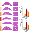
Prevention of histopathological liver damage by CAS administration in a mouse model of APAP-induced hepatotoxicity. (A) Representative H&E images of liver structural damage in the blank, control, and CAS groups. White scale bar = 1000 µm, yellow scale bar = 100 µm. (B,C) Quantitative analysis of the serum AST and ALT levels in blood from each group of mice (n = 8). The data are expressed as the mean ± SEM and were analyzed via ordinary ANOVA with Tukey’s post-hoc analysis. Statistical differences are indicated as follows: #### p < 0.0001 vs. blank group; **** p < 0.0001 vs. APAP group.
Figure 4
Figure 4
Anti-inflammatory activity of CAS in APAP-induced hepatic inflammation in mice. (AC) ELISA results showing the expression of the inflammation-related cytokines, (A) IL-6, (B) TNFα, and (C) IL-1β in the liver tissues of each group (n = 6). (D) ELISA results of IL-10 expression in the liver tissues of each group (n = 6). The data are expressed as the mean ± SEM and were analyzed via one-way ANOVA with Tukey’s post-hoc test. Statistical differences are indicated as follows: # p < 0.05 and #### p < 0.0001 vs. blank group; * p < 0.05, ** p < 0.01, and **** p < 0.0001 vs. APAP group.
Figure 5
Figure 5
Hepatic antioxidant effect of CAS against APAP-induced oxidative stress in mice via modulating Sirt1/HO-1 mRNA expression. (A) The hepatic GSH content in the liver tissues of each group (n = 6). (B) The ROS level in the liver tissues of each group (n = 6). (CF) RT-qPCR results of antioxidant-related mRNA expression (iNOS, COX-2, Sirt1, and HO-1) in the liver tissues of mice after CAS and APAP administration (n = 6). The data are expressed as the mean ± SEM and were analyzed via one-way ANOVA with Tukey’s post-hoc test. Significant differences are indicated as follows: ## p < 0.01, ### p < 0.001, and #### p < 0.0001 vs. blank group; * p < 0.05, ** p < 0.01, *** p < 0.001, and **** p < 0.0001 vs. APAP group.

Similar articles

Cited by

References

    1. Jaeschke H. Acetaminophen: Dose-Dependent Drug Hepatotoxicity and Acute Liver Failure in Patients. Dig. Dis. 2015;33:464–471. doi: 10.1159/000374090. - DOI - PMC - PubMed
    1. Mossanen J.C., Tacke F. Acetaminophen-induced acute liver injury in mice. Lab. Anim. 2015;49:30–36. doi: 10.1177/0023677215570992. - DOI - PubMed
    1. Larson A.M., Polson J., Fontana R.J., Davern T.J., Lalani E., Hynan L.S., Reisch J.S., Schiodt F.V., Ostapowicz G., Shakil A.O., et al. Acetaminophen-induced acute liver failure: Results of a United States multicenter, prospective study. Hepatology. 2005;42:1364–1372. doi: 10.1002/hep.20948. - DOI - PubMed
    1. Reddy K.R., Ellerbe C., Schilsky M., Stravitz R.T., Fontana R.J., Durkalski V., Lee W.M., Acute Liver Failure Study Group Determinants of outcome among patients with acute liver failure listed for liver transplantation in the United States. Liver Transpl. 2016;22:505–515. doi: 10.1002/lt.24347. - DOI - PMC - PubMed
    1. Du K., Ramachandran A., Jaeschke H. Oxidative stress during acetaminophen hepatotoxicity: Sources, pathophysiological role and therapeutic potential. Redox Biol. 2016;10:148–156. doi: 10.1016/j.redox.2016.10.001. - DOI - PMC - PubMed