. 2023 Feb 2;12(2):240.
doi: 10.3390/pathogens12020240.

The DarT/DarG Toxin-Antitoxin ADP-Ribosylation System as a Novel Target for a Rational Design of Innovative Antimicrobial Strategies

Affiliations

The DarT/DarG Toxin-Antitoxin ADP-Ribosylation System as a Novel Target for a Rational Design of Innovative Antimicrobial Strategies

Giuliana Catara et al. Pathogens. .

Abstract

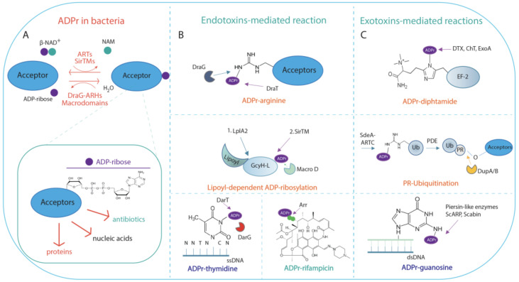
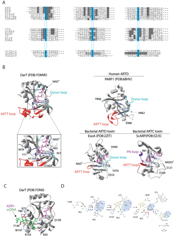
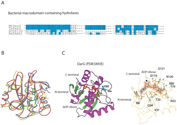
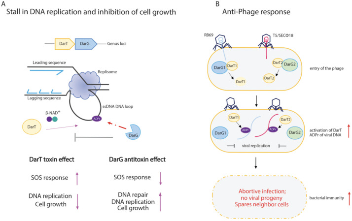
The chemical modification of cellular macromolecules by the transfer of ADP-ribose unit(s), known as ADP-ribosylation, is an ancient homeostatic and stress response control system. Highly conserved across the evolution, ADP-ribosyltransferases and ADP-ribosylhydrolases control ADP-ribosylation signalling and cellular responses. In addition to proteins, both prokaryotic and eukaryotic transferases can covalently link ADP-ribosylation to different conformations of nucleic acids, thus highlighting the evolutionary conservation of archaic stress response mechanisms. Here, we report several structural and functional aspects of DNA ADP-ribosylation modification controlled by the prototype DarT and DarG pair, which show ADP-ribosyltransferase and hydrolase activity, respectively. DarT/DarG is a toxin-antitoxin system conserved in many bacterial pathogens, for example in Mycobacterium tuberculosis, which regulates two clinically important processes for human health, namely, growth control and the anti-phage response. The chemical modulation of the DarT/DarG system by selective inhibitors may thus represent an exciting strategy to tackle resistance to current antimicrobial therapies.

Keywords: ADP-ribosylation; DNA modification; DarT/DarG; antimicrobial resistance; cell growth; phage defence; toxin–antitoxin.


Conflict of interest statement

The authors declare no conflict of interest.

Figures

Figure 1
Figure 1
Schematic representation of ADP-ribosylation reaction in bacterial meta-cell. (A) ADP-ribosylation (ADPr) reaction is catalysed by NAD+-dependent diphtheria toxin-like ARTs ARTDs, the cholera toxin-like ARTs ARTCs, or SirTMs, which transfer a single ADP-ribose unit on acceptors. Macrodomain-containing hydrolases (Macrodomains) or DraG-related ADP-ribosylhydrolases reverse the reaction by generating free ADP-ribose and unmodified acceptor. Inset: ADP-ribose moiety linked to acceptor substrates, which can be proteins, nucleic acids, or antibiotics. (B) Endotoxin-mediated reaction. Endotoxins can modify proteins, nucleic acids, or antibiotics. ARTDs, ARTCs, and SirTM modify endogenous bacterial substrates on different residues as indicated. ADP-ribosylated (ADPr)-arginine: MARylation of arginine residue is performed by the DraT enzyme, and is reversed by the cognate DraG ADP-ribosylhydrolase. Further examples are provided in the text. Lipoyl-dependent MARylation is carried out by SirTM and is dependent on prior lipoylation of the lipoyl-carrier protein GcyH-L, by the lipoate-protein ligase A (LplA2). The modification is reversed by the MacroD hydrolase, which is encoded within the same SirTM operon; ADP-ribosylated (ADPr)-thymidine: the reaction is performed by the endotoxin DarT that modifies thymidine base on ssDNA; the cognate DarG antitoxin reverses the modification; ADP-ribosylated (ADPr)-rifampicin: MARylation of the rifampin antibiotic is catalysed by Arr toxin. (C) Exotoxin-mediated reactions. ARTDs and ARTCs modify host targets on different residues as indicated. ADP-ribosylated (ADPr)-diphthamide: the reaction is catalysed by the toxins DTX, ChT, and ExoA, which irreversibly transfer ADP-ribose on the residue diphthamide on the elongation factor 2; PR-Ubiquitination. SdeA toxin catalyses the ADP-ribosylation (ADPr)-dependent ubiquitination of host proteins in a two-step reaction as detailed in the text. The reaction is reversed by the phosphodiesterases DubA/B; ADP-ribosylated (ADPr)-guanosine. The irreversible ADP-ribosylation on guanosine in dsDNA is performed by the pierisin-like enzymes ScARP and Scabin.
Figure 2
Figure 2
Comparison of amino acid sequences and 3D structures of representative ARTs. (A) Alignment of the partial sequences of the bacterial ARTs. ARTD members, which harbour the H-Y-E catalytic residues, include: DTX, diphtheria toxin from C. diphtheriae; ETA, exotoxin A from P. aeruginosa; Ch toxin, cholix toxin from V. cholerae; SrADP, toxin from Streptomyces roseifaciens; Arr-Ms, rifamycin ADP-ribosylation toxin from Mycobacterium smegmatis; Cd-PARP, toxin from Clostridium perfringens CD 160. ARTC members, which enclose R-S-E catalytic residues, include: CTX, cholera toxin from V. cholerae; ScARP, toxin from S. coelicolor; Scabin from S. scabies; IT, iota toxin from C. perfringens; DraT, dinitrogenase reductase ADP-ribosyltransferase from R. rubrum; SdeA, ADP-ribosylation-dependent ubiquitination toxin from L. pneumophila. Divergent enzymes include: CtTpt1, Tpt1 RNA-phosphotransferase enzyme from Clostridium thermocellum; ParT, ADP-ribosylating toxin of ParT/ParS TA system from Sphingobium sp. YBL2; TaDarT, DNA ADP-ribosylating toxin of DarT/DarG TA system from T. aquaticus. The residues involved in catalysis are boxed on a light blue background. Identities or accepted amino acid substitutions are indicated in dark and light grey, respectively. (B) Cartoon–stick model of Thermus sp. 2.9 DarT(E160A) (PDB:7OMW, [78]) showing the NAD+ binding site in complex with NAD+, the amino acid residues involved in the catalytic activity (blue), the regulatory ARTT-loop (red) and the donor-loop (light blue). Inset: the catalytic site residues R51, H65, Y71, M78, H119, and E160A localise in proximity of nicotinamide in the active site. Cartoon–stick models of the 3D structure of the human ARTD PARP1 (PDB:6BHV, [113]), the bacterial ARTD-toxin ExoA (PDB:2ZIT, [114]), and the bacterial ARTC-toxin ScARP (PDB:5ZJ5, [115]) are shown as exemplars. (C) Cartoon–stick model of Thermus sp. 2.9 DarT(E160A) (PDB:7ON0, [78]) in complex with ADP-ribosylated ssDNA showing the residues (dark red) involved in the interaction with ssDNA (green). (D) Catalytic mechanism proposed for DarT-mediated ADP-ribosylation reaction of DNA.
Figure 3
Figure 3
Comparison of amino acid sequences and 3D structures of macrodomain-containing hydrolases belonging to ALC1-like group. (A) Alignment of partial sequences of ALC1-like hydrolases from bacteria. MtDarG, DarG from M. tuberculosis; TaDarG, DarG from T. aquaticus; SCO6735, macrodomain-containing hydrolase from S. coelicolor; FmTARG1, TARG1 homologue from F. mortiferum. Identities are indicated in light blue. (B) Structural comparison between DarG from T. aquaticus in complex with ADP-ribose (yellow line, PDB: 5M3E, [38]), human TARG1 in complex with ADP-ribose (blue line, PDB:4J5S, [55]) and SCO6735 (red line, PDB:5E3B, [119]). (C) DarG from T. aquaticus (cartoon) in complex with ADP-ribose (ball and stick). The catalytic K80 is shown in light blue (left panel). Close up of the T. aquaticus DarG active site showing the residues involved in ADP-ribose binding (right panel).
Figure 4
Figure 4
Schematic representation of DarT/DarG TA system biological functions. (A) DarT/DarG system in the regulation of bacterial cell growth. DarT-mediated ADP-ribosylation of ssDNA on thymidine found in consensus sequences causes a stall of DNA replication and concomitant arrest of cell growth. The activity of the DarG antitoxin counteracts DarT activity through the removal of ADP-ribose from the marked thymidine on ssDNA: DarG-mediated removal of ADP-ribose enables the replication to proceed and cell growth to re-establish. (B) The DarT/DarG system and the anti-phage response. Upon entry of the phages, the DarT1 and DarT2 endotoxins ADP-ribosylate viral DNA, which is unable to replicate. The overall downregulation of cell metabolic processes triggers the abortive infection programme, which leads to the host cell death and prevents viral progeny spreading in order to protect the bacterial cell population.

Similar articles

Cited by

References

    1. Perina D., Mikoč A., Ahel J., Ćetković H., Žaja R., Ahel I. Distribution of Protein Poly(ADP-Ribosyl)Ation Systems across All Domains of Life. DNA Repair. 2014;23:4–16. doi: 10.1016/j.dnarep.2014.05.003. - DOI - PMC - PubMed
    1. Aravind L., Zhang D., de Souza R.F., Anand S., Iyer L.M. The Natural History of ADP-Ribosyltransferases and the ADP-Ribosylation System. Curr. Top. Microbiol. Immunol. 2015;384:3–32. doi: 10.1007/82_2014_414. - DOI - PMC - PubMed
    1. Palazzo L., Mikoč A., Ahel I. ADP-Ribosylation: New Facets of an Ancient Modification. FEBS J. 2017;284:2932–2946. doi: 10.1111/febs.14078. - DOI - PMC - PubMed
    1. Mikolčević P., Hloušek-Kasun A., Ahel I., Mikoč A. ADP-Ribosylation Systems in Bacteria and Viruses. Comput. Struct. Biotechnol. J. 2021;19:2366–2383. doi: 10.1016/j.csbj.2021.04.023. - DOI - PMC - PubMed
    1. Jørgensen R., Purdy A.E., Fieldhouse R.J., Kimber M.S., Bartlett D.H., Merrill A.R. Cholix Toxin, a Novel ADP-Ribosylating Factor from Vibrio Cholerae. J. Biol. Chem. 2008;283:10671–10678. doi: 10.1074/jbc.M710008200. - DOI - PubMed

LinkOut - more resources