. 2023 Jan 17;15(2):314.
doi: 10.3390/pharmaceutics15020314.

Synergistic Antitumor Potency of a Self-Assembling Cyclodextrin Nanoplex for the Co-Delivery of 5-Fluorouracil and Interleukin-2 in the Treatment of Colorectal Cancer

Affiliations

Synergistic Antitumor Potency of a Self-Assembling Cyclodextrin Nanoplex for the Co-Delivery of 5-Fluorouracil and Interleukin-2 in the Treatment of Colorectal Cancer

Safiye Akkın et al. Pharmaceutics. .

Abstract

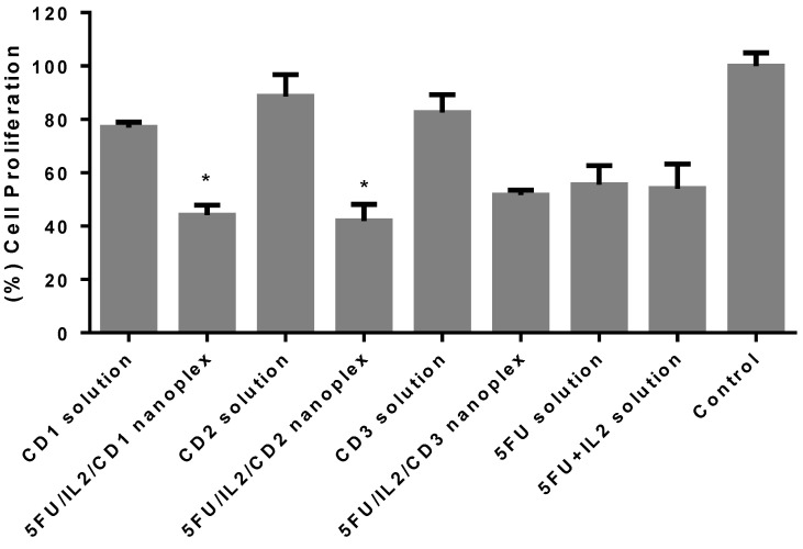
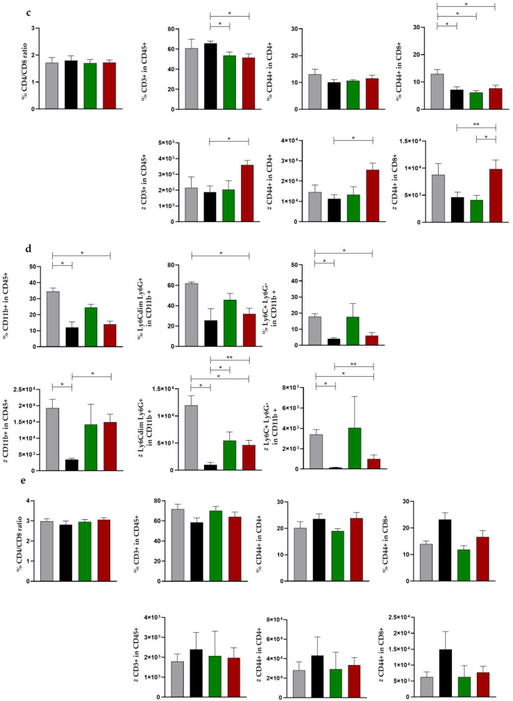
Chemotherapy is the most used method after surgery in the treatment of colon cancer. Cancer cells escape the recognition mechanism of immune system cells to survive and develop chemoresistance. Therefore, the use of immunotherapy in combination with chemotherapy can increase the effectiveness of the treatment. Nanoparticles have been used clinically to increase the accumulation of therapeutics in target tissues and reduce toxicity. In this paper, nanoplexes were formed via cationic cyclodextrin polymer, 5-Fluorouracil, and Interleukin-2 based on the opposite charge interaction of macromolecules without undergoing any structural changes or losing the biological activity of Interleukin-2. Anticancer activities of nanoplexes were determined in two-dimensional and three-dimensional cell culture setups. The dual drug-loaded cyclodextrin nanoplexes diffused deeper into the spheroids and accelerated apoptosis when compared with 5-FU solutions. In the colorectal tumor-bearing animal model, survival rate, antitumor activity, metastasis, and immune response parameters were assessed using a cyclodextrin derivative, which was found to be safe based on the ALT/AST levels in healthy mice. Histomorphometric analysis showed that the groups treated with the nanoplex formulation had significantly fewer initial tumors and lung foci when compared with the control. The dual drug-loaded nanoplex could be a promising drug delivery technique in the immunochemotherapy of colorectal cancer.

Keywords: 3D cell culture; 5-Fluorouracil; Interleukin-2; chemoimmunotherapy; colon cancer; cyclodextrin polymer; in vivo model; nanoplex.


Conflict of interest statement

The authors declare no conflict of interest. The funders had no role in the design of the study; in the collection, analyses, or interpretation of data; in the writing of the manuscript; or in the decision to publish the results.

Figures

Figure 1
Figure 1
Cell index values of CT26 cells applied with 5FU/IL2/CD nanoplexes, 5-FU solution, 5-FU+IL-2 solution, and control, assessed by xCELLigence RTCA. The control group was treated with DMEM and was considered as 100% (n = 4, ± SD).
Figure 2
Figure 2
Graph of the duplication time of CT26 cells due to different formulations applied, by xCELLigence Real-Time cell analysis. The control group was treated with the DMEM and was considered as 100% (n = 4) (* p < 0.05 compared with 5-FU solution).
Figure 3
Figure 3
Cell viability analysis results after treatment was applied to the 3D CT-26 tumor model. The assays were performed with WST-1 after 72 h. The control group was treated with DMEM and was considered as 100% (n = 6 ± SD) (* p < 0.05 compared with 5-FU solution).
Figure 4
Figure 4
Determination of apoptosis in 3D CT26 spheroids incubated with 5FU/IL2/CD1 nanoplex (a), 5FU/IL2/CD2 nanoplex (b), 5FU/IL2/CD3 nanoplex (c), 5-FU solution (d), IL-2 solution (e) and culture medium (f). DNA strand breaks in apoptotic cells were stained green using fluoresceine-conjugated d-UTP.
Figure 5
Figure 5
Representative micrographs of semi-thin section samples taken from the spheroids of the formulation and control groups; cells losing their integrity (**), cells undergoing apoptosis (↘).
Figure 6
Figure 6
The representative micrographs of colon cancer spheroids labeled with CD44 and IL-2. Nucleus: (DAPI) ×1000.
Figure 7
Figure 7
Dose dependent effect of CD polymers and naked IL-2 and IL-2 released from CD nanoplexes on splenocyte cell proliferation (n = 2, AVG ± SEM).
Figure 8
Figure 8
Tumor size observed at the end of the period according to the treatment applied (a), the change in weight over time in mice with colon cancer (b), and change in survival over time (c).
Figure 9
Figure 9
The number of metastasis foci in mice at the end of the treatment period (* p < 0.05).
Figure 10
Figure 10
The immune cell profile in various tissues as a result of the treatments. Changes in the proportions and numbers of T lymphocytes in tumor tissue (a), change in myeloid cell percentages and numbers in tumor tissue (b), T lymphocyte numbers and percentages in the spleen (c), percentages and quantities of myeloid cells in the spleen (d), T lymphocyte numbers and percentages in the lymph node (e), (* p < 0.05, ** p < 0.01).
Figure 10
Figure 10
The immune cell profile in various tissues as a result of the treatments. Changes in the proportions and numbers of T lymphocytes in tumor tissue (a), change in myeloid cell percentages and numbers in tumor tissue (b), T lymphocyte numbers and percentages in the spleen (c), percentages and quantities of myeloid cells in the spleen (d), T lymphocyte numbers and percentages in the lymph node (e), (* p < 0.05, ** p < 0.01).
Figure 11
Figure 11
The analysis of T lymphocyte-related proinflammatory cytokine level in serum (* p < 0.05, ** p < 0.01, *** p < 0.001).
Figure 12
Figure 12
The primary tumor focal area in the treated groups with PBS, 5-FU solution, 5-FU+IL-2 solution and 5FU/IL2/CD2 nanoplex (* p < 0.05).
Figure 13
Figure 13
Colorectal tumor (A,D,G,J), liver (B,E,H,K) and lung (C,F,I,L) micrographs of the groups treated with PBS, 5-FU solution, 5-FU+IL-2 solution, and 5FU/IL2/CD2 nanoplex. Arrow indicates orthotopic tumor tissue and peritumoral tissue (*), metastatic tumor tissue located in the liver parenchyma (formula image
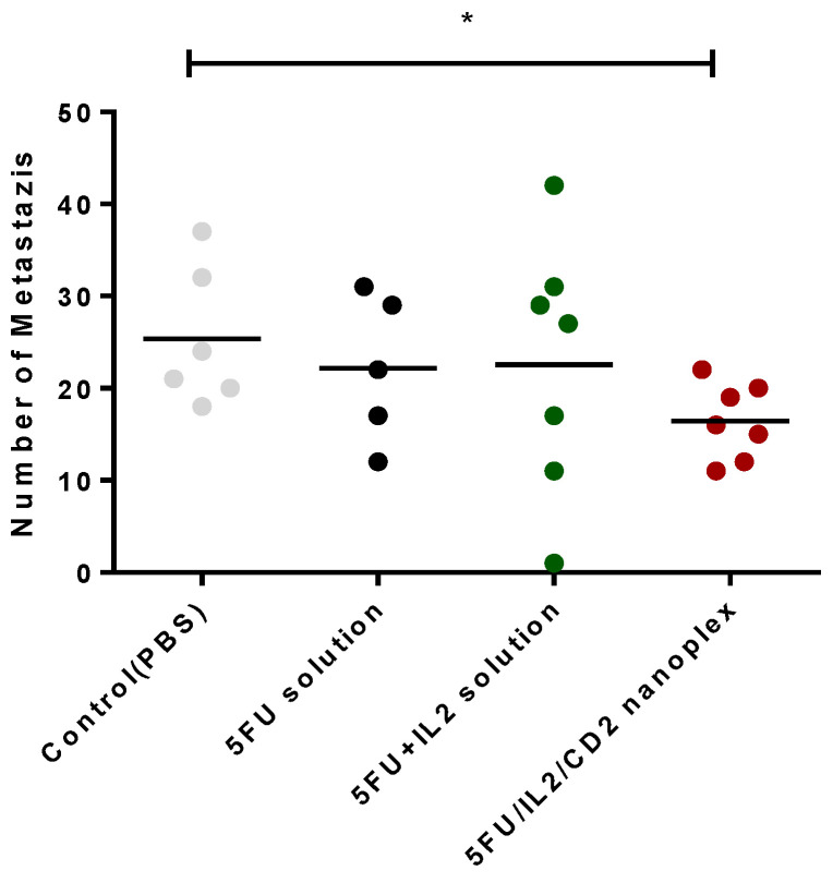
), and metastatic tumor tissue located in the lung parenchyma (**).
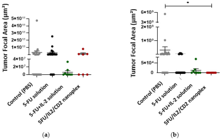
Figure 14
Figure 14
The metastatic areas (*) in liver (a), and lung (b), in treatment groups treated with PBS, 5-FU solution, 5-FU+IL-2 solution and 5FU/IL2/CD2 nanoplex (* p < 0.05).

Similar articles

Cited by

References

    1. Labadie J.D., Savas S., Harrison T.A., Banbury B., Huang Y., Buchanan D.D., Campbell P.T., Gallinger S.J., Giles G.G., Gunter M.J., et al. Genome-wide association study identifies tumor anatomical site-specific risk variants for colorectal cancer survival. Sci. Rep. 2022;12:127. doi: 10.1038/s41598-021-03945-x. - DOI - PMC - PubMed
    1. Akkin S., Varan G., Bilensoy E. A Review on Cancer Immunotherapy and Applications of Nanotechnology to Chemoimmunotherapy of Different Cancers. Molecules. 2021;26:3382. doi: 10.3390/molecules26113382. - DOI - PMC - PubMed
    1. Feng M., Zhao Z., Yang M., Ji J., Zhu D. T-cell-based immunotherapy in colorectal cancer. Cancer Lett. 2021;498:201–209. doi: 10.1016/j.canlet.2020.10.040. - DOI - PubMed
    1. Unal S., Can Ozturk S., Bilgic E., Yanik H., Korkusuz P., Aktas Y., Benito J.M., Esendagli G., Bilensoy E. Therapeutic efficacy and gastrointestinal biodistribution of polycationic nanoparticles for oral camptothecin delivery in early and late-stage colorectal tumor-bearing animal model. Eur. J. Pharm. Biopharm. 2021;169:168–177. doi: 10.1016/j.ejpb.2021.10.010. - DOI - PubMed
    1. Ahmed M. Colon Cancer: A Clinician’s Perspective in 2019. Gastroenterol. Res. 2020;13:1–10. doi: 10.14740/gr1239. - DOI - PMC - PubMed

LinkOut - more resources