Review
. 2023 Jan 20;15(2):348.
doi: 10.3390/pharmaceutics15020348.

Saponins: Research Progress and Their Potential Role in the Post-COVID-19 Pandemic Era

Affiliations
Review

Saponins: Research Progress and Their Potential Role in the Post-COVID-19 Pandemic Era

Daniel Mieres-Castro et al. Pharmaceutics. .

Abstract

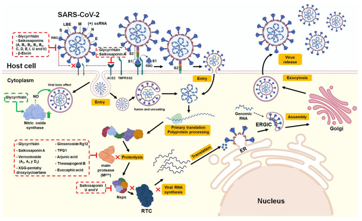
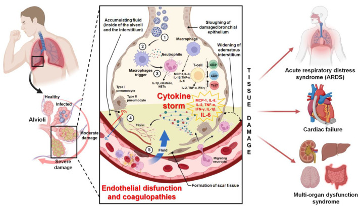
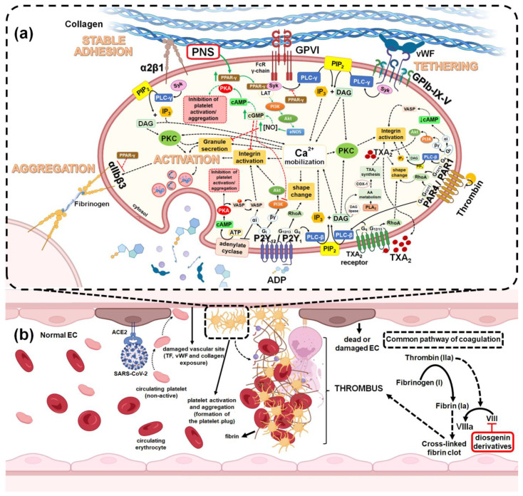
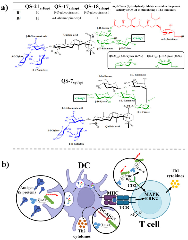
In the post-COVID-19 pandemic era, the new global situation and the limited therapeutic management of the disease make it necessary to take urgent measures in more effective therapies and drug development in order to counteract the negative global impacts caused by severe acute respiratory syndrome coronavirus 2 (SARS-CoV-2) and its new infectious variants. In this context, plant-derived saponins-glycoside-type compounds constituted from a triterpene or steroidal aglycone and one or more sugar residues-may offer fewer side effects and promising beneficial pharmacological activities. This can then be used for the development of potential therapeutic agents against COVID-19, either as a therapy or as a complement to conventional pharmacological strategies for the treatment of the disease and its prevention. The main objective of this review was to examine the primary and current evidence in regard to the therapeutic potential of plant-derived saponins against the COVID-19 disease. Further, the aim was to also focus on those studies that highlight the potential use of saponins as a treatment against SARS-CoV-2. Saponins are antiviral agents that inhibit different pharmacological targets of the virus, as well as exhibit anti-inflammatory and antithrombotic activity in relieving symptoms and clinical complications related to the disease. In addition, saponins also possess immunostimulatory effects, which improve the efficacy and safety of vaccines for prolonging immunogenicity against SARS-CoV-2 and its infectious variants.

Keywords: COVID-19; adjuvant; anti-inflammatory; antithrombotic; antiviral; immunostimulatory; saponin.


Conflict of interest statement

The authors declare no conflict of interest.

Figures

Figure 1
Figure 1
Chemical structure diversity in saponin aglycones and main saponins with anti-SARS-CoV-2 activity. (a) Glycyrrhizin; (b) β-Escin; (c) Saikosaponin A; (d) Saikosaponin B2; (e) Saikosaponin C; (f) Saikosaponin D; (g) Saikosaponin B4; (h) Saikosaponin U; (i) Saikosaponin V; (j) Vernonioside A2; (k) Vernonioside A4; (l) Vernonioside D2; (m) XGG-pentahydroxycycloartane; (n) Ginsenoside Rg12; (o) TPG1; (p) arjunic acid; (q) Theasapogenil B; and (r) euscaphic acid.
Figure 2
Figure 2
Viral replication cycle of SARS-CoV-2 and the effects of saponins on the viral pharmacological targets. According to Mieres-Castro et al. [66], the structure of SARS-CoV-2 consists of a positive-sense single-stranded RNA genome (+ssRNA), a lipid bilayer envelope (LBE), and different structural proteins including spike or S protein, nucleoprotein (N), membrane protein (M), envelope protein (E), and hemagglutinin esterase (not shown in Figure). The +ssRNA is encapsulated by N, while M and E are incorporated during the viral assembly process. The replication cycle begins with the arrival of the SARS-CoV-2 to the target cell. The S protein binds to ACE2 and its receptor on the host cell; it is, then, cleaved by the cell surface serine protease TMPRSS2, forming two subunits, the S1 subunit containing the receptor-binding domain (RBD), and the S2 subunit containing the peptide for binding to the membrane-bound fusion protein of the host cell. This allows entry of the virus into the host cell, either by the formation of an endosome or by fusion of the viral envelope. After the fusion of the membranes of the virus and the host cell, viral RNA is uncoated and released into the cytoplasm in order to initiate the primary translation of co-terminal polyproteins (pp1a/ab) that perform the viral genome replication. After translation, the homodimeric cysteine protease Mpro self-cleaves in order to cleave the polyproteins into Nsps. Different Nsps proteins interact with Nsp12 (also called RNA-dependent RNA polymerase (RdRp)) in order to form the replicase-transcriptase complex (RTC), which synthesizes the full-length viral genome (replication) and subgenomic RNA (transcription). The mRNAs of the viral structural proteins are translated and moved to the endoplasmic reticulum (ER). Genomic RNA is encapsulated with protein N and translocates with structural proteins in the ER-Golgi intermediate compartment (ERGIC) in order to form new virions. Finally, the new viruses are exocytosed from the infected cell and released into the extracellular space in order to infect other cells. The boxes indicate the effects of saponins on the different viral pharmacological targets (red boxes and arrows: inhibition; green boxes and arrows: stimulation). The antiviral mechanisms of action include: inhibiting viral spike glycoprotein and ACE2 in the host cell, thus preventing the binding and entry of the virus; stimulating nitric oxide synthase (iNOS) activity and increasing nitric oxide (NO) in order to cause toxic effects on the virus; and inhibiting Mpro (binding to the active site), thus inhibiting the proteolysis of viral polyproteins that are necessary for virus replication, as well as binding to Nsp15 and the inhibition of its activity in the RTC complex for viral RNA synthesis and replication.
Figure 3
Figure 3
The pathophysiological mechanism of COVID-19 related to clinical complications, such as cytokine storm and thromboembolic coagulopathies. According to Ortega-Paz et al. [68] and Bhaskar et al. [69] the pathophysiological mechanism is characterized by: (1) The viral binding of SARS-CoV-2 to the ACE2 receptor present in type II pneumocytes and vascular endothelial cells of the pulmonary alveoli; (2) immune cells, including macrophages, that identify the virus, become activated and produce acute response chemokines and cytokines; (3) chemokines and cytokines that attract and activate more immune cells, in particular the activation of neutrophils, macrophages, and Th17 cells (and cause the downregulation of CD4+ and CD8+ T cells) and that causes an exacerbated increase in the production of proinflammatory cytokines, thereby creating a cycle of inflammation or a hyperinflammatory response, such as the cytokine storm. This is then characterized by a marked increase in the monocyte chemoattractant protein-1 (MCP-1), interleukin (IL)-8, IL-2, tumor necrosis factor alpha (TNF-α), Interferon gamma (IFN-γ), IL-1β and in particular IL-6; (4) hyper-elevated proinflammatory cytokines could promote and cause cell death, not only of pneumocytes, but also of the endothelial cells of adjacent blood vessels, thereby causing the release of tissue factors (TF), the exposure of subendothelial components (Von Willebrand factor (VWF) and collagen), and fibrin formation. These events stimulate endothelial dysfunction and coagulation (extrinsic and intrinsic pathways), which leads to intravascular thrombosis and, finally, to the development of thromboembolic coagulopathies; (5) weakened and damaged vascular endothelium allows protein-rich fluid to leak and fill the lung cavities (into the alveoli and alveolar interstitium), leading to the damage and death of the bronchial epithelium, formation of scar tissue, and failure of respiratory system (decreased gas exchange), which leads to different pathologies and clinical complications. These are also initially generated at the pulmonary level, but which later led to the heart and other tissues of the body causing multi-organ dysfunction syndrome and death.
Figure 4
Figure 4
Main anti-inflammatory mechanisms of action of saponins in models of pulmonary inflammation. Both triterpene (e.g., Ginsenoside Rh2) and steroidal saponins (e.g., Dioscin) have been shown to reduce histopathological changes associated with inflammation in lung tissue by inhibiting the expression (mRNA and protein) and secretion of proinflammatory cytokines (TNF-α, TGF-β1R, IL-1b, IL-6 and IL-8) and increased expression of anti-inflammatory cytokines (TGF-β1 and IL-10). According to Passos et al. [26], Hsieh et al., [81], and Wang, et al., [104] the anti-inflammatory molecular mechanism of saponins commonly described for pulmonary inflammatory models involves the inactivation of the TLR2 and TLR4 signaling and expression pathway, since either inhibition of the TLR4/MYD88/PI3K/Akt signaling pathway or the TLR2/MYD88/MAPK pathway, thereby inhibiting the activation of NF-κB and AP-1, and consequently inhibiting gene expression associated with proinflammatory (proinflammatory enzymes and cytokines) and apoptotic responses. On the other hand, triterpene and steroidal saponins positively regulate the Nrf2 signaling pathways, promoting the expression of antioxidant genes, causing the expression of antioxidant enzymes and molecules (HO-1, SOD, CAT, GSH-px, GSH), decreasing cell death associated with oxidative stress by the inflammatory process. Red boxes and arrows indicate inhibition; Green boxes and arrows indicate stimulation.
Figure 5
Figure 5
Main antiplatelet-antithrombotic mechanisms of action of saponins in models of pulmonary coagulopathies. (a) Antiplatelet mechanism of saponins. According to Olas et al., [21], Lee et al., [47], and Gómez-Mesa et al., [114], after damage and death of vascular endothelial cells (EC) occurs due to the SARS-CoV-2 infection, the circulating (non-active) platelet recognizes the exposure of components of the subendothelium (TF, vWF and collagen) at the damaged vascular site, promoting different events, including: the initial “anchoring or tethering” of the platelet, characterized by transient interactions between vWF immobilized in the subendothelial matrix and to the GP Ib-IX-V glycoprotein complex present on the platelet surface. This union between “agonists” and their respective platelet receptors triggers different signaling pathways that culminate in “platelet activation and aggregation”, an event that promotes the formation of the “platelet plug”. Platelet activation mediated by “extracellular matrix agonists” (such as vWF and collagen) involves the participation of different glycoproteins (GP Ib-IX-V, GPVI and α2β1, respectively) and their signaling pathway is characterized by the recruitment and activation of Syk, which promotes the activation of the enzyme phospholipase c (PLC)-γ that hydrolyzes membrane phospholipids (Phosphatidylinositol 4,5-bisphosphate, PIP2), producing the mediators diacylglycerol (DAG) and inositol 1,4,5-trisphosphate (IP3) that promote the mobilization of intracellular Ca2+. The increase in intracellular Ca2+ induces the activation of signaling pathways that converge in the activation of the αIIbβ3 integrin, and also promotes changes at the cytoskeleton level, which converge in changes in platelet shape, such as the extension of filopodia and lamellipodia until the complete spreading or full cell spreadin. DAG can be sequentially hydrolyzed by diglyceride lipase and promote the formation of arachidonic acid for the synthesis of Thromboxane A2 (TXA2). Furthermore, the increase in DAG and Ca2+ promote the activation of the protein kinase C (PKC) enzyme, which participates in different signaling pathways, among them it initiates the αIIbβ3 integrin activation cascade to produce platelet aggregation, and also activates signaling pathways that cause the mobilization and secretion of granules (dense and alpha) that contain “soluble agonists” that stimulate and promotes the activation of other platelets. Platelet activation mediated by “soluble agonists” (such as ADP, thrombin and TXA2) involves the participation of different G protein-coupled receptors, which ensure and promotes the amplification of the platelet activation and aggregation response through different signaling pathways. ADP acts as an agonist at P2Y1 (coupled to Gq and G12/13) and P2Y12 (coupled to Gi) receptors. Thrombin acts as an agonist of receptors activated by proteases (PAR) of the PAR1 (coupled to Gq, G12/13 and Gi) and PAR4 (coupled to Gq and G12/13) type. TXA2 acts as an agonist of the TXA2 receptor (coupled to Gq and G12/13). Gq-associated signal transduction is characterized by regulation of intracellular Ca2+ levels and activation of PKC through the PLC-β→IP3/DAG pathway to promote shape change, integrin activation, granule secretion and TXA2 synthesis. G12/13-associated signal transduction is characterized by activation of the GTPase Rho isoform A (RhoA), which promotes platelet shape change by regulating actin cytoskeletal dynamics. Gi-associated signal transduction is characterized by the action of its two subunits, which through different pathways promote the stabilization of platelet aggregation through the activation of the αIIbβ3 integrin. The αi subunit promotes the inhibition of adenylate cyclase and the consequent inhibition of VASP phosphorylation. The βγ subunit promotes the PI3K/Akt signaling pathway. Most studies related to models of pulmonary coagulopathies have been carried out with the main saponins of Panax notoginseng (PNS) (e.i. ginsenoside Rg1, ginsenoside Rb1 and notoginsenoside R1) (red box) [110,112,113]. PNS inhibit collagen-induced platelet activation by promoting a downregulation of signaling downstream of the GPVI receptor. This occurs through an overexpression of PPAR-γ, and upregulation of the PPAR-γ-dependent PI3K/Akt/eNOS pathway, which results in increased nitric oxide (NO) synthesis and consequent increase in cGMP levels, which prevents the activation of platelets through different mechanisms: the indirect increase in cAMP levels by inhibition of phosphodiesterase 3 (PDE-3); the increase in cAMP levels acts synergistically with that of cGMP to inhibit platelet aggregation; inhibits the activation of phosphatidylinositol-3 kinase (PI3K) which leads to the activation of integrin αIIbβ3; and produces phosphorylation of the TXA2 receptor and inhibits its function. In addition, and independently of cGMP production, NO inhibits exocytosis of platelet granules. (b) Mechanism of antithrombotic action of saponins. The antithrombotic activity of saponins on models of pulmonary coagulopathies derives from the ability to inhibit platelet aggregation and formation of the platelet plug (primary hemostasis) and, consequently, prevent the cross-linked fibrin clot and thrombus (secondary hemostasis). However, diosgenin derivatives also have the capacity to inhibit the factor VIII of the common pathway of the coagulation cascade [111]. Factor VIII is activated by thrombin (IIa) and promotes the formation of covalent bonds that cross-link the fibrin polymers (formed from activated monomers) leading to the thrombus formation. Inhibition of factor VIII by diosgenin derivatives (red box) prevents the common pathway of the coagulation cascade and fibrin thrombus formation.
Figure 6
Figure 6
Structure-activity relationship of saponins from Quillaja saponaria and adjuvant mechanism of action of QS-21. According to Wang et al. [121] and Marciani et al. [123], QS-21 is characterized by having a triterpene aglycone nucleus, quillaic acid (black), which contains an aldehyde group attached to C4 (carbonyl C23), a hydroxyl group attached to C16, and a carboxyl group at C28. The quillaic acid core is substituted with a branched trisaccharide (blue: β-D-GlcA, β-D-Xyl β-D-Gal) that is connected via a beta-glucosidic ether bond to the hydroxyl group at C3. It also has a reducing end composed of linear tetrasaccharide (green: β-D-Fuc, β-D-Rha, β-D-Xyl, β-D-Xyl/β-D-Api) beginning with a fucose residue attached by a beta glycosidic ether bond at C28. QS-21 (as well as QS-7, QS-17, and QS-18) represents a mixture of the xylose and apiose substituted variants (65:35). The fucopyranosyl residue at position C28 is in turn connected to an acyl chain with a terminal arabinose (red) through a hydrolytically labile ester. The main structural difference between QS-21 and QS-17/18 is in the C28 oligosaccharide domain. Instead of having a linear tetrasaccharide as in QS-21, QS-17/18 has an additional β-D-glucopyranosyl unit (R1 group) connected to the α-L-rhamnopyranosyl unit at its 3-O position. QS-17 differs from QS-18 only in the R2 group of the acyl side chain, i.e., QS-17 has a disaccharide unit while QS-18 has a monosaccharide unit at the other end of the side chain. On the other hand, QS-7 is characterized by not having an acyl chain in the C28 oligosaccharide domain and the latter with 2 more sugar residues than QS-21 (one β-D-glucopyranosyl unit in position R1 and α unit -L-rhamnopyranosyl connected in position 3-O of the fucose residue). Although QS have a similar adjuvant activity profile (can stimulate a Th1/Th2 response), they have different toxicity profiles (QS-17/18 > QS-21 > QS-7) [121,123]. However, the immunostimulatory activity and toxicity of QS are probably determined by the specific molecular structure of each individual saponin, rather than by the presence/absence of a certain structural feature in the saponin or by its amphipathicity [121,123,130]. On the other hand, considering that a Th1 response is always followed by a Th2 type, and that Th2 can exist as a single type of immunity [123], the structure-activity relationship of QS-21 has shown that acyl chain removal it leads to loss of the ability to stimulate a Th1 response (production of cytotoxic T lymphocyte (CTL)) [121,123,130]. However, when the loss of the acyl chain occurs, the fucosyl unit at the reducing end of the C28 oligosaccharide is exposed for its interaction with C-type lectin receptors (CLRs), specifically, with dendritic cell-specific intercellular adhesion molecule3-grabbing non-integrin (DC-SIGN) receptors, skewing the DCs to a single Th2 immunity [121,123,133]. It has also been described that the aldehyde group in position C4 of the saponin can form an imine with the ɛ-amino group of the T lymphocyte receptor (most likely CD2) triggering the activation of the MAPK signaling pathway, changes in transport channels of K+ and Na+, and finally skewing the activated T cells to a Th1 immunity with increased production of Th1 cytokines [49,122,124]. (b) Immunostimulatory mechanism of action for QS-21. According to Zhang et al., [138], the mechanism of action for QS-21-based adjuvants (AS01, ISCOM, ISCOMATRIX, and Matrix M) is characterized by the following: DCs arrive at the site where the vaccine was inoculated, and through endocytosis, they incorporate into their interior the exogenous protein antigens (e.g., S protein from SARS-CoV-2) and QS-21. Following QS-21-mediated endosomal membrane disruption (attributed to acyl chain action), protein antigens are degraded by the proteasome into smaller peptide fragments. Protein fragments are transported to the ER by carrier molecules, where chaperones facilitate their binding to newly synthesized MHC-I molecules for vesicular migration through the Golgi apparatus to the cell surface. Finally, surface-exposed peptide epitopes of DC in association with MHC-I molecules are presented to naive CD8+ T cells (cross-presentation) via the T cell receptor (TCR) stimulating a Th1 response. QS-21 can act on both DCs and T cells. Oligosaccharide chains of saponins can activate DC by binding to surface-expressed DC-SING, skewing the DC to a Th2 response. The aldehyde group of quillaic acid forms an imine with an amino group of a T-cell surface receptor (CD2), which sends an intracellular signal that activates MAPK/ERK2 and with changes in cellular K+ and Na+ transport, skewing the T cells to a Th1 response.

Similar articles

Cited by

References

    1. Oleszek M., Oleszek W. Saponins in Food. In: Xiao J., Sarker S.D., Asakawa Y., editors. Handbook of Dietary Phytochemicals. 1st ed. Volume 1. Springer; Singapore: 2019. pp. 1–40. - DOI
    1. Vincken J.-P., Heng L., de Groot A., Gruppen H. Saponins, classification and occurrence in the plant kingdom. Phytochemistry. 2007;68:275–297. doi: 10.1016/j.phytochem.2006.10.008. - DOI - PubMed
    1. Sparg S.G., Light M.E., van Staden J. Biological activities and distribution of plant saponins. J. Ethnopharmacol. 2004;94:219–243. doi: 10.1016/j.jep.2004.05.016. - DOI - PubMed
    1. Yang Y., Laval S., Yu B. Chemical Synthesis of Saponins. Adv. Carbohydr. Chem. Biochem. 2014;71:137–226. doi: 10.1016/b978-0-12-800128-8.00002-9. - DOI - PubMed
    1. Osbourn A. Saponins and plant defence—a soap story. Trends Plant Sci. 1996;1:4–9. doi: 10.1016/S1360-1385(96)80016-1. - DOI

LinkOut - more resources