

Choosing an Optimal Solvent Is Crucial for Obtaining Cell-Penetrating Peptide Nanoparticles with Desired Properties and High Activity in Nucleic Acid Delivery
- PMID: 36839718
- PMCID: PMC9963036
- DOI: 10.3390/pharmaceutics15020396
Choosing an Optimal Solvent Is Crucial for Obtaining Cell-Penetrating Peptide Nanoparticles with Desired Properties and High Activity in Nucleic Acid Delivery
Abstract
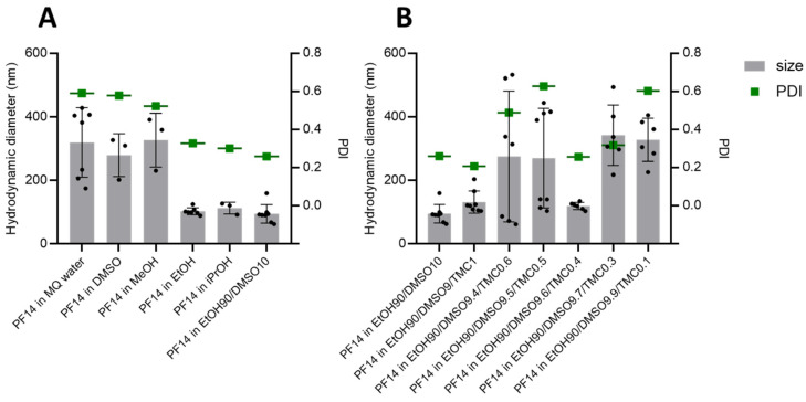
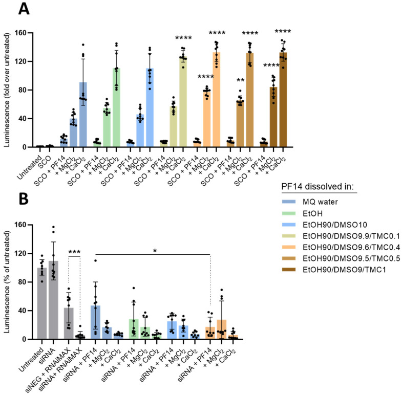
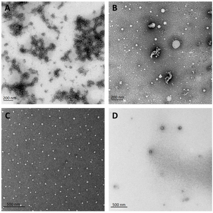
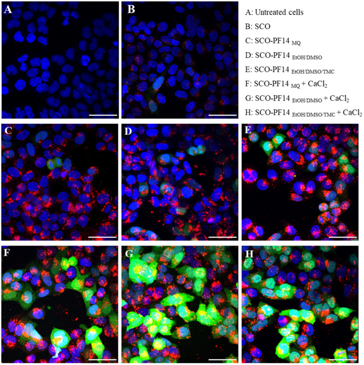
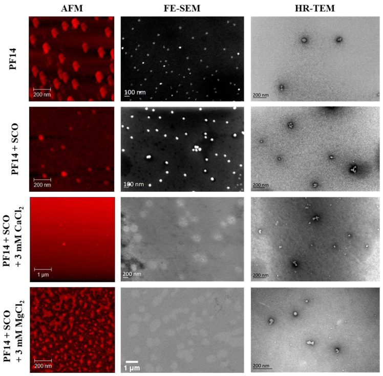
Cell-penetrating peptides (CPPs) are highly promising transfection agents that can deliver various compounds into living cells, including nucleic acids (NAs). Positively charged CPPs can form non-covalent complexes with negatively charged NAs, enabling simple and time-efficient nanoparticle preparation. However, as CPPs have substantially different chemical and physical properties, their complexation with the cargo and characteristics of the resulting nanoparticles largely depends on the properties of the surrounding environment, i.e., solution. Here, we show that the solvent used for the initial dissolving of a CPP determines the properties of the resulting CPP particles formed in an aqueous solution, including the activity and toxicity of the CPP-NA complexes. Using different biophysical methods such as dynamic light scattering (DLS), atomic force microscopy (AFM), transmission and scanning electron microscopy (TEM and SEM), we show that PepFect14 (PF14), a cationic amphipathic CPP, forms spherical particles of uniform size when dissolved in organic solvents, such as ethanol and DMSO. Water-dissolved PF14, however, tends to form micelles and non-uniform aggregates. When dissolved in organic solvents, PF14 retains its α-helical conformation and biological activity in cell culture conditions without any increase in cytotoxicity. Altogether, our results indicate that by using a solvent that matches the chemical nature of the CPP, the properties of the peptide-cargo particles can be tuned in the desired way. This can be of critical importance for in vivo applications, where CPP particles that are too large, non-uniform, or prone to aggregation may induce severe consequences.
Keywords: cell-penetrating peptides; nanoparticle formation; nucleic acid delivery; solvent.
Conflict of interest statement
The funders had no role in the design of the study; in the collection, analyses, or interpretation of data; in the writing of the manuscript; or in the decision to publish the results. Authors A.G., J.J., M.P., M.M. and S.K.T.S.W. have applied for patent PCT/SE2022/050657 based on some of the results presented in this manuscript. This patent application is publicly available on the wipo website. Authors A.G., J.J. and S.K.T.S.W. are shareholders in the company CellPept Sweden AB. This company had no role in the design of the study; in the collection, analyses, or interpretation of data; in the writing of the manuscript; or in the decision to publish the results.
Figures
Similar articles
-
Engineered PepFect14 analog for efficient cellular delivery of oligonucleotides.Biomed Pharmacother. 2025 Mar;184:117872. doi: 10.1016/j.biopha.2025.117872. Epub 2025 Feb 1. Biomed Pharmacother. 2025. PMID: 39891949
-
Peptide vectors for the nonviral delivery of nucleic acids.Acc Chem Res. 2012 Jul 17;45(7):1048-56. doi: 10.1021/ar2002304. Epub 2012 Mar 28. Acc Chem Res. 2012. PMID: 22455499
-
Genetic, cellular, and structural characterization of the membrane potential-dependent cell-penetrating peptide translocation pore.Elife. 2021 Oct 29;10:e69832. doi: 10.7554/eLife.69832. Elife. 2021. PMID: 34713805 Free PMC article.
-
Cell penetration: scope and limitations by the application of cell-penetrating peptides.J Pept Sci. 2014 Oct;20(10):760-84. doi: 10.1002/psc.2672. Epub 2014 Aug 11. J Pept Sci. 2014. PMID: 25112216 Review.
-
Recent in vivo advances in cell-penetrating peptide-assisted drug delivery.Expert Opin Drug Deliv. 2016;13(3):373-87. doi: 10.1517/17425247.2016.1125879. Epub 2015 Dec 22. Expert Opin Drug Deliv. 2016. PMID: 26634750 Review.
Cited by
-
PepFect14 mediates the delivery of mRNA into human primary keratinocytes and in vivo.Front Pharmacol. 2023 Jul 13;14:1219761. doi: 10.3389/fphar.2023.1219761. eCollection 2023. Front Pharmacol. 2023. PMID: 37521463 Free PMC article.
References
-
- U.S. Food and Drug Administration What Is Gene Therapy? [(accessed on 1 January 2023)]; Available online: https://www.fda.gov/vaccines-blood-biologics/cellular-gene-therapy-produ....
Grants and funding
LinkOut - more resources
Full Text Sources
Miscellaneous
