. 2023 Jan 25;15(2):407.
doi: 10.3390/pharmaceutics15020407.

Nanostructured Lipid Carriers for Enhanced Transscleral Delivery of Dexamethasone Acetate: Development, Ex Vivo Characterization and Multiphoton Microscopy Studies

Affiliations

Nanostructured Lipid Carriers for Enhanced Transscleral Delivery of Dexamethasone Acetate: Development, Ex Vivo Characterization and Multiphoton Microscopy Studies

Felipe M González-Fernández et al. Pharmaceutics. .

Abstract

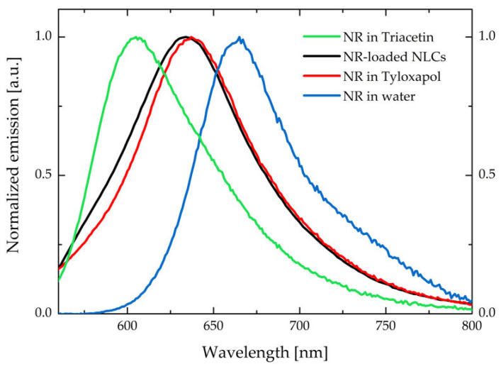
Corticosteroids, although highly effective for the treatment of both anterior and posterior ocular segment inflammation, still nowadays struggle for effective drug delivery due to their poor solubilization capabilities in water. This research work aims to develop nanostructured lipid carriers (NLC) intended for periocular administration of dexamethasone acetate to the posterior segment of the eye. Pre-formulation studies were initially performed to find solid and liquid lipid mixtures for dexamethasone acetate solubilization. Pseudoternary diagrams at 65 °C were constructed to select the best surfactant based on the macroscopic transparency and microscopic isotropy of the systems. The resulting NLC, obtained following an organic solvent-free methodology, was composed of triacetin, Imwitor® 491 (glycerol monostearate >90%) and tyloxapol with Z-average = 106.9 ± 1.2 nm, PDI = 0.104 ± 0.019 and zeta potential = -6.51 ± 0.575 mV. Ex vivo porcine sclera and choroid permeation studies revealed a considerable metabolism in the sclera of dexamethasone acetate into free dexamethasone, which demonstrated higher permeation capabilities across both tissues. In addition, the NLC behavior once applied onto the sclera was further studied by means of multiphoton microscopy by loading the NLC with the fluorescent probe Nile red.

Keywords: NLC; dexamethasone; dexamethasone acetate; ex vivo; multiphoton microscopy; ocular delivery; posterior segment; tyloxapol.


Conflict of interest statement

The authors declare no conflict of interest. Felipe M. González-Fernández and Paolo Gasco are from Nanovector S.r.l.: the company had no role in the design of the study; in the collection, analysis, or interpretation of data; in the writing of the manuscript, or in the decision to publish the results.

Figures

Figure 1
Figure 1
Solubility of dexamethasone acetate in diverse lipid matrices, liquid (l) or solid (s) at room temperature. Mean values are displayed ± standard deviation (n = 3).
Figure 2
Figure 2
Pseudoternary phase diagrams for the selected lipid mix (80% GMS: 20% triacetin)/surfactant/water mixtures at 65 °C: (a) Tween® 20, (b) Tween® 80, and (c) tyloxapol. Green points represent the studied mixtures that yield macroscopically transparent systems defining an area (indicated in grey) of possible microemulsion templates for the NLC production.
Figure 3
Figure 3
Pseudoternary phase diagrams for the water/tyloxapol/lipid mix pseudoternary systems when lipid mix was composed of (a) 80% GMS:20% triacetin and (b) 60% GMS:40% triacetin. Note how the large gel-like region (light blue) in (a) disappeared once GMS content was reduced (b).
Figure 4
Figure 4
Pseudoternary phase diagram corrected after microscopic characterization under polarized light (left) and different micrographs of the observed anisotropic structures (right).
Figure 5
Figure 5
Pseudoternary phase diagram (a) with the composition of the six studied formulation candidates and (b) size and PDI values of the obtained NLC at t = 24 h after production. Data values are mean ± standard deviation (n = 3).
Figure 6
Figure 6
Comparison between emission spectra of NR in triacetin, NLC (aqueous suspension), tyloxapol (2.7 mg/mL in water), and water (with <1% DMSO). The spectra have been collected exciting at 500 nm.
Figure 7
Figure 7
Percentage recovery of DexAc (orange) and Dex (grey) from fresh (a) and thawed (b) isolated sclera and choroid. Data were collected by applying either DexAc or Dex to ocular tissues.
Figure 8
Figure 8
MPM images of porcine sclera (excitation wavelength: 1100 nm) after 2 h of contact with NR-loaded NLC (0.1 μg/mg triacetin), at a depth of 40 μm (a,b,c) and 150 μm (d,e,f) from the tissue surface, respectively: (a and d) Signal detected in the green channel, mainly due to the SHG of collagen fibers; (b and e) Signal collected in the red channel, which is attributed to NR emission; (c and f) Corresponding channels overlay, showing NR distribution (red) between collagen fibers (green). Images size: 170 μm × 170 μm.
Figure 9
Figure 9
MPM images of porcine sclera after 2 h contact with NR-loaded NLC (0.1 μg/mg triacetin). (a) Volume rendering of the tissue reconstructed from the Z-stack (Z-step: 1 µm, total depth: 142 µm), (b) image of the tissue surface (size: 82 μm × 82 μm), (c) YZ slice extracted from the Z-stack reported in panel a (image size: 512 μm × 142 μm), (d) image acquired 30 μm below the surface of the tissue (size: 92 μm × 92 μm). The excitation wavelength was set to 1100 nm for all the collected images.
Figure 10
Figure 10
MPM images of porcine sclera (depth of 40 μm, image size: 512 μm × 512 μm and excitation wavelength of 1100 nm) after 2 h of contact with NLC: (a) blank NLC, (b) NR-loaded NLC (starting from 0.1 µg/mg NR), (c) NR-loaded NLC (starting from 0.5 µg/mg NR). Panels (d and e) report the TPEF (broad band)/SHG (intense sharp peak; peak maximum is not shown) profiles obtained with the spectral detector in correspondence of images (a,b,c) focal planes, exciting at 1080 nm. The spectra in panel e have been normalized and compared to the emission signal collected with the microscope from a NR-loaded NLC aqueous suspension. Images were acquired in the same experimental conditions (detector gains and laser power), as well as emission spectra.

Similar articles

Cited by

References

    1. Fung A.T., Tran T., Lim L.L., Samarawickrama C., Arnold J., Gillies M., Catt C., Mitchell L., Symons A., Buttery R., et al. Local delivery of corticosteroids in clinical ophthalmology: A review. Clin. Exp. Ophthalmol. 2020;48:366–401. doi: 10.1111/ceo.13702. - DOI - PMC - PubMed
    1. Gaballa S.A., Kompella U.B., Elgarhy O., Alqahtani A.M., Pierscionek B., Alany R.G., Abdelkader H. Corticosteroids in ophthalmology: Drug delivery innovations, pharmacology, clinical applications, and future perspectives. Drug Deliv. Transl. Res. 2021;11:866–893. doi: 10.1007/s13346-020-00843-z. - DOI - PubMed
    1. Villanueva J.R., Villanueva L.R., Navarro M.G. Pharmaceutical technology can turn a traditional drug, dexamethasone into a first-line ocular medicine. A global perspective and future trends. Int. J. Pharm. 2017;516:342–351. doi: 10.1016/j.ijpharm.2016.11.053. - DOI - PubMed
    1. Usayapant A., Karana A.H., Narurkar M.M. Effect of 2-hydroxypropyl-β-cyclodextrin on the ocular absorption of dexamethasone and dexamethasone acetate. Pharm. Res. 1991;8:1495–1499. doi: 10.1023/A:1015838215268. - DOI - PubMed
    1. Leibowitz H.M., Kupferman A., Stewart R.H., Kimbrough R.L. Evaluation of Dexamethasone Acetate as a Topical Ophthalmic Formulation. Am. J. Ophthalmol. 1978;86:418–423. doi: 10.1016/0002-9394(78)90249-0. - DOI - PubMed

LinkOut - more resources