Review
. 2023 Jan 26;15(2):413.
doi: 10.3390/pharmaceutics15020413.

Biomaterials Based on Organic Polymers and Layered Double Hydroxides Nanocomposites: Drug Delivery and Tissue Engineering

Affiliations
Review

Biomaterials Based on Organic Polymers and Layered Double Hydroxides Nanocomposites: Drug Delivery and Tissue Engineering

Vera Regina Leopoldo Constantino et al. Pharmaceutics. .

Abstract

The development of biomaterials has a substantial role in pharmaceutical and medical strategies for the enhancement of life quality. This review work focused on versatile biomaterials based on nanocomposites comprising organic polymers and a class of layered inorganic nanoparticles, aiming for drug delivery (oral, transdermal, and ocular delivery) and tissue engineering (skin and bone therapies). Layered double hydroxides (LDHs) are 2D nanomaterials that can intercalate anionic bioactive species between the layers. The layers can hold metal cations that confer intrinsic biological activity to LDHs as well as biocompatibility. The intercalation of bioactive species between the layers allows the formation of drug delivery systems with elevated loading capacity and modified release profiles promoted by ion exchange and/or solubilization. The capacity of tissue integration, antigenicity, and stimulation of collagen formation, among other beneficial characteristics of LDH, have been observed by in vivo assays. The association between the properties of biocompatible polymers and LDH-drug nanohybrids produces multifunctional nanocomposites compatible with living matter. Such nanocomposites are stimuli-responsive, show appropriate mechanical properties, and can be prepared by creative methods that allow a fine-tuning of drug release. They are processed in the end form of films, beads, gels, monoliths etc., to reach orientated therapeutic applications. Several studies attest to the higher performance of polymer/LDH-drug nanocomposite compared to the LDH-drug hybrid or the free drug.

Keywords: anionic clays; composite biomaterials; drug delivery system; hydrotalcite; intercalation compounds; layered double hydroxides; layered materials; nano-based drug carrier; nanocomposites; tissue engineering.


Conflict of interest statement

The authors declare no conflict of interest.

Figures

Figure 1
Figure 1
Classification of biomaterials by chemical composition. Materials having two or more phases are nominated (nano)composites. Biomaterials can have several properties guiding a particular application.
Figure 2
Figure 2
Biomaterials-based nanocomposites focused on this work: 2D nanomaterials and organic polymers.
Figure 3
Figure 3
Chemical structures of some natural and synthetic organic polymers are used to develop biomaterial-based delivery systems and biomaterials for tissue engineering.
Figure 4
Figure 4
(A) Variable chemical and physical parameters considered in the design of the nanoparticles; (B) Nanoparticles can have different chemical natures, shapes, porosity, dimensionality etc.
Figure 5
Figure 5
Experimental approaches to produce nanocomposites based on organic polymer and inorganic nanofillers: in situ methods (left side) and ex situ methods (right side).
Figure 6
Figure 6
Multilayer composites are obtained by methods that alternate de phase’s deposition to form an object.
Figure 7
Figure 7
LDH structural aspects: (A) [M(OH)6] units, (B) edge-sharing among octahedra, (C) sequence of the layers stacking and (D) face-to-face arrangement of the layers. Uppercase symbols A, B, and C represent hydroxide ion positions; P indicates a prismatic site.
Figure 8
Figure 8
LDH materials can be intercalated with inorganic or organic anions to neutralize the layer’s charge.
Figure 9
Figure 9
Schematic representation of methods used for (A) preparation of PAM/MgRAl-LDH hydrogel by in situ polymerization; AM: acrylamide; APS: ammonium peroxydisulfate; TEMED: N,N,N′,N′-tetramethyl-ethylenediamine (reprinted from Wu and Chen [120] with permission of Wiley-VCH GmbH) and (B) PEGylation of LDH nanolayers (reprinted from Cao et al. [121] with permission of Elsevier Inc.).
Figure 10
Figure 10
Suitable LDH properties for therapeutic and/or diagnosis purposes.
Figure 11
Figure 11
Schematic illustration of (A) LDH tablet implanted in the intramuscular abdominal region of rats and (B) microcirculation monitoring by SDF videomicroscopy image (both figures reprinted from Constantino et al. [152] with permission of World Scientific Publishing Co.); (C) Iron histochemistry findings following A-Mg4Al2-Cl LDH, B-Zn4Al2-Cl LDH, C-Mg4FeAl-Cl LDH and D-Zn4FeAl-Cl LDH tablets implants between abdominal wall intermuscular spaces, after 28th P.O. The white dotted line with the blue arrow indicates the macroscopic appearance of the implanted LDH tablet and the cut line for histological processing. LDH tablet (*); positive staining (→ arrow black); positive staining in fibroblast (→ arrow green); positive staining in ECM matrix (→ yellow); perimysium between abdominal wall muscle layers (►). Prussian blue staining (reprinted from Figueiredo et al. [143] with permission of American Chemical Society).
Figure 12
Figure 12
Strategies to modify the LDH surfaces through electrostatic or covalent linkages: (A) hydrophobization of surfaces by anionic surfactants; (B) hydrophobization of surfaces by anionic drugs having hydrophobic groups; (C) surfaces modification by silanization reaction; (D) surface modification with neutral polymers having hydrophobic and hydrophilic segments; (E) surface modification with charged polymers; and (F) surface functionalization with targeting molecules.
Figure 13
Figure 13
Polymer/LDH nanocomposites: (A) microcapsules and microspheres-based delivery systems; (B) intercalated and exfoliated nanocomposites.
Figure 14
Figure 14
Classification of the Drug Delivery Systems considering the administration route.
Figure 15
Figure 15
Mechanisms for the drug delivery from DDS based on polymer/LDH nanocomposites.
Figure 16
Figure 16
(A) Amoxicillin drug release profile at in vitro simulated gastrointestinal conditions (2 h at pH 1.2, 2 h at pH 6.8 and 4 h at pH 7.4, simulating the gastric juice and the first and second zone of intestinal fluid, respectively) from LDH-amoxicillin and CMC-zein coated LDH-amoxicillin systems (reprinted from Rebitski et al. [229] with permission of Elsevier) and (B) Cumulative DOX release profile at pH 5.5 and 7.4 in the absence and presence of NIR radiation from the LDH-based chemophotothermal nanocomposite system (reprinted from Anirudhan and Chithra-Sekhar [230] with permission of Elsevier).
Figure 17
Figure 17
Progression of wound healing: some characteristics of each step of wound healing (top description) and the corresponding possible contribution of polymeric matrices and LDH (bottom description).
Figure 18
Figure 18
(A) Preparation of the composite membranes and (B) NAP anions release percentage as a function of time for the different composites (reprinted from Figueiredo et al. [73] with permission of Elsevier).
Figure 19
Figure 19
Representative photographs of wounds treated by control, CMC-PEO, and IBU/CMC-PEO, LDH-VAN/CMC-PEO, and LDH-VAN/IBU/CMC-PEO groups on days 0, 3, 7, 10, 14, and 19. (reprinted from Yoosefi et al. [269] with permission of Elsevier).
Figure 20
Figure 20
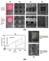
(A) Structural characterization of the PCL-based scaffolds: (a) macroscopic images; (b) top view (scale bar: 500 μm), (c) cross-sectional view (scale bar: 500 μm), and (d) top view of a pore of a 3D-printed grid (scale bar: 200 μm), and electrospun nanofibers (scale bar: 20 μm); (B) In vitro drug release: (a) cumulative release of Pam and (b) images of PCL/LDH-Pam fibers in the scaffold at the water penetration and degradation phases (scale bar: 200 μm), after 28 days of immersing in PBS solution (reprinted from Belgheisi et al. [280] with permission of Elsevier).

References

    1. [(accessed on 18 November 2022)]. Available online: https://sustainabledevelopment.un.org/outcomedocuments/agenda21.
    1. Ghasemi-Mobarakeh L., Kolahreez D., Ramakrishna S., Williams D. Key Terminology in Biomaterials and Biocompatibility. Curr. Opin. Biomed. Eng. 2019;10:45–50. doi: 10.1016/j.cobme.2019.02.004. - DOI
    1. Williams D.F. There Is No Such Thing as a Biocompatible Material. Biomaterials. 2014;35:10009–10014. doi: 10.1016/j.biomaterials.2014.08.035. - DOI - PubMed
    1. IUPAC . In: Compendium of Chemical Terminology. 2nd ed. McNaught A.D., Wilkinson A., editors. Blackwell Scientific Publications; Oxford, UK: 1997. the “Gold Book”.
    1. Han X., Alu A., Liu H., Shi Y., Wei X., Cai L., Wei Y. Biomaterial-Assisted Biotherapy: A Brief Review of Biomaterials Used in Drug Delivery, Vaccine Development, Gene Therapy, and Stem Cell Therapy. Bioact. Mater. 2022;17:29–48. doi: 10.1016/j.bioactmat.2022.01.011. - DOI - PMC - PubMed

LinkOut - more resources