Review
. 2023 Jan 27;15(2):423.
doi: 10.3390/pharmaceutics15020423.

Development of Janus Particles as Potential Drug Delivery Systems for Diabetes Treatment and Antimicrobial Applications

Affiliations
Review

Development of Janus Particles as Potential Drug Delivery Systems for Diabetes Treatment and Antimicrobial Applications

Kei Xian Tan et al. Pharmaceutics. .

Abstract

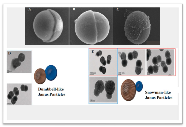
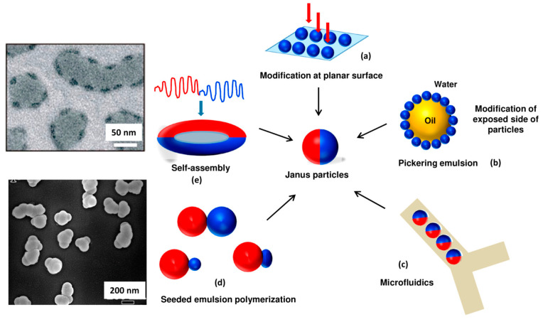
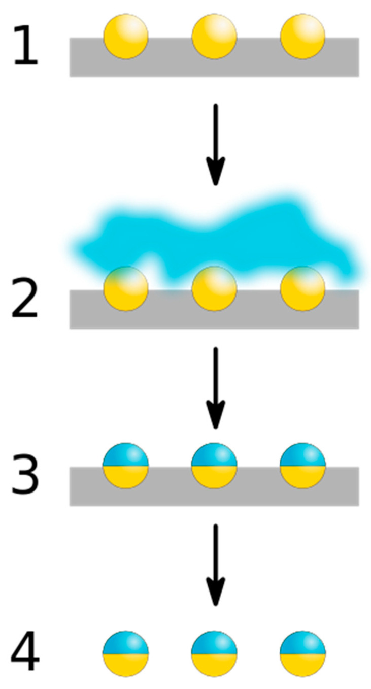
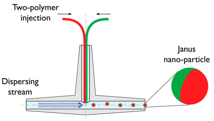
Janus particles have emerged as a novel and smart material that could improve pharmaceutical formulation, drug delivery, and theranostics. Janus particles have two distinct compartments that differ in functionality, physicochemical properties, and morphological characteristics, among other conventional particles. Recently, Janus particles have attracted considerable attention as effective particulate drug delivery systems as they can accommodate two opposing pharmaceutical agents that can be engineered at the molecular level to achieve better target affinity, lower drug dosage to achieve a therapeutic effect, and controlled drug release with improved pharmacokinetics and pharmacodynamics. This article discusses the development of Janus particles for tailored and improved delivery of pharmaceutical agents for diabetes treatment and antimicrobial applications. It provides an account of advances in the synthesis of Janus particles from various materials using different approaches. It appraises Janus particles as a promising particulate system with the potential to improve conventional delivery systems, providing a better loading capacity and targeting specificity whilst promoting multi-drugs loading and single-dose-drug administration.

Keywords: Janus particle; drug delivery; formulation; pharmacodynamics; pharmacokinetics.


Conflict of interest statement

The authors declare no conflict of interest.

Figures

Figure 1
Figure 1
Anisotropic Janus particles with a variety of structural conformations. (AC) SEM images of various spherical Janus particles and (D,E) TEM images of dumbbell-like and snowman-like Janus particles. Reproduced with permission from [19], © Wiley (2017) and © Royal Society of Chemistry (2010).
Figure 2
Figure 2
Interparticle interaction of stable Janus particles in fluid interfaces. (a) Surface group dissociation leading to dipole-dipole-based repulsive interactions; (b) Oil phase-based contact of particle surface for water entrapment leading to long-range repulsive interactions; (c) Generate attractive capillary interactions via gravitational forces deformed interface and (d) Surface roughness improved by interface undulations to enhance the stability of Janus particles via capillary interaction. Reproduced with permission from Correia et al. (2021), © MDPI, 2021 [21] (Open access).
Figure 3
Figure 3
Synthesis and characterization of a specific Janus particle. (a) Schematics of glucose oxidase (GOx)-coated Janus particle formation, where (i) streptavidin-coated colloids with a magnet, (ii) Modification of the surface exposed to the solvent with biotinylated enzymes, (iii) Janus particles partially covered with enzymes after removal of the magnet, (iv) Biotinylated arginine-glycine-aspartic acid (RGDS) peptides; Fluorescence microscope image of (b) bovine serum albumin and asymmetric arrangement of GOx on the Janus particle surface; and (c) Janus particle modified only with GOx. Reproduced with permission from Rucinskaite et al. (2017), © Royal Society of Chemistry (RSC), 2017 [34].
Figure 4
Figure 4
Nanocorals in Janus structure as multifunctional targeting, sensing, and drug delivery nanoprobe; Inset: Scanning electron microscope (SEM) images of Janus structured nano-coral probes. Reproduced with permission from Wu et al. (2010), © Wiley, 2010 [42].
Figure 5
Figure 5
Overview of production approaches of Janus particles. (a) Modification and fixation on solid substrates, (b) Pickering emulsion method, (c) Microfluidic method, (d) Seeded emulsion polymerization approach, (e) Self-assembly approach. Reproduced with permission from Agarwal and Agarwal (2019), © American Chemical Society, 2019 [53].
Figure 6
Figure 6
Schematic representation of the crucial steps in the masking technique to form Janus particles. (1) Exposure of a hemisphere of homogeneous nanoparticles; (2) Application of masking techniques; (3) Chemical modification of particle properties via masking process; and (4) Removal of the masking agent, resulting in Janus particle formation.
Figure 7
Figure 7
Self-assembly of Janus particles into clusters and chains at a patch ratio of 0.3. (a) Bright-field images above and simulation plots below show self-assembled structures of Janus particles at different temperatures; (b) Proportion in clusters of different sizes as a function of temperature. For cluster sizes greater than 20, histogram bars integrate over ten or more bins: 21–30, 31–40, 41–50, and >50. (c) Snapshots and (d) Figures show the formation of chain structures by collective polymerization through cluster addition and reconfiguration. Red arrow represents the formation of chain structure. Reproduced with permission from Oh et al. (2019), ©Nature Communication, 2019 [63].
Figure 8
Figure 8
Schematic of the one-step fluidic nanoprecipitation system to fabricate Janus particles. Reproduced with permission from [64], © American Chemical Society, 2012.
Figure 9
Figure 9
Schemes showing the thermally induced phase separation for the formation of Janus droplets. Reproduced with permission from Zhang et al. (2022), ©American Chemical Society (ACS), 2022 [70].
Figure 10
Figure 10
Schematic Representation of magnesium/platinum-based Janus micromotor assisted glucose biosensor in human serum using screen printed electrode. Reproduced with permission from Kong et al. (2019), ©American chemical society (ACS), 2019 [105].
Figure 11
Figure 11
Illustrations of (A) composite dressing structure with modified Janus membrane and (B) absorption of massive wound exudates from the wound bed. SAP—sodium polyacrylate superabsorbent particles; BG—silicate bioglass; PU—polyurethane; dCA—deacetylated cellulose acetate layer. Reprinted with permission from [116], © Wiley, 2020.
Figure 12
Figure 12
Silver-silicon dioxide Janus nanoparticles with distinct functional groups, such as amine, thiol, and epoxy synthesized via the Pickering emulsion method. (a) Mechanistic attachment of amine-thiol-epoxy functional ATE-silver (Ag) Janus on cotton fabric, (b) amine functionalized silver Janus interacts with cotton fabric via electrostatic attraction and (c) attachment of amine functionalized silver Janus on epoxy functionalized cotton fabric. Reproduced with permission from [122], ©Elsevier (2018).
Figure 13
Figure 13
Schematics of optical biosensing platform based on the LAMP assay. (A) Synthesis of amplified LAMP product and (B) Preparation of non-spectroscopic signaling probe. Reproduced with permission from Chun et al. (2018), ©American Chemical Society, 2018.

References

    1. Perro A., Reculusa S., Ravaine S., Bourgeat-Lami E., Duguet E. Design and synthesis of Janus micro-and nanoparticles. J. Mater. Chem. 2005;15:3745–3760. doi: 10.1039/b505099e. - DOI
    1. Auschra S. Von der Fakultät für Physik und Geowissenschaften. der Universität Leipzig; Borna, Germany: 1988. On the Properties of Self-Thermophoretic Janus Particles.
    1. De Gennes P.-G. Soft matter. Science. 1992;256:495–497. doi: 10.1126/science.256.5056.495. - DOI - PubMed
    1. Rahiminezhad Z., Tamaddon A.M., Borandeh S., Abolmaali S.S. Janus nanoparticles: New generation of multifunctional nanocarriers in drug delivery, bioimaging and theranostics. Appl. Mater. Today. 2020;18:100513. doi: 10.1016/j.apmt.2019.100513. - DOI
    1. Tran L.-T.-C., Lesieur S., Faivre V. Janus nanoparticles: Materials, preparation and recent advances in drug delivery. Expert Opin. Drug Deliv. 2014;11:1061–1074. doi: 10.1517/17425247.2014.915806. - DOI - PubMed

LinkOut - more resources