. 2023 Feb 7;15(2):554.
doi: 10.3390/pharmaceutics15020554.

Cannabidiol-Loaded Extracellular Vesicles from Human Umbilical Cord Mesenchymal Stem Cells Alleviate Paclitaxel-Induced Peripheral Neuropathy

Affiliations

Cannabidiol-Loaded Extracellular Vesicles from Human Umbilical Cord Mesenchymal Stem Cells Alleviate Paclitaxel-Induced Peripheral Neuropathy

Anil Kumar Kalvala et al. Pharmaceutics. .

Erratum in

Abstract

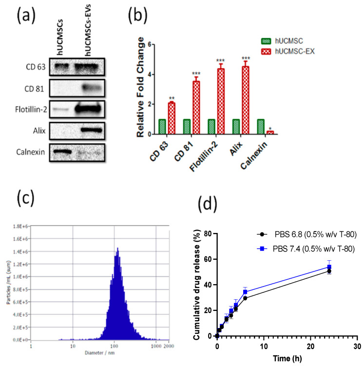
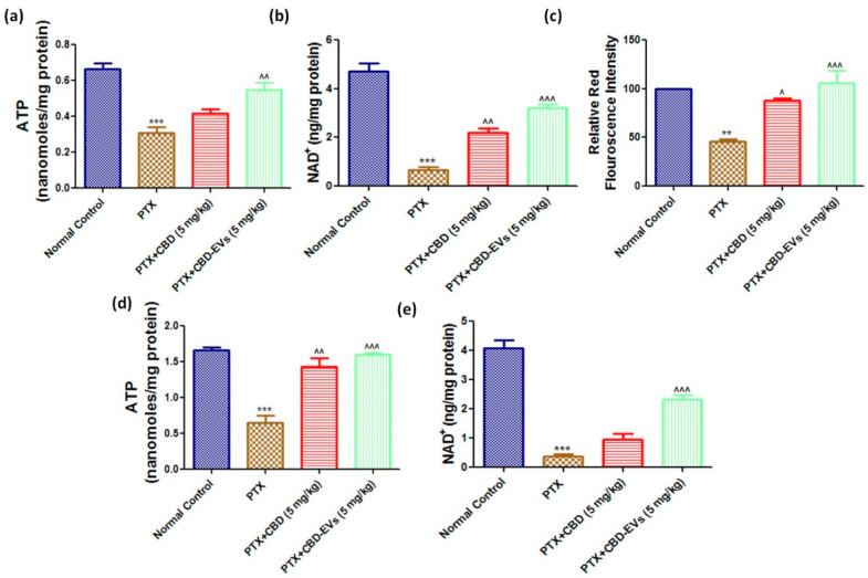
In cancer patients, chronic paclitaxel (PTX) treatment causes excruciating pain, limiting its use in cancer chemotherapy. The neuroprotective potential of synthetic cannabidiol (CBD) and CBD formulated in extracellular vesicles (CBD-EVs) isolated from human umbilical cord derived mesenchymal stem cells was investigated in C57BL/6J mice with PTX-induced neuropathic pain (PIPN). The particle size of EVs and CBD-EVs, surface roughness, nanomechanical properties, stability, and release studies were all investigated. To develop neuropathy in mice, PTX (8 mg/kg, i.p.) was administered every other day (four doses). In terms of decreasing mechanical and thermal hypersensitivity, CBD-EVs treatment was superior to EVs treatment or CBD treatment alone (p < 0.001). CBD and CBD-EVs significantly reduced mitochondrial dysfunction in dorsal root ganglions and spinal homogenates of PTX-treated animals by modulating the AMPK pathway (p < 0.001). Studies inhibiting the AMPK and 5HT1A receptors found that CBD did not influence the neurobehavioral or mitochondrial function of PIPN. Based on these results, we hypothesize that CBD and CBD-EVs mitigated PIPN by modulating AMPK and mitochondrial function.

Keywords: AMPK; CBD-EVs; Young’s modulus; atomic force microscopy; extracellular vesicles; hUCMSCs; morphology.


Conflict of interest statement

The authors declare that there is no conflict of interest.

Figures

Figure 1
Figure 1
Isolation and characterization of CBD-loaded EVs (hUCMSCs): (a). Western blots, and (b). densitometric analysis of exosomal markers CD 63, CD 81, Flotillin-2, and stem cell marker; calnexin in human umbilical cord derived stem cells (hUCMSCs) lysates and extracellular vesicles protein lysates derived from hUCMSC (hUCMSCs-EVs). (c). Histogram represents the mean particle size distribution of CBD-EVs and (d). representative line plot showing % cumulative release of CBD from extracellular vesicles in PBS at pH 7.4 and 6.8, respectively, at different time points. (Statistical significance: *; p < 0.05, **; p < 0.01 and ***; p < 0.001).
Figure 2
Figure 2
Morphology and corresponding quantification of human mesenchymal stem cell (hUCMSC) EVs under various conditions. Representative images of plain mica (a) Height image (b) Peak force error image. Representative images of APTES: DIPEA modified mica (c) Height image (d) Peak force error image. Representative images of hUCMSC-derived EVs (e) Height image (f) Peak force error image. Representative images of hUCMSC EVs post sonication (control) (g) Height image (h) Peak force error image. Representative images of hUCMSC EVs loaded with CBD (i) Height image (j) Peak force error image. (Scale bar: 1 µm; color bar for height image: −65 nm to 65 nm; color bar for peak force error image: −300 pN to 300 pN). Morphology quantification (k) Average height (l) Average surface roughness. (Statistical significance: ns; not significant. *, p < 0.05 and **** p < 0.0001).
Figure 3
Figure 3
Nanomechanical characteristics of human mesenchymal stem cell (hUCMSC) EVs under various conditions. A representative force-separation curve displaying adhesion value corresponding to EVs derived from (a) hUCMSC, (b) hUCMSC post sonication (control), (c) CBD loaded hUCMSC. Nanomechanical attributes displaying (d) Young’s modulus, (e) Deformation, and (f) Adhesion. (Statistical significance: ns; not significant. ****; p < 0.0001).
Figure 4
Figure 4
Effect of CBD and CBD-EVS on neurobehavior of PTX-induced neuropathic mice. Bar graphs represent (a) Hot immersion test (b) Hargreaves plantar test, (c) Vonfrey test, and (d) Randall selitto test; values are expressed as mean ± SEM (n = 3). *** p < 0.001 vs. NC, ^ p < 0.05, ^^ p < 0.01 and ^^^ p < 0.001 vs. PTX (8 mg/kg), $ p < 0.05 and $$ p < 0.01 vs. PTX + CBD (5 mg/kg). NC: untreated age-matched mice, PTX: mice received (8 mg/kg/day) on alternate days for four injections cumulatively, PTX + CBD (5 mg/kg): 5 mg/kg CBD (i.p.) injected twice a week for six weeks after the last PTX injection, PTX + CBD-EVs: 5 mg/kg of CBD loaded in hUCMSCs-derived EVs (i.p.) injected twice a week for six weeks after the last dose of PTX injection, PTX + WAY + CBD: 10 mg/kg/day, i.p., WAY100135 (5HT1A receptor inhibitor) was given to mice for four weeks and three hours before administering CBD, PTX + CBD + RIMO: 3 mg/kg/day, i.p., rimonabant (CB1 receptor blocker) was given to mice for four weeks and three hours before administering CBD.
Figure 5
Figure 5
Effect of CBD and CBD-EVs on AMPK-SIRT1-NRF1/2 Axis. (a) Western blots of DRG homogenates from PTX-treated mice show treatment with CBD (5 mg/kg) and CBD-EVs (5 mg/kg) for six weeks after last dose of PTX administration. (b) Bar graphs represent the respective western blots quantification. Values are expressed as mean ± SEM (n = 3). ** p < 0.01 and *** p < 0.001 vs. Normal control, ^ p < 0.05, ^^ p < 0.01 and ^^^ p < 0.001 vs. PTX (8 mg/kg). NC: untreated age-matched mice, PTX: mice received (8 mg/kg/day) on alternate days for four injections cumulatively, PTX + CBD (5 mg/kg): 5 mg/kg CBD (i.p.) injected twice a week for six weeks after the last PTX injection, PTX + CBD − EVs: 5 mg/kg of CBD loaded in hUCMSCs-derived EVs (i.p.) injected twice a week for six weeks after the last dose of PTX injection.
Figure 6
Figure 6
Effect of CBD and CBDEVs on AMPK-SIRT1-NRF1/2 Axis in spinal homogenates and effect of CBD on CB1 and 5HT1A receptors expression in DRG homogenates of PTX induced neuropathic mice. (a) Western blots of spinal homogenates show treatment with CBD (5 mg/kg) and CBD-EVs (5 mg/kg) and (c) Western blots of DRG homogenates show treatment with CBD (5 mg/kg) in PTX-treated mice for six weeks after last dose of PTX administration. (b,d) Bar graphs represent the respective western blots quantification. Values are expressed as mean ± SEM (n = 3). * p < 0.05, ** p < 0.01 and *** p < 0.001 vs. Normal control, ^^ p < 0.01 and ^^^ p < 0.001 vs. PTX (8 mg/kg). NC: untreated age-matched mice, PTX: mice received (8 mg/kg/day) on alternate days for four injections cumulatively, PTX + CBD (5 mg/kg): 5 mg/kg CBD (i.p.) injected twice a week for six weeks after the last PTX injection, PTX + CBD-EVs: 5 mg/kg of CBD loaded in hUCMSCs-derived EVs (i.p.) injected twice a week for six weeks after the last dose of PTX injection.
Figure 7
Figure 7
Effect of CBD and CBD-EVs on Mitochondrial Function and mitochondrial membrane potential (ΔΨm). (a) Representative bar graphs showing the ATP levels in Fresh DRG homogenates and (d) spinal homogenates of PTX-treated mice, (b) bar graph showing NAD+ levels in DRG homogenates and (e) in spinal homogenates of PTX-treated mice, (c) Bar graph represents the JC1 trimer aggregates relative red fluorescence intensity. Values are expressed as mean ± SEM (n = 3). ** p < 0.01 and *** p < 0.001 vs. Normal control, ^ p < 0.05, ^^ p < 0.01 and ^^^ p < 0.001 vs. PTX (8 mg/kg). NC: untreated age-matched mice, PTX: mice received (8 mg/kg/day) on alternate days for four injections cumulatively, PTX + CBD (5 mg/kg): 5 mg/kg CBD (i.p.) injected twice a week for six weeks after the last PTX injection, PTX + CBD-EVs: 5 mg/kg of CBD loaded in hUCMSCs-derived EVs (i.p.) injected twice a week for six weeks after the last dose of PTX injection.
Figure 8
Figure 8
Immunoexpressions of the AMPK pathway in cultured DRG primary cells (a) Representative confocal microscope images showing the upper panel nuclear (DAPI) staining, middle panel: immuno expression of p-AMPK (Thr 172) labeled with rhodamine, and lower panel showing the merge of p-AMPK and DAPI (b) Western blots of cultured DRG lysates show treatment with 12 μM either of CBD and CBD-EVs in PTX-treated neuronal cells for 48 h (c) Bar graphs represent the respective western blots quantification. ** p < 0.01 and *** p < 0.001 vs. NC, ^ p < 0.05, ^^ p < 0.01 and ^^^ p < 0.001 vs. PTX (8 mg/kg). $ p < 0.05, $$ p < 0.01 and $$$ p < 0.001 vs. PTX + CBD. NC: Untreated adult rat DRG primary cells, PTX: The primary cells (DRG cultures) were grown and treated with 3 µM paclitaxel for 48 h, PTX + CBD: The primary cells (DRG cultures) were grown and treated with 3 µM paclitaxel followed by post-treatment with 12 µM of CBD for 48 h, PTX + CBD-EVs: The primary cells (DRG cultures) were grown and treated with 3 µM paclitaxel followed by post-treatment with 12 µM of CBD-EVs for 48 h, PTX + CBD + CC: The primary cells (DRG cultures) were grown and treated with 3 µM paclitaxel followed by post-treatment with 12 µM of CBD and 10 µM of compound C for 48 h.

References

    1. Nehate C., Jain S., Saneja A., Khare V., Alam N., Dubey R., Gupta P. Paclitaxel Formulations: Challenges and Novel Delivery Options. Curr. Drug Deliv. 2014;11:666–686. doi: 10.2174/1567201811666140609154949. - DOI - PubMed
    1. Patel A.R., Chougule M.B., Ian T., Patlolla R., Wang G., Singh M. Efficacy of Aerosolized Celecoxib Encapsulated Nanostructured Lipid Carrier in Non-small Cell Lung Cancer in Combination with Docetaxel. Pharm. Res. 2013;30:1435–1446. doi: 10.1007/s11095-013-0984-9. - DOI - PMC - PubMed
    1. Ferdous A.J., Stembridge N.Y., Singh M. Role of monensin PLGA polymer nanoparticles and liposomes as potentiator of ricin A immunotoxins in vitro. J. Control. Release. 1998;50:71–78. doi: 10.1016/S0168-3659(97)00116-8. - DOI - PubMed
    1. Rugo H.S., Barry W.T., Moreno-Aspitia A., Lyss A.P., Cirrincione C., Leung E., Mayer E.L., Naughton M., Toppmeyer D., Carey L.A., et al. Randomized phase III trial of paclitaxel once per week compared with nanoparticle albumin-bound nab-paclitaxel once per week or ixabepilone with bevacizumab as first-line chemotherapy for locally recurrent or metastatic breast cancer: CALGB 40502/NCCTG N063H (Alliance) J. Clin. Oncol. 2015;33:2361. - PMC - PubMed
    1. Zajączkowska R., Kocot-Kępska M., Leppert W., Wrzosek A., Mika J., Wordliczek J. Mechanisms of Chemotherapy-Induced Peripheral Neuropathy. Int. J. Mol. Sci. 2019;20:1451. doi: 10.3390/ijms20061451. - DOI - PMC - PubMed

LinkOut - more resources