. 2023 Feb 9;15(2):584.
doi: 10.3390/pharmaceutics15020584.

Semi-Synthetic Dihydrotestosterone Derivatives Modulate Inherent Multidrug Resistance and Sensitize Colon Cancer Cells to Chemotherapy

Affiliations

Semi-Synthetic Dihydrotestosterone Derivatives Modulate Inherent Multidrug Resistance and Sensitize Colon Cancer Cells to Chemotherapy

Ferenc István Nagy et al. Pharmaceutics. .

Abstract

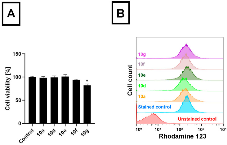
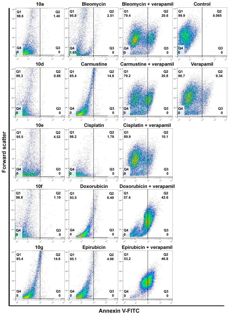
Multidrug resistance (MDR) is a serious hurdle to successful cancer therapy. Here, we examined the efficiency of novel semi-synthetic dihydrotestosterone derivatives, more specifically androstano-arylpyrimidines in inhibiting the efflux activity of ATP-binding cassette (ABC) transporters and sensitizing inherently MDR colon cancer cells to various chemotherapy drugs. Using the Rhodamine123 accumulation assay, we evaluated the efflux activity of cancer cells following treatments with androstano-arylpyrimidines. We found that acetylated compounds were capable of attenuating the membrane efflux of inherently MDR cells; however, deacetylated counterparts were ineffective. To delineate the possible molecular mechanisms underlying these unique activities of androstano-arylpyrimidines, the degree of apoptosis induction was assessed by AnnexinV-based assays, both upon the individual as well as by steroid and chemotherapy agent combination treatments. Five dihydrotestosterone derivatives applied in combination with Doxorubicin or Epirubicin triggered massive apoptosis in MDR cells, and these combinations were more efficient than chemotherapy drugs together with Verapamil. Furthermore, our results revealed that androstano-arylpyrimidines induced significant endoplasmic reticulum stress (ER stress) but did not notably modulate ABC transporter expression. Therefore, ER stress triggered by acetylated androstano-arylpyrimidines is probably involved in the mechanism of efflux pump inhibition and drug sensitization which can be targeted in future drug developments to defeat inherently multidrug-resistant cancer.

Keywords: ABC transporters; combination therapy; drug-resistant cancer; endoplasmic reticulum stress; multidrug resistance; pyrimidine-fused androgens.


Conflict of interest statement

The authors declare no conflict of interest.

Figures

Figure 1
Figure 1
Chemical structures of androstane-derived A-ring-fused arylpyrimidines 10a, 10d, 10e, 10f, 10g, 11a, 11d, 11e, 11f, and 11g differing in their C-17 functionality.
Figure 2
Figure 2
Acetylated androstano-arylpyrimidines attenuate the efflux transporter activity in multidrug-resistant Colo 320 cells. (A) Retention of Rhodamine 123 in Colo 320 cells treated with acetylated (10a, 10d, 10e, 10f, or 10g) and deacetylated (11a, 11d, 11e, 11f, or 11g) androstano-arylpyrimidines (at 20 μM concentration for 24 h) or with Verapamil (40 μM, 2 h treatment). Rhodamine 123 fluorescence of at least 10,000 cells/sample was measured by flow cytometry. (B) The viability of Colo 320 multidrug-resistant colon cancer cells treated with acetylated androstane compounds 10a, 10d, 10e, 10f, or 10g (at 20 μM, for 24 h, 104 cells/well density) was assessed by MTT cell viability assay. Bar graphs represent mean ± SD values. Fisher’s LSD test, *: p < 0.05.
Figure 3
Figure 3
Androstano-arylpyrimidine 17-acetates do not cause Rhodamine 123 accumulation in Colo 205 cells. (A) Viability of Colo 205 drug-sensitive colon cancer cells following exposure to acetylated androstane compounds 10a, 10d, 10e, 10f, or 10g (20 μM, 24 h treatment, 104 cells/well density) assessed by MTT cell viability assay. Bar graphs represent mean ± SD values. Fisher’s LSD test, *: p < 0.05. (B) Rhodamine 123 retentions of Colo 205 drug-sensitive colon cancer cells treated with acetylated androstane compounds 10a, 10d, 10e, 10f, or 10g (at 20 μM concentration for 24 h). Rhodamine 123 fluorescence of at least 10,000 cells/sample was measured by flow cytometry.
Figure 4
Figure 4
The degree of apoptosis induction in multidrug-resistant Colo 320 cells upon exposure to androstano-arylpyrimidines or various chemotherapy drugs. Annexin V-fluorescein isothiocyanate fluorescence of 10,000 cells/sample was measured with flow cytometry and plotted against forward scatter. Cells were treated either with compound 10a, 10d, 10e, 10f, or 10g alone (in 20 μM concentration for 24 h) or with chemotherapy drugs alone (Bleomycin, Carmustine, Cisplatin, Doxorubicin or Epirubicin at various concentrations, please refer to Materials and methods and Supplementary Material Table S2) for 24 h. Apoptosis induction was assessed upon Verapamil (in 4 μM for 24 h) as well as following Verapamil and chemotherapy drug combination treatments. The numbers in the Q2 quadrants represent the percentage of Annexin V-positive apoptotic cells. In the color density plots each dot represents a single detected event.
Figure 5
Figure 5
Androstano-arylpyrimidine 17-acetates sensitize Colo 320 cells to chemotherapy drug-induced apoptosis. Annexin V-fluorescein isothiocyanate fluorescence of 10,000 cells/sample were measured with flow cytometry and plotted against forward scatter. Cells were treated either with selected combinations prepared from compound 10a, 10d, 10e, 10f, or 10g (in 20 μM concentration for 24 h) and chemotherapy drugs (Bleomycin, Carmustine, Cisplatin, Doxorubicin, or Epirubicin at various concentrations, please refer to Materials and methods and Supplementary Material Table S2) for 24 h. The numbers in the Q2 quadrants represent the percentage of Annexin V-positive apoptotic cells. In the color density plots each dot represents a single detected event.
Figure 6
Figure 6
Acetylated androstano-arylpyrimidines do not suppress the efflux activity of multidrug-resistant cells by attenuating the expression of ABC transporters. (A) Relative mRNA levels of selected ABC transporters in Colo 320 cells treated with acetylated androstano-arylpyrimidines 10a, 10d, 10e, 10f, and 10g (in 20 μM concentration for 24 h) determined by RT-qPCR measurements and subsequent analyses by the ΔΔCt method using GAPDH as the reference gene. For primer sequences and concentrations, please refer to Supplementary Material Table S3. (B) Representative Western blot images and densitometric analysis of ABCB1 protein levels in Colo 320 cells following exposure to androstano-arylpyrimidine acetates (treatment with steroid compounds at 20 μM for 24 h) or Tunicamycin (600 nM, 24 h). The equal loading of protein samples was verified by probing the membrane with an anti-actin antibody. C = Control, Tun = Tunicamycin. In both panels, bar graphs represent mean ± SD values. Fisher’s LSD test, *: p < 0.05, **: p < 0.01, ***: p < 0.001, ****: p < 0.0001 ns: non-significant.
Figure 7
Figure 7
Acetylated androstano-arylpyrimidines induce endoplasmic reticulum stress in multidrug-resistant Colo 320 cells. (A) Bar graphs of the relative mRNA levels of various endoplasmic reticulum stress markers in Colo 320 cells treated with steroid derivatives 10a, 10d, 10e, 10f, and 10g (in 20 μM concentration for 24 h) determined by RT-qPCR measurements and data analysis by the ΔΔCt method using GAPDH as the reference gene. For primer sequences and concentrations, please refer to Supplementary Material Table S3. (B) Representative Western blot images and densitometric analysis of the ER stress marker protein BIP in Colo 320 cells upon exposure to androstano-arylpyrimidine acetates (treatment with steroid compounds 10a, 10d, 10e, 10f, and 10g in 20 μM, 24 h) or Tunicamycin (600 nM, 24 h). Equal loading of samples was verified by probing the membrane with an anti-actin antibody. C = Control, Tun = Tunicamycin. In both panels of figure (A, B), bar graphs represent mean ± SD values. Fisher’s LSD test, *: p < 0.05, **: p < 0.01, ****: p < 0.0001.

Similar articles

References

    1. Naghavi M., Abajobir A.A., Abbafati C., Abbas K.M., Abd-Allah F., Abera S.F., Aboyans V., Adetokunboh O., Ärnlöv J., Afshin A., et al. Global, Regional, and National Age-Sex Specific Mortality for 264 Causes of Death, 1980–2016: A Systematic Analysis for the Global Burden of Disease Study 2016. Lancet. 2017;390:1151–1210. doi: 10.1016/S0140-6736(17)32152-9. - DOI - PMC - PubMed
    1. Bukowski K., Kciuk M., Kontek R. Mechanisms of Multidrug Resistance in Cancer Chemotherapy. Int. J. Mol. Sci. 2020;21:3233. doi: 10.3390/ijms21093233. - DOI - PMC - PubMed
    1. Li Y.J., Lei Y.H., Yao N., Wang C.R., Hu N., Ye W.C., Zhang D.M., Chen Z.S. Autophagy and Multidrug Resistance in Cancer. Chin. J. Cancer. 2017;36:52. doi: 10.1186/s40880-017-0219-2. - DOI - PMC - PubMed
    1. Mansoori B., Mohammadi A., Davudian S., Shirjang S., Baradaran B. The Different Mechanisms of Cancer Drug Resistance: A Brief Review. Adv. Pharm. Bull. 2017;7:339. doi: 10.15171/apb.2017.041. - DOI - PMC - PubMed
    1. Emran T.B., Shahriar A., Mahmud A.R., Rahman T., Abir M.H., Siddiquee M.F.R., Ahmed H., Rahman N., Nainu F., Wahyudin E., et al. Multidrug Resistance in Cancer: Understanding Molecular Mechanisms, Immunoprevention and Therapeutic Approaches. Front. Oncol. 2022;12:2581. doi: 10.3389/fonc.2022.891652. - DOI - PMC - PubMed

LinkOut - more resources