Review
. 2023 Feb 9;15(2):587.
doi: 10.3390/pharmaceutics15020587.

A Systematic Overview of Eudragit® Based Copolymer for Smart Healthcare

Affiliations
Review

A Systematic Overview of Eudragit® Based Copolymer for Smart Healthcare

Aniket Nikam et al. Pharmaceutics. .

Abstract

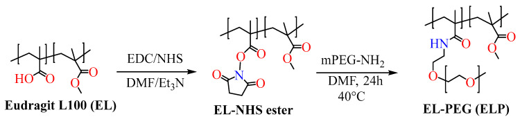
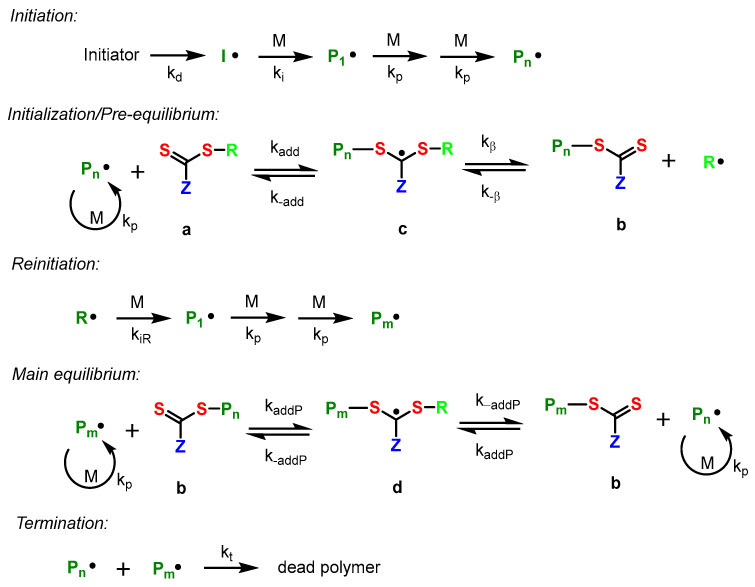
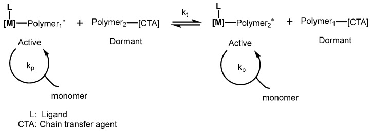
Eudragit, synthesized by radical polymerization, is used for enteric coating, precise temporal release, and targeting the entire gastrointestinal system. Evonik Healthcare Germany offers different grades of Eudragit. The ratio of methacrylic acid to its methacrylate-based monomers used in the polymerization reaction defines the final product's characteristics and consequently its potential range of applications. Since 1953, these polymers have been made to use in a wide range of healthcare applications around the world. In this review, we reviewed the "known of knowns and known of unknowns" about Eudragit, from molecule to material design, its characterization, and its applications in healthcare.

Keywords: Eudragit classification; Eudragit synthesis; biosensor; drug delivery; nanoparticles.


Conflict of interest statement

The authors declare no conflict of interest.

Figures

Figure 1
Figure 1
X-ray diffractogram of various Eudragit polymers, Eudragit L 100, S 100, Eudragit RL, and Eudragit RS (Adapted from Ref. [6]).
Figure 2
Figure 2
Temperature dependencies of the FT-IR peak intensity for the various IR bands of Eudragits L, S, and L30D. (A) Eudragit L; (B) Eudragit S; (C) Eudragit L30D (adapted with permission from Ref. [8] Copyright).
Figure 3
Figure 3
Three-dimensional plots of reflectance FT-IR spectra of Eudragits L, S, and respect to temperature. (A) Eudragit L; (B) Eudragit S; (C) Eudragit L30D (adapted with permission from Ref [9] Copyright).
Figure 3
Figure 3
Three-dimensional plots of reflectance FT-IR spectra of Eudragits L, S, and respect to temperature. (A) Eudragit L; (B) Eudragit S; (C) Eudragit L30D (adapted with permission from Ref [9] Copyright).
Scheme 1
Scheme 1
Synthesis of preactivated thiolated Eudragit L100-55.
Scheme 2
Scheme 2
One-step synthetic scheme for the preparation of thiolated Eudragit.
Scheme 3
Scheme 3
Synthesis of dye-anchored Eudragit polymers.
Scheme 4
Scheme 4
Scheme for the synthesis of PEGylated Eudragit L100 polymer.
Scheme 5
Scheme 5
Synthesis of anisamide-based Eudragit polymer.
Scheme 6
Scheme 6
Synthesis of Acrylated Eudragit polymer.
Scheme 7
Scheme 7
General mechanism for transition-metal-catalyzed ATRP.
Scheme 8
Scheme 8
Mechanism of basic equilibria process involved during RAFT polymerization.
Scheme 9
Scheme 9
Process involving chain transfer polymerization. * Scheme represented the Coordinative Chain Transfer Polymerization a process involving a dynamical equilibrium between propagating and dormant species.
Figure 4
Figure 4
(a) Hydrogels containing nanocapsules of Eudragit S100 were examined using scanning electron microscopy (HG-HEC), as were, (b) hydrogels containing nanocapsules of Eudragit RS100, as well as hydrogels containing poly(ε-caprolactone) nanocapsules, (c) hydrogels with nanocapsules of poly(ε-caprolactone) had an uneven surface and were devoid of spheres (HG-NC-RS), (d) hydrogels of hydroxyethylcellulose containing nanocapsules of poly(ε-caprolactone) (HG-NC-PCL). (Adapted with permission from Ref. [25] Copyright).
Figure 5
Figure 5
Wound pH-dependent release system schematic illustration (adapted with permission from Ref. [37] Copyright Elsevier 2021).
Figure 6
Figure 6
SEM images illustrating the fiber morphology of (a) pure polyurethane, (b) pure Eudragit L100-55, (c) pure polyurethane with Paclitaxel, and (df) polyurethane/Eudragit L composite mats (3:1. 2:1, and 1:1). Pure polyurethane and pure polyurethane with Paclitaxel have an average diameter of 850 nm and 790 nm, pure Eudragit L100-55 has an average diameter of 250 nm. Combining the two polymers yielded nanofibers with varying fiber diameters. Polyurethane/Eudragit L 1:1 formed nanofibers with an average diameter of 590 nm, while Polyurethane/Eudragit 2:1 and Polyurethane/Eudragit 3:1 both have an average diameter of 637 nm and 714 nm, respectively (adapted with permission from Ref. [43] Copyright Elsevier 2015).
Figure 7
Figure 7
SEM images of drug-loaded zein/Eudragit S100 composite nanofibers. The inset in the picture is an SEM image of the same fibers taken at a magnification of 10,000× (adapted with permission from Ref. [44] Copyright Elsevier 2012).
Figure 8
Figure 8
Confocal laser scanning microscopy images of HeLa cells 1 h, 3 h, and 5 h after the incubation with calcium phosphate/FITC-BSA/Eudragit nanoparticles (green; (A1A3)) and calcium phosphate/siRNA/THPP/Eudragit nanoparticles (red; (B1B3)). The scale bar corresponds to 20 mm (adapted with permission from Ref. [60] Copyright Royal Society of Chemistry 2014).
Figure 9
Figure 9
Vibrio cholerae cumulative release profiles from Eudragit L30D-55-VC and Eudragit FS-30D-VC microparticles (adapted with permission from Ref. [64] Copyright Elsevier 2011).
Figure 10
Figure 10
BSA that was extracted from the as prepared ‘Pollens + Eudragit Matrix’ formulation into PBS showed fluorescence spectra and intensity similar to that of native BSA solution in PBS. This shows that the formulation conditions did not adversely affect the tertiary structure of BSA (Adapted with permission from Ref. [66] Copyright Elsevier 2018).
Figure 11
Figure 11
Schematic preparation, administration, and in vivo release behavior of TBSS-DRM (adapted with permission from Ref. [68] Copyright Royal Society of Chemistry 2016).
Figure 12
Figure 12
Scanning electron micrographs of insulin-loaded Eudragit S100 microspheres (adapted from Ref. [76]).

Similar articles

Cited by

References

    1. Shah H., Jain A., Laghate G., Prabhudesai D. Remington: The Science and Practice of Pharmacy. Academic Press; Cambridge, MA, USA: 2020. Pharmaceutical Excipients; pp. 633–643. - DOI
    1. Patra C.N., Priya R., Swain S., Kumar Jena G., Panigrahi K.C., Ghose D. Pharmaceutical Significance of Eudragit: A Review. Futur. J. Pharm. Sci. 2017;3:33–45. doi: 10.1016/j.fjps.2017.02.001. - DOI
    1. Thakral S., Thakral N.K., Majumdar D.K. Eudragit®: A Technology Evaluation. Expert Opin. Drug Deliv. 2013;10:131–149. doi: 10.1517/17425247.2013.736962. - DOI - PubMed
    1. Wen H., Park K. Oral Controlled Release Formulation Design and Drug Delivery: Theory to Practice. John Wiley & Sons; Hoboken, NJ, USA: 2010. - DOI
    1. Lin S.Y., Chen K.S., Run-Chu L. Organic Esters of Plasticizers Affecting the Water Absorption, Adhesive Property, Glass Transition Temperature and Plasticizer Permanence of Eudragit Acrylic Films. J. Control. Release. 2000;68:343–350. doi: 10.1016/S0168-3659(00)00259-5. - DOI - PubMed

LinkOut - more resources