. 2023 Feb 13;15(2):624.
doi: 10.3390/pharmaceutics15020624.

The Ability of Some Polysaccharides to Disaggregate Lysozyme Amyloid Fibrils and Renature the Protein

Affiliations

The Ability of Some Polysaccharides to Disaggregate Lysozyme Amyloid Fibrils and Renature the Protein

Olga Makshakova et al. Pharmaceutics. .

Abstract

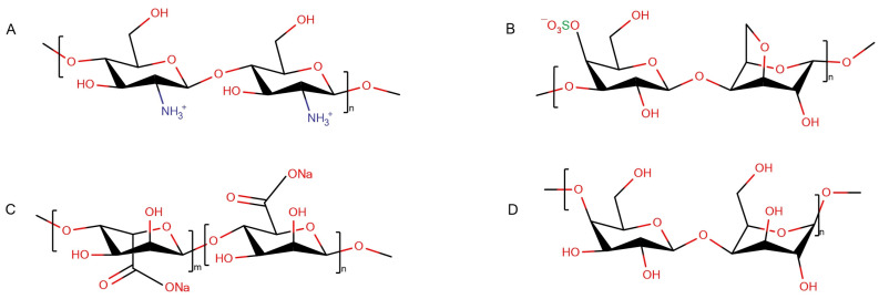
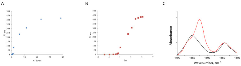
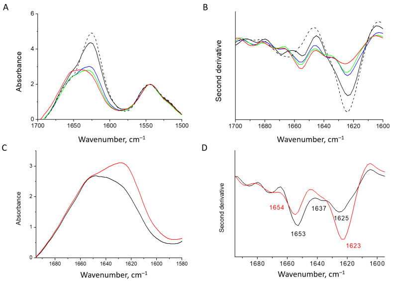
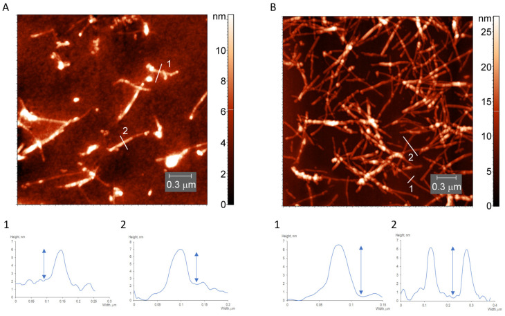
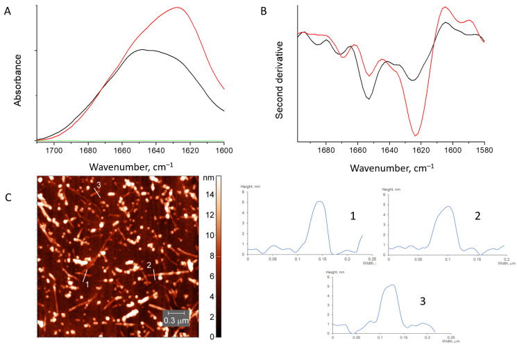
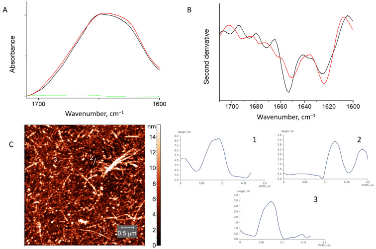
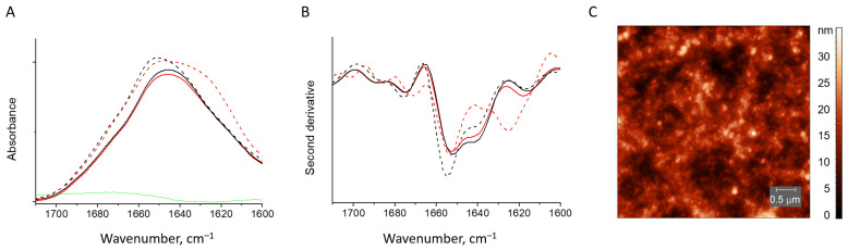
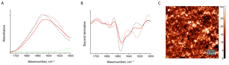
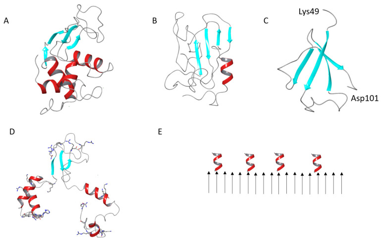
The deposition of proteins in the form of insoluble amyloid fibril aggregates is linked to a range of diseases. The supramolecular architecture of such deposits is governed by the propagation of β-strands in the direction of protofilament growth. In the present study, we analyze the structural changes of hen egg-white lysozyme fibrils upon their interactions with a range of polysaccharides, using AFM and FTIR spectroscopy. Linear anionic polysaccharides, such as κ-carrageenan and sodium alginate, are shown to be capable to disaggregate protofilaments with eventual protein renaturation. The results help to understand the mechanism of amyloid disaggregation and create a platform for both the development of new therapeutic agents for amyloidose treatment, and the design of novel functional protein-polysaccharide complex-based nanomaterials.

Keywords: alginate; amyloid fibrils; atomic force microscopy; carrageenan; chitosan; disaggregation; galactan; infrared spectroscopy; lysozyme; polysaccharides.


Conflict of interest statement

The authors declare no conflict of interest.

Figures

Figure 1
Figure 1
Structural scheme of polysaccharides: chitosan (A), κ-carrageenan (B), sodium alginate (C), β-(1,4)-galactan (D).
Figure 2
Figure 2
The kinetic curve of the HEWL fibril growth (at pH = 1.9 and T = 65 °C) followed by ThT fluorescence intensity (A,B). Relative FTIR absorbance spectra show the conversion of native HEWL (black) into fibrils (red) (C).
Figure 3
Figure 3
Absorbance spectra (A) and second derivative spectra (B) of the HEWL fibrils at 25 °C: dashed line—initial mature fibrils; black, blue, green and red—fibrils dialyzed against water for 2, 4, 6 and 24 h, respectively. Absorbance spectra (C) and second derivative spectra (D) of two fractions of the HEWL fibrils separated by centrifugation: sediment—red and supernatant—black.
Figure 4
Figure 4
AFM images and height profiles of the light (A) and heavy (B) fractions of desalted lysozyme fibrils.
Figure 5
Figure 5
Absorbance spectra (A) and second derivative spectra (B) of HEWL fibrils mixed with chitosan (red), initial HEWL fibrils (black) and pure chitosan (green). AFM image of HEWL fibrils mixed with chitosan (C).
Figure 6
Figure 6
Absorbance spectra (A) and second derivative spectra (B) of HEWL fibrils mixed with β-(1,4)-galactan (red) and pure HEWL fibrils (black). The spectrum of β-(1,4)-galactan is in green. AFM image of HEWL fibrils mixed with β-(1,4)-galactan (C).
Figure 7
Figure 7
Absorbance spectra (A) and second derivative spectra (B) of HEWL fibrils (dashed red line) and those mixed with κ-carrageenan (solid red line), native HEWL mixed with κ-carrageenan (solid black line), native HEWL (dashed black line). AFM image of the trace from the gel formed by HEWL fibril and κ-carrageenan mixture (C).
Figure 8
Figure 8
Absorbance spectra (A) and second derivative spectra (B) of HEWL fibrils (dashed red line), those mixed with sodium alginate (solid red line), native HEWL (black dashed line) and pure sodium alginate (green). AFM image of HEWL fibrils mixed with sodium alginate (C).
Figure 9
Figure 9
Cartoon representation of the native HEWL (A), a model of the β-structure-rich conformer of denatured HEWL (B), a model of the β-structure-rich conformer of amyloidogenic HEWL fragment (C), a model of the partially desaturated HEWL globule, positively charged residues are shown in sticks (D), a scheme of the deposition of helices on the HEWL fibril surface (E).

Similar articles

Cited by

References

    1. Boersema P.J., Melnik A., Hazenberg B.P.C., Rezeli M., Marko-Varga G., Kamiie J., Portelius E., Blennow K., Zubarev R.A., Polymenidou M., et al. Biology/Disease-Driven Initiative on Protein-Aggregation Diseases of the Human Proteome Project: Goals and Progress to Date. J. Proteome Res. 2018;17:4072–4084. doi: 10.1021/acs.jproteome.8b00401. - DOI - PubMed
    1. Debnath K., Sarkar A.K., Jana N.R., Jana N.R. Inhibiting Protein Aggregation by Small Molecule-Based Colloidal Nanoparticles. Acc. Mater. Res. 2022;3:54–66. doi: 10.1021/accountsmr.1c00193. - DOI
    1. Wu L., Velander P., Brown A.M., Wang Y., Liu D., Bevan D.R., Zhang S., Xu B. Rosmarinic Acid Potently Detoxifies Amylin Amyloid and Ameliorates Diabetic Pathology in a Transgenic Rat Model of Type 2 Diabetes. ACS Pharmacol. Transl. Sci. 2021;4:1322–1337. doi: 10.1021/acsptsci.1c00028. - DOI - PMC - PubMed
    1. Azzam S.K., Jang H., Choi M.C., Alsafar H., Lukman S., Lee S. Inhibition of Human Amylin Aggregation and Cellular Toxicity by Lipoic Acid and Ascorbic Acid. Mol. Pharm. 2018;15:2098–2106. doi: 10.1021/acs.molpharmaceut.7b01009. - DOI - PubMed
    1. Tran C.H., Saha R., Blanco C., Bagchi D., Chen I.A. Modulation of A-Synuclein Aggregation in Vitro by a DNA Aptamer. Biochemistry. 2022;61:1757–1765. doi: 10.1021/acs.biochem.2c00207. - DOI - PMC - PubMed

LinkOut - more resources