

Production and Utility of Extracellular Vesicles with 3D Culture Methods
- PMID: 36839984
- PMCID: PMC9961751
- DOI: 10.3390/pharmaceutics15020663
Production and Utility of Extracellular Vesicles with 3D Culture Methods
Abstract
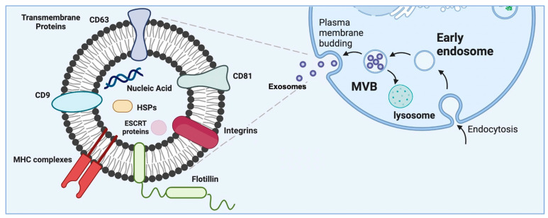
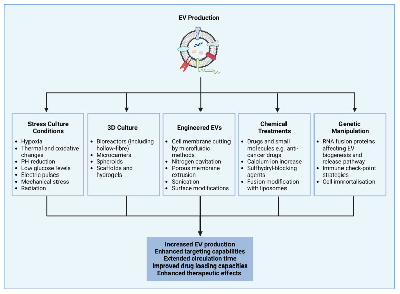
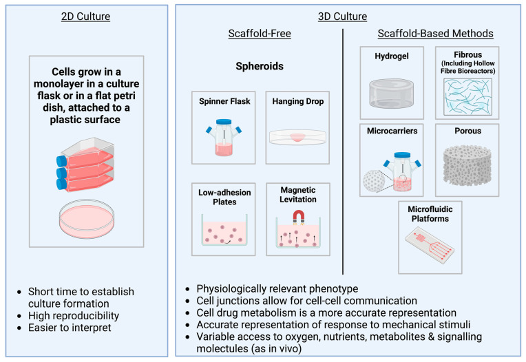
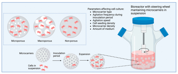
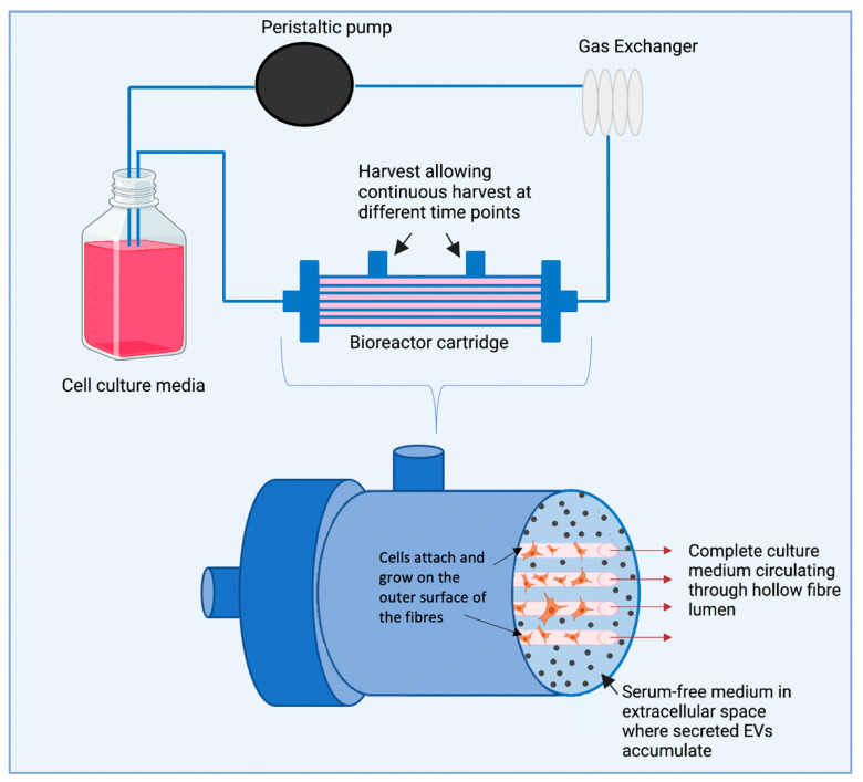
In recent years, extracellular vesicles (EVs) have emerged as promising biomarkers, cell-free therapeutic agents, and drug delivery carriers. Despite their great clinical potential, poor yield and unscalable production of EVs remain significant challenges. When using 3D culture methods, such as scaffolds and bioreactors, large numbers of cells can be expanded and the cell environment can be manipulated to control the cell phenotype. This has been employed to successfully increase the production of EVs as well as to enhance their therapeutic effects. The physiological relevance of 3D cultures, such as spheroids, has also provided a strategy for understanding the role of EVs in the pathogenesis of several diseases and to evaluate their role as tools to deliver drugs. Additionally, 3D culture methods can encapsulate EVs to achieve more sustained therapeutic effects as well as prevent premature clearance of EVs to enable more localised delivery and concentrated exosome dosage. This review highlights the opportunities and drawbacks of different 3D culture methods and their use in EV research.
Keywords: 3D culture; bioreactors; extracellular vesicles; scaffolds; spheroids.
Conflict of interest statement
The authors declare no conflict of interest. The funders had no role in the design of the study, in the writing of the manuscript, or in the decision to publish the study.
Figures
References
Publication types
Grants and funding
LinkOut - more resources
Full Text Sources
Other Literature Sources
