. 2023 Jan-Dec:32:9636897231156215.
doi: 10.1177/09636897231156215.

In Situ-Formed Fibrin Hydrogel Scaffold Loaded With Human Umbilical Cord Mesenchymal Stem Cells Promotes Skin Wound Healing

Affiliations

In Situ-Formed Fibrin Hydrogel Scaffold Loaded With Human Umbilical Cord Mesenchymal Stem Cells Promotes Skin Wound Healing

Lvzhong Hu et al. Cell Transplant. 2023 Jan-Dec.

Abstract

Healing of full-thickness skin wounds remains a major challenge. Recently, human umbilical cord mesenchymal stem cells (hUC-MSCs) were shown to possess an extraordinary potential to promote skin repair in clinical settings. However, their low survival rate after transplantation limits their therapeutic efficiency in treating full-thickness skin wounds. Hydrogels are considered an ideal cell transplantation vector owing to their three-dimensional mesh structure, good biosafety, and biodegradation. The objective of this study was to investigate the skin wound healing effect of a fibrin hydrogel scaffold loaded with hUC-MSCs. We found that the fibrin hydrogel had a three-dimensional mesh structure and low cytotoxicity and could prolong the time of cell survival in the peri-wound area. The number of green fluorescent protein (GFP)-labeled hUC-MSCs was higher in the full-thickness skin wound of mice treated with hydrogel-hUC-MSCs than those of mice treated with cell monotherapy. In addition, the combination therapy between the hydrogel and hUC-MSCs speed up wound closure, its wound healing rate was significantly higher than those of phosphate-buffered saline (PBS) therapy, hydrogel monotherapy, and hUC-MSCs monotherapy. Furthermore, the results showed that the combination therapy between hydrogel and hUC-MSCs increased keratin 10 and keratin 14 immunofluorescence staining, and upregulated the relative gene expressions of epidermal growth factor (EGF), transforming growth factor-β1 (TGF-β1), and vascular endothelial growth factor A (VEGFA), promoting epithelial regeneration and angiogenesis. In conclusion, the fibrin hydrogel scaffold provides a relatively stable sterile environment for cell adhesion, proliferation, and migration, and prolongs cell survival at the wound site. The hydrogel-hUC-MSCs combination therapy promotes wound closure, re-epithelialization, and neovascularization. It exhibits a remarkable therapeutic effect, being more effective than the monotherapy with hUC-MSCs or hydrogel.

Keywords: fibrin; human umbilical cord mesenchymal stem cells; hydrogel; skin wound healing.


Conflict of interest statement

The author(s) declared no potential conflicts of interest with respect to the research, authorship, and/or publication of this article.

Figures
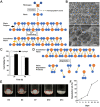
Figure 1.
Figure 1.
Synthesis and characterization of fibrin hydrogel scaffold. (A) Schematic diagram of fibrinogen and thrombin combining to form the fibrin hydrogel. (B) Representative field emission scanning electron microscopy image of fibrin hydrogel. (C) Cytotoxicity of hydrogel on human umbilical cord mesenchymal stem cells incubated in DMEM/F12 at 24 and 48 h (D) Schematic diagram of hydrogel degradation. (E) In vitro degradation behavior and (F) degradation rate of hydrogel in phosphate buffered saline over 14 days. Black arrow shows non-covalent bond. Yellow arrow shows covalent bond. Scale bar = 10 μm. Data are presented as mean ± standard error of the mean. DMEM/F12: Dulbecco’s Modified Eagle Medium and Ham’s F-12.
Figure 2.
Figure 2.
hUC-MSCs possess the characteristics of stem cells and multidirectional differentiation potential. (A) Microscopic morphology of hUC-MSCs. (B) Oil red O staining showed formation of lipid droplets (blue arrowheads). (C) Alizarin red staining showed formation of mineralized nodules (green arrowheads). (D–G) Flow cytometric analysis of surface antigens in hUC-MSCs. The blue curves represent isotypic controls. Scale bar = 200 μm. hUC-MSCs: Human umbilical cord mesenchymal stem cells.
Figure 3.
Figure 3.
Fibrin hydrogel scaffold prolongs the survival time of hUC-MSCs at the wound site. Skin wounds were treated with GFP-labeled hUC-MSCs or hydrogel–GFP-labeled hUC-MSCs. In vivo testing—fluorescence tracer results on (A, B) day 3 after surgery (C, D) day 7 after surgery. (E) Comparison of GFP fluorescence intensity between the hUC-MSCs monotherapy and hydrogel–hUC-MSCs combination groups on days 3 and 7 after surgery. Scale bar = 100 μm. Data are presented as mean ± standard error of the mean. hUC-MSCs: human umbilical cord mesenchymal stem cells; DAPI: 4’,6-diamidino-2-phenylindole; GFP: green fluorescent protein. ***P < 0.001.
Figure 4.
Figure 4.
Hydrogel–hUC-MSCs combination therapy promotes wound closure. (A) The flow diagram of animal protocol and treatment. (B) Wound healing of mice in the control, hydrogel monotherapy, hUC-MSCs monotherapy, and hydrogel–hUC-MSCs combination groups on days 3, 7, 10, and 14 after surgery. (C) Comparison of wound healing rates among the control, hydrogel monotherapy, hUC-MSCs monotherapy group, and hydrogel–hUC-MSCs combination groups on days 3, 7, 10, and 14 after surgery. (D) H&E staining of the wound in the hydrogel–hUC-MSCs combination group on day 3 after surgery. (E) H&E staining of the wound in the hydrogel–hUC-MSCs combination group on day 7 after surgery. (F) H&E staining of the wound in the hydrogel–hUC-MSCs combination group on day 14 after surgery. (G–I) The ratio of wound re-epithelialization length of the control, hydrogel monotherapy, hUC-MSCs monotherapy and hydrogel–hUC-MSCs combination groups on days 3, 7, and 14 after surgery. Green dotted line shows the edge of the wound. Scale bar = 500 μm. Data are presented as mean ± standard error of the mean. H&E: hematoxylin and eosin; hUC-MSCs: human umbilical cord mesenchymal stem cells; W: wound area; N: normal area. *P < 0.05; **P < 0.01; ***P < 0.001.
Figure 5.
Figure 5.
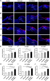
Hydrogel–hUC-MSCs combination therapy promotes re-epithelialization. (A, B) Keratin 10 immunofluorescence results of the wounds in the control, hydrogel monotherapy, hUC-MSCs monotherapy, and hydrogel–hUC-MSCs combination groups on days 3 and 7 after surgery. (C, D) Keratin 14 immunofluorescence results of the wounds in the control, hydrogel monotherapy, hUC-MSCs monotherapy, and hydrogel–hUC-MSCs combination groups on days 3 and 7 after surgery. (E–G) Levels of expression of EGF in the in lesioned tissue of control, hydrogel monotherapy, hUC-MSCs monotherapy, and hydrogel–hUC-MSCs combination groups on days 3, 7, and 14 after surgery. (H–J) Levels of expression of TGF-β1 in the in lesioned tissue of control, hydrogel monotherapy, hUC–MSCs monotherapy and hydrogel–hUC-MSCs combination groups on days 3, 7, and 14 after surgery. White dotted line shows the edge of the wound. Scale bar = 500 μm. Data are presented as mean ± standard error of the mean. hUC-MSCs: human umbilical cord mesenchymal stem cells; EGF: epidermal growth factor; TGF-β1: transforming growth factor-β1; W: wound area; N: normal area. *P < 0.05; **P < 0.01; ***P < 0.001.
Figure 6.
Figure 6.
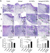
Hydrogel–hUC-MSCs combination therapy promotes neovascularization). (A–C) H&E staining of the wounds in the control, hydrogel monotherapy, hUC-MSCs monotherapy, and hydrogel–hUC-MSCs combination groups on days 3, 7, and 14 after surgery. (D–F) Levels of expression of VEGFA in lesioned tissue of the control, hydrogel monotherapy, hUC-MSCs monotherapy, and hydrogel–hUC-MSCs combination groups on days 3, 7, and 14 after surgery. Black dotted line shows the edge of the wound. Green arrowhead shows the blood vessel. Scale bar = 200 μm. Data are presented as mean ± standard error of the mean. H&E: hematoxylin and eosin; hUC-MSCs: human umbilical cord mesenchymal stem cells; VEGFA: vascular endothelial growth factor A; W: wound area; N: normal area. *P < 0.05; **P < 0.01; ***P < 0.001.

Similar articles

Cited by

References

    1. Zheng Z, Bian S, Li Z, Zhang Z, Liu Y, Zhai X, Pan H, Zhao X. Catechol modified quaternized chitosan enhanced wet adhesive and antibacterial properties of injectable thermo-sensitive hydrogel for wound healing. Carbohydr Polym. 2020;249:116826. - PubMed
    1. Lei Z, Singh G, Min Z, Shixuan C, Xu K, Pengcheng X, Xueer W, Yinghua C, Lu Z, Lin Z. Bone marrow-derived mesenchymal stem cells laden novel thermo-sensitive hydrogel for the management of severe skin wound healing. Mater Sci Eng C Mater Biol Appl. 2018;90:159–67. - PubMed
    1. Sadiq A, Shah A, Jeschke MG, Belo C, Qasim Hayat M, Murad S, Amini-Nik S. The role of serotonin during skin healing in post-thermal injury. Int J Mol Sci. 2018;19(4):1034. - PMC - PubMed
    1. Gurtner G, Werner S, Barrandon Y, Longaker M. Wound repair and regeneration. Nature. 2008;453(7193):314–21. - PubMed
    1. Walter MNM, Wright KT, Fuller HR, MacNeil S, Johnson WEB. Mesenchymal stem cell-conditioned medium accelerates skin wound healing: an in vitro study of fibroblast and keratinocyte scratch assays. Exp Cell Res. 2010;316(7):1271–81. - PubMed

Publication types

Substances