. 2023 Aug 3;25(8):1474-1486.
doi: 10.1093/neuonc/noad033.

Clinical, histological, and molecular features of gliomas in adults with neurofibromatosis type 1

Affiliations

Clinical, histological, and molecular features of gliomas in adults with neurofibromatosis type 1

Carlos G Romo et al. Neuro Oncol. .

Erratum in

Abstract

Background: People with NF1 have an increased prevalence of central nervous system malignancy. However, little is known about the clinical course or pathologic features of NF1-associated gliomas in adults, limiting clinical care and research.

Methods: Adults (≥18 years) with NF1 and histologically confirmed non-optic pathway gliomas (non-OPGs) at Johns Hopkins Hospital, Memorial Sloan Kettering Cancer Center, and Washington University presenting between 1990 and 2020 were identified. Retrospective data were collated, and pathology was reviewed centrally.

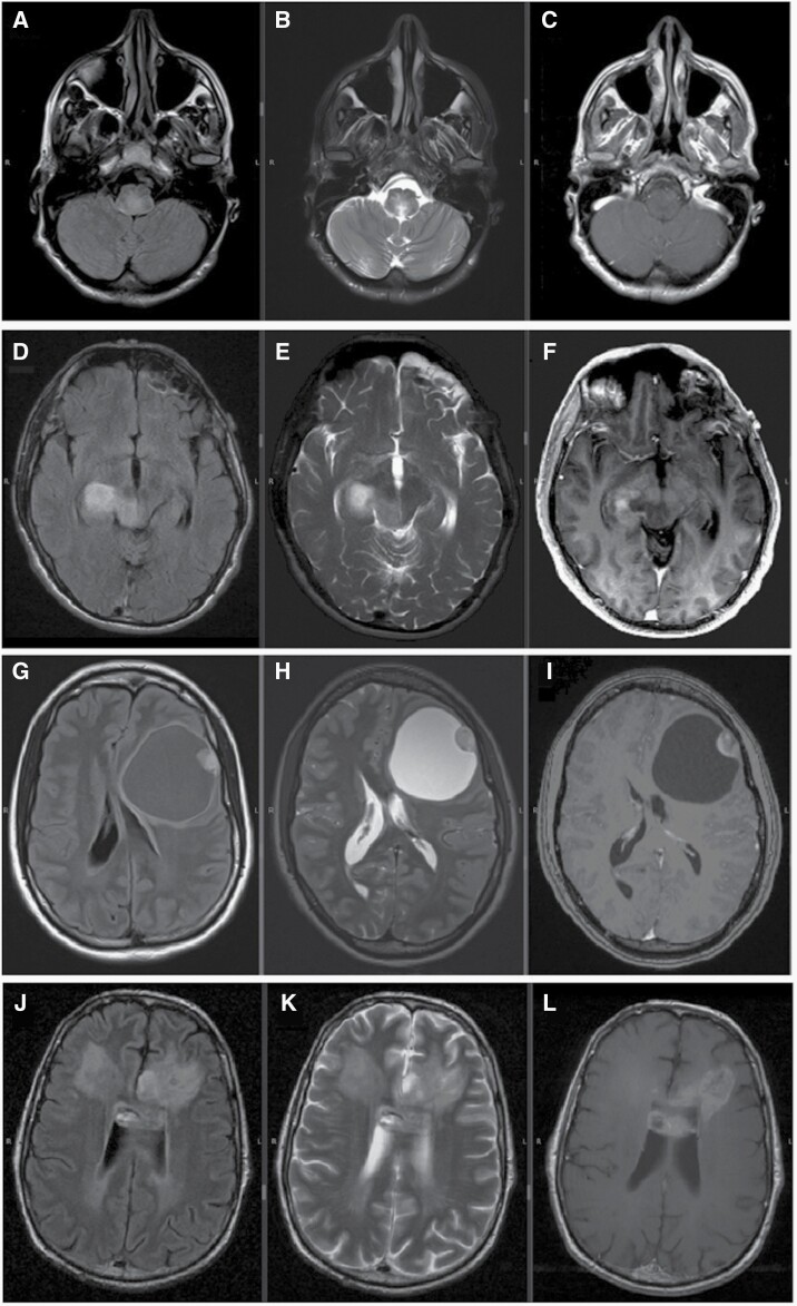
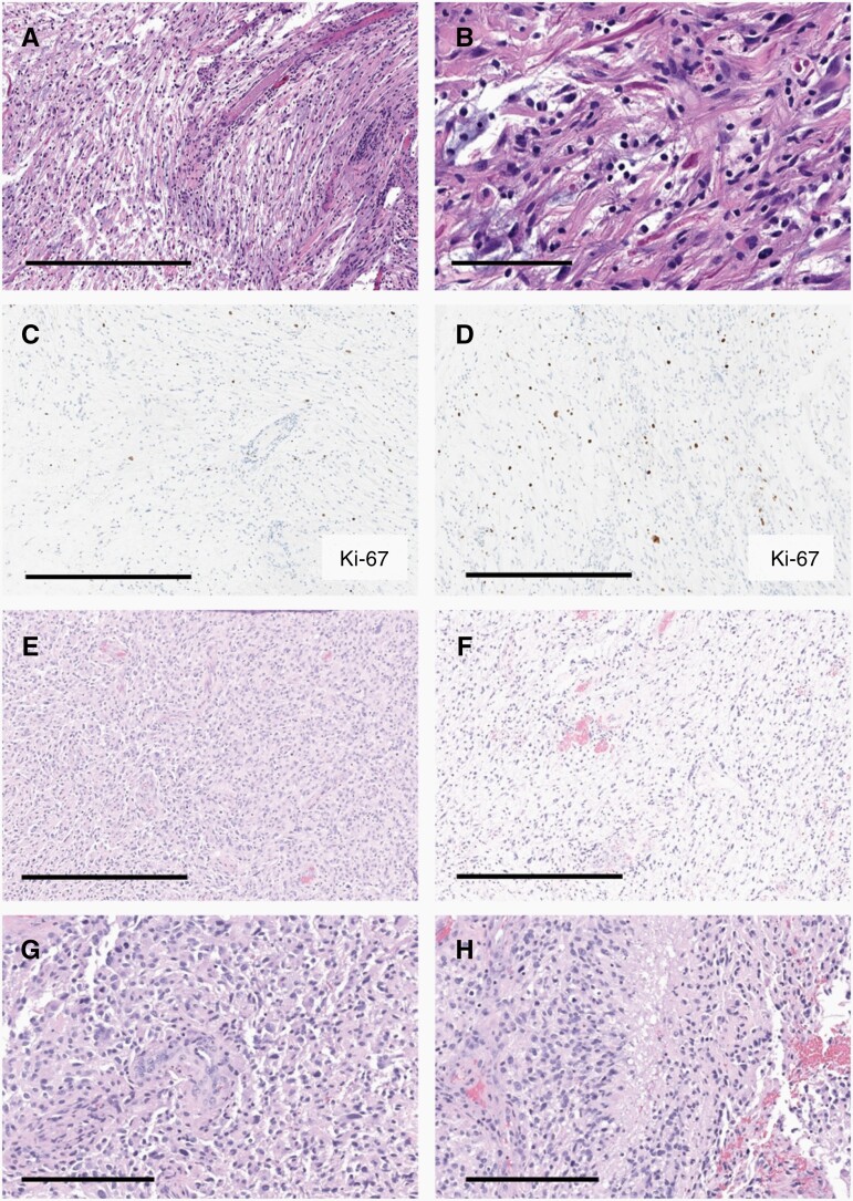
Results: Forty-five patients, comprising 23 females (51%), met eligibility criteria, with a median of age 37 (18-68 years) and performance status of 80% (30%-100%). Tissue was available for 35 patients. Diagnoses included infiltrating (low-grade) astrocytoma (9), glioblastoma (7), high-grade astrocytoma with piloid features (4), pilocytic astrocytoma (4), high-grade astrocytoma (3), WHO diagnosis not reached (4) and one each of gliosarcoma, ganglioglioma, embryonal tumor, and diffuse midline glioma. Seventy-one percent of tumors were midline and underwent biopsy only. All 27 tumors evaluated were IDH1-wild-type, independent of histology. In the 10 cases with molecular testing, the most common genetic variants were NF1, EGFR, ATRX, CDKN2A/B, TP53, TERT, and MSH2/3 mutation. While the treatments provided varied, the median overall survival was 24 months [2-267 months] across all ages, and 38.5 [18-109] months in individuals with grade 1-2 gliomas.

Conclusions: Non-OPGs in adults with NF1, including low-grade tumors, often have an aggressive clinical course, indicating a need to better understand the pathobiology of these NF1-associated gliomas.

Keywords: Neurofibromatosis type 1; Non-optic pathway glioma; glioma; overall survival; treatment outcome.


Conflict of interest statement

The authors have no conflicting interests to disclose.

Figures

Figure 1.
Figure 1.
Magnetic resonance images, axial sections from 4 selected cases. In each row, from right to left, FLAIR, T2, and T1 post-gadolinium sequences. (A–C) 30-year-old woman with an infiltrating (low-grade) astrocytoma (WHO grade 2) involving the medulla. Images revealed an infiltrative T2/FLAIR hyperintense lesion without contrast enhancement. Survival was 33 months after multiple treatments that included carboplatin as monotherapy, bevacizumab in combination with everolimus, and proton radiation therapy in combination with bevacizumab. (D–F) Infiltrating (low-grade) astrocytoma (WHO grade 2), which despite its low-grade classification on histology, demonstrated contrast enhancement and significant peritumoral edema in the midbrain with extension to the right thalamus. (G–I) Low-grade glioma with focal neuronal differentiation in a 20-year-old woman. The tumor was predominantly cystic and contained an enhancing mural nodule in the left frontal lobe with associated mass effect. (JL) Images of a 40-year-old man with glioblastoma revealed an ill-defined mass with heterogeneous enhancement involving the left greater than right frontal lobes and anterior corpus callosum, with ependymal extension and perilesional edema.
Figure 2.
Figure 2.
Representative histologic features. Photomicrographs of patients with a cerebellar mass initially consistent with pilocytic astrocytoma, including bipolar hair-like (pilocytic) cells (A) and abundant Rosenthal fibers (B). The overall tumor Ki67 index was low (<3%, C) with moderate focal elevated indices (5%–10%, D). Four years after initial diagnosis, recurrent tumor biopsy demonstrated malignant transformation with predominately dense cellularity and cytologic atypia (E), and only small foci resembling the original low-grade tumor (F). High-grade features including pseudo-palisading necrosis (G) and microvascular proliferation (H) are readily identified. (Scale bars: 500 μm [A, C–F]; 100 μm [B]; 200 μm [G, H]).
Figure 3.
Figure 3.
Kaplan–Meier survival analysis. (A) The median OS for the entire cohort was 24 months (2–267 months). There was no significant association between age at diagnosis, Karnofsky Performance Status and death. (B) Overall survival is based on WHO grade. Those with WHO grade 4 had worse OS; however, survival for patients with WHO grade 1–3 was not statically different. (C) OS between tumors categorized as high- versus low-grade. When analyzed in a dichotomous fashion, a high-grade classification more reliably predicted worse outcome. (D) OS based on histologic classification.

References

    1. Anderson JL, Gutmann DH.. Neurofibromatosis type 1. Handb Clin Neurol. 2015;132:75–86. - PubMed
    1. Ratner N, Miller S.. A RASopathy gene commonly mutated in cancer: the neurofibromatosis type 1 tumour suppressor. Nat Rev Cancer. 2015;15(5):290–301. - PMC - PubMed
    1. Uusitalo E, Rantanen M, Kallionpaa R, et al. . Distinctive cancer associations in patients with neurofibromatosis type 1. J Clin Oncol. 2016;34(17):1978–1986. - PubMed
    1. Seminog OO, Goldacre MJ.. Risk of benign tumours of nervous system, and of malignant neoplasms, in people with neurofibromatosis: population-based record-linkage study. Br J Cancer. 2013;108(1):193–198. - PMC - PubMed
    1. Byrne S, Connor S, Lascelles K, et al. . Clinical presentation and prognostic indicators in 100 adults and children with neurofibromatosis 1 associated non-optic pathway brain gliomas. J Neurooncol. 2017;133(3):609–614. - PMC - PubMed

Publication types

LinkOut - more resources