. 2023 Apr 24;51(7):e41.
doi: 10.1093/nar/gkad098.

Development of a selection assay for small guide RNAs that drive efficient site-directed RNA editing

Affiliations

Development of a selection assay for small guide RNAs that drive efficient site-directed RNA editing

Juan Felipe Diaz Quiroz et al. Nucleic Acids Res. .

Abstract

A major challenge confronting the clinical application of site-directed RNA editing (SDRE) is the design of small guide RNAs (gRNAs) that can drive efficient editing. Although many gRNA designs have effectively recruited endogenous Adenosine Deaminases that Act on RNA (ADARs), most of them exceed the size of currently FDA-approved antisense oligos. We developed an unbiased in vitro selection assay to identify short gRNAs that promote superior RNA editing of a premature termination codon. The selection assay relies on hairpin substrates in which the target sequence is linked to partially randomized gRNAs in the same molecule, so that gRNA sequences that promote editing can be identified by sequencing. These RNA substrates were incubated in vitro with ADAR2 and the edited products were selected using amplification refractory mutation system PCR and used to regenerate the substrates for a new round of selection. After nine repetitions, hairpins which drove superior editing were identified. When gRNAs of these hairpins were delivered in trans, eight of the top ten short gRNAs drove superior editing both in vitro and in cellula. These results show that efficient small gRNAs can be selected using our approach, an important advancement for the clinical application of SDRE.


Figures

Figure 1.
Figure 1.
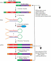
Selection assay. A DNA substrate was assembled by annealing the Target oligo containing a T7 promoter (green), a universal forward primer (P1, orange), the target sequence (red) and the connecting loop (green); and the Guide antisense oligo containing the connecting loop sequence (green), the randomized guides pool (striped purple) and a universal reverse primer (P2, orange). After annealing and amplification (Step 1), the DNA substrate was transcribed (Step 2) into RNA hairpins. The hairpins were incubated with HEK293T cell extracts overexpressing human ADAR2 (Step 3) leading to two possible outcomes, unedited or edited hairpins. The products of the editing reaction were reverse transcribed (Step 4) to obtain cDNA. The cDNA was then used as substrate for a Non-Selective PCR to monitor editing efficiency by Sanger sequencing (lower dotted arrow). The cDNA was also used as a substrate for a Selective PCR (ARMS PCR) (Step 5) to amplify only the substrates that were edited. From the products of the Selective PCR, the guide regions of the edited cDNAs were amplified (Step 6) and used (Guide oligo) to regenerate the substrate and start a new cycle of the assay. The regenerated substrate was also sent for MiSeq sequencing to monitor the diversity of the guide region (upper dotted arrow).
Figure 2.
Figure 2.
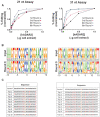
Evaluation of the selection assay. (A) Percent editing of the hairpin pool at different hADAR2 concentrations for rounds 1, 3, 6 and 9 (products after step 4 in Figure 1). For comparison purposes, percent editing was normalized to the highest value, and hADAR2 concentrations were normalized to the amount of cell extract that produced the highest editing per substrate per round. n = 1. (B) Logo plots showing the base preference at each position of the Guide region in the pool of hairpins (products after step 6 in Figure 1). Input refers to the starting material prior to the first round of editing. Position 0 marks the position complementary to the target adenosine. (C) Sequences of the Guide region for the top 10 most abundant hairpins after nine rounds of selection. Bases in red mark mismatches except for the C mismatch opposite the target A, which is marked in orange. Left panels correspond to the assay run with the 21 nt substrate and the right panels correspond to the assay run with the 31 nt substrate.
Figure 3.
Figure 3.
Testing top hairpins in vitro. (A) A cartoon showing in vitro editing reactions using individual hairpins containing one of the 10 most abundant guide sequences and with purified hADAR2. (B) Percent editing of the top 10 most abundant hairpins using 40nM (pink) or 5nM (black) concentrations of hADAR2. Asterisks denote a statistically significant difference in comparison to the control hairpin (two-way ANOVA, **** P < 0.0001, *** P < 0.001, for 21 nt control n = 6, for all other samples n = 3). Error bars represent S.D. (C) Reaction kinetics for two of the most abundant hairpins and the control hairpin under single turnover conditions. Values in parenthesis are the respective rate constants in s−1. For comparison purposes, percent editing was normalized to the highest value for each hairpin. Data were fit to a one-phase association curve (see methods). Error bars represent S.D., n = 3.
Figure 4.
Figure 4.
Testing top guide RNAs in vitro. (A) A cartoon showing in vitro editing reactions using individual gRNAs (in trans) of the 10 most abundant guide sequences with purified hADAR2 and full length mCherry_P2A_eGFP W58X (UAG) mRNA. (B) Percent editing of the top 10 most abundant gRNAs at a 5nM concentration of hADAR2. Asterisks denote a statistically significant difference in comparison to the control gRNA (one way ANOVA, **** P < 0.0001, ** P < 0.01, *P < 0.05, n = 3). Error bars represent S.D. (C) Reaction kinetics for two of the most abundant gRNAs and the control gRNA. Values in parenthesis are the respective rate constants in s−1. For comparison purposes percent editing was normalized to the highest value for each gRNA. Data were fit to a one-phase association curve (see methods). Error bars represent S.D., n = 3.
Figure 5.
Figure 5.
Testing top guide RNAs in cellula. (A) A map of the chemical modifications within the stabilized 31 nt ASOs of the 10 most abundant gRNAs as well as the control gRNA sequences. ASOs (100 nM) were transfected into HEK293T cells transiently expressing mCherry_P2A_eGFP W58X (UAG) and hADAR1-p110, hADAR1-p150 or hADAR2. (B) Percent editing of the top 10 31 nt gRNAs from the selection assay in comparison to the control. Asterisks denote statistically significant differences in comparison to the control gRNA (Two-way ANOVA, **** P < 0.0001, *** P < 0.001, ** P < 0.01, *P < 0.05, n.s. not significant). Error bars represent S.D., and for all data points, n = 8.
Figure 6.
Figure 6.
Dose response of the stabilized 31 nt ASOs of the 10 most abundant gRNAs in cellula. (A) Percent editing at different concentrations of the top 10 most abundant gRNAs in comparison to the control after HEK293T cells co-transfection with plasmids encoding mCherry_P2A_eGFP W58X (UAG) and hADAR2. A four-parameter non-linear plot was used to fit the data. For all data points, n = 4. (B) GFP fluorescence at different concentrations of transfected gRNAs shown in (A). A four-parameter non-linear plot was used to fit the data. For all data points, n = 4. (C) Linear regression showing the correlation between percent editing and GFP fluorescence.

References

    1. Havens M.A., Hastings M.L.. Splice-switching antisense oligonucleotides as therapeutic drugs. Nucleic Acids Res. 2016; 44:6549. - PMC - PubMed
    1. Juliano R.L. The delivery of therapeutic oligonucleotides. Nucleic Acids Res. 2016; 44:6518. - PMC - PubMed
    1. Crooke S.T., Witztum J.L., Bennett C.F., Baker B.F.. RNA-targeted therapeutics. Cell Metab. 2018; 27:714–739. - PubMed
    1. Sahin U., Karikó K., Türeci Ö.. MRNA-based therapeutics-developing a new class of drugs. Nat. Rev. Drug Discov. 2014; 13:759–780. - PubMed
    1. Magadum A., Kaur K., Zangi L.. mRNA-based protein replacement therapy for the heart. Mol. Ther. 2019; 27:785–793. - PMC - PubMed

Publication types

Substances