. 2023 Apr 11;51(6):2586-2601.
doi: 10.1093/nar/gkad100.

Red light-emitting short Mango-based system enables tracking a mycobacterial small noncoding RNA in infected macrophages

Affiliations

Red light-emitting short Mango-based system enables tracking a mycobacterial small noncoding RNA in infected macrophages

Oksana S Bychenko et al. Nucleic Acids Res. .

Abstract

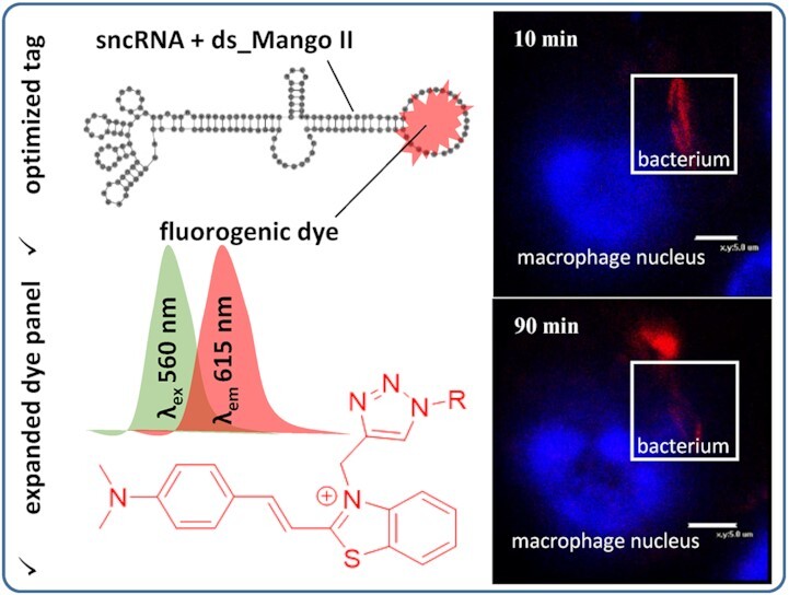
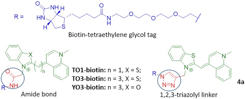
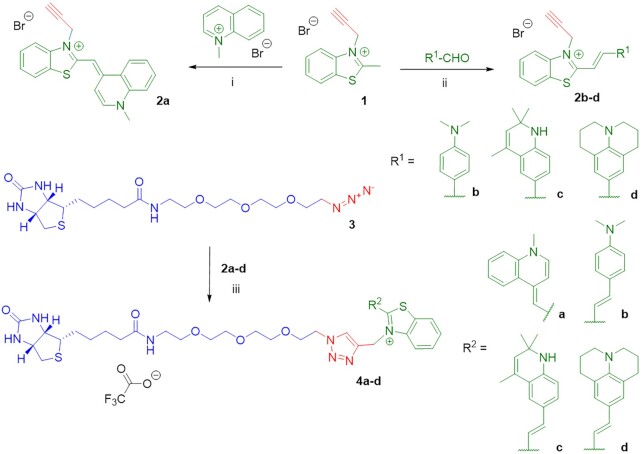
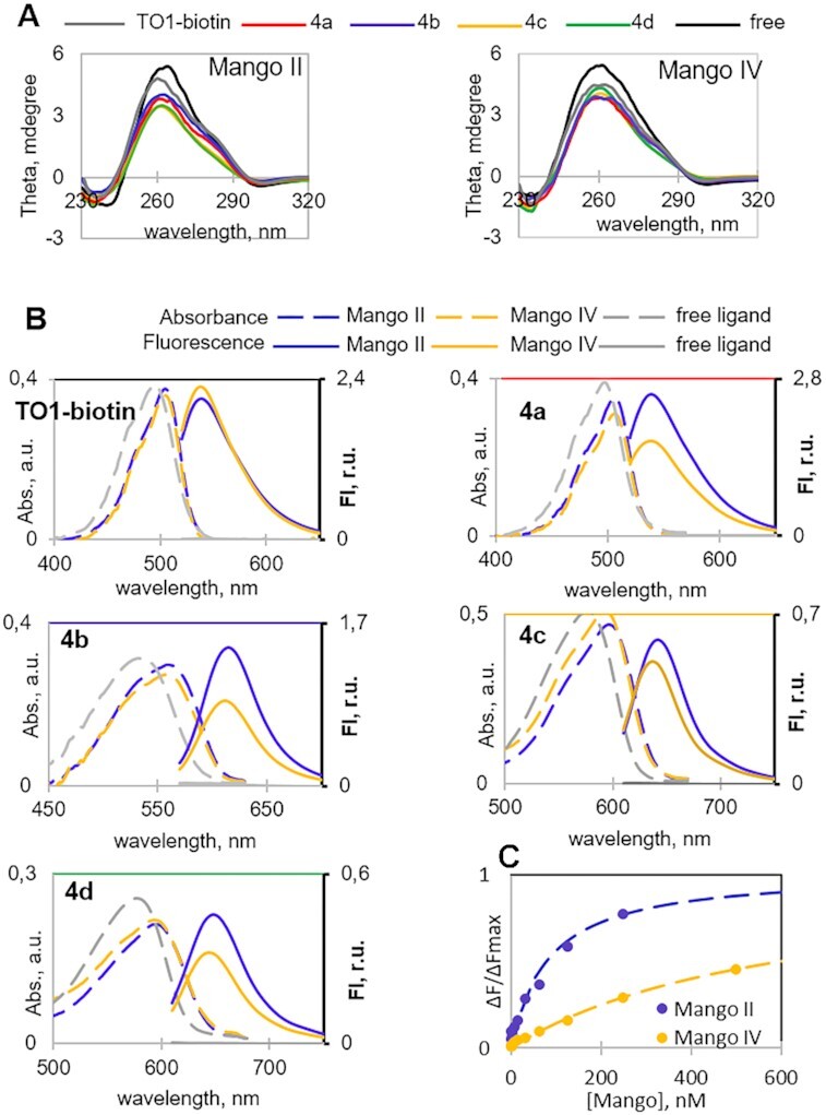
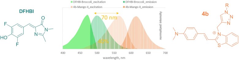
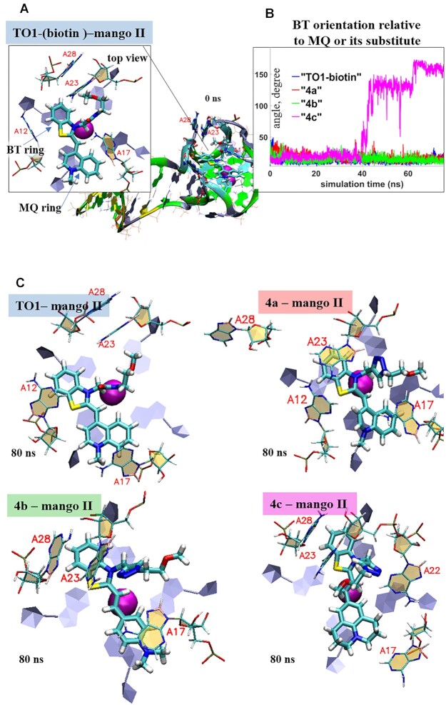
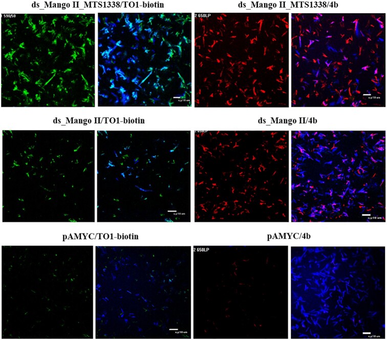
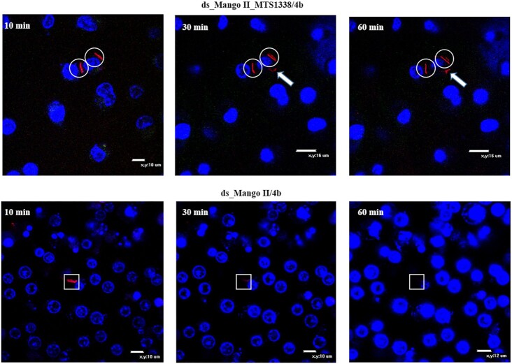
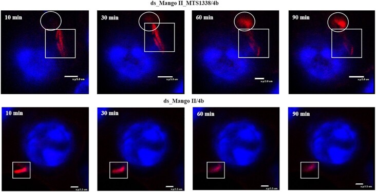
Progress in RNA metabolism and function studies relies largely on molecular imaging systems, including those comprising a fluorogenic dye and an aptamer-based fluorescence-activating tag. G4 aptamers of the Mango family, typically combined with a duplex/hairpin scaffold, activate the fluorescence of a green light-emitting dye TO1-biotin and hold great promise for intracellular RNA tracking. Here, we report a new Mango-based imaging platform. Its key advantages are the tunability of spectral properties and applicability for visualization of small RNA molecules that require minimal tag size. The former advantage is due to an expanded (green-to-red-emitting) palette of TO1-inspired fluorogenic dyes, and the truncated duplex scaffold ensures the latter. To illustrate the applicability of the improved platform, we tagged Mycobacterium tuberculosis sncRNA with the shortened aptamer-scaffold tag. Then, we visualized it in bacteria and bacteria-infected macrophages using the new red light-emitting Mango-activated dye.


Figures

Graphical Abstract
Graphical Abstract
Tag-activated fluorogenic dyes with tunable spectral properties were obtained by a new synthetic approach. A very short aptamer-based tag for labeling short RNA was constructed. The developed fluorogenic dye-tag system was used to visualize target RNA in bacteria internalized by macrophages.
Figure 1.
Figure 1.
The structures of reported fluorogenic dyes TO1-biotin, TO3-biotin and YO3-biotin and triazolyl-linked TO1-biotin analog 4a.
Scheme 1.
Scheme 1.
Synthesis of TO1-biotin analogs. Reagents and conditions: (i) TEA, CH2Cl2; (ii) Ac2O, reflux; (iii) CuI, TBTA, DIPEA, CH3OH/CH3CN, rt, then preparative HPLC.
Figure 2.
Figure 2.
TO1-biotin and new dyes 4a–d in complexes with Mango II and IV aptamers: spectral properties and binding affinity. (A) Verification of dye impact on Mango secondary structure by CD spectroscopy. (B) Absorption and fluorescence emission spectra of the free dyes and their complexes. (C) 4b-Mango binding assay. Conditions in A and B: 5 μM Mango aptamer, 5 μM dye, 10 mM Tris–HCl, pH 7.5 and 140 mM КСl. Conditions in C: 10 nM dye.
Figure 3.
Figure 3.
Excitation and emission spectra of DFHBI and 4b in complex with Broccoli and Mango II aptamers, respectively.
Figure 4.
Figure 4.
Dynamics of Mango II complexes with TO1-biotin and its analogs: planarity versus rotational mobility of dye residues. (A) Initial model of TO1-biotin-Mango II complex (0 ns MD simulation snapshot) illustrating starting positions of benzothiazolium (BT) and methylquinolinyl (MQ) moieties, their stacking with external Mango II G4 tetrad and orientation relative to key neighboring Mango II residues (A12, A17, A23 and A28). Biotin residue is not shown and was excluded from the analysis. (B) Evolution of the angle between the planes of BT ring and MQ ring (TO1-biotin and 4a) or its substitute (4b–c). (C) End-point conformations of the complexes (80 ns MD simulation snapshots), top views. Color labeling of aptamer residues in TO1-biotin-Mango II full structure (a) G, green; A, grey; T, orange; C, cyan. Aptamer residues in complex fragment, top view: C, beige; G, grey. Ligand atoms: C, cyan; N, blue; O, red; S, yellow.
Figure 5.
Figure 5.
Visualization of the genetically encoded modular RNA ds_Mango II_MTS1338 and ds_Mango II in M. smegmatis in solution using TO1-biotin (in a green channel) and 4b (in a red channel). Bacteria transformed by pAMYC without insertion were used as a negative control. Bacteria were stained with Hoechst 33258 (in a blue channel).
Figure 6.
Figure 6.
Tracking of the red fluorescent signal for 1 h (10, 30 and 60 min), using the genetically encoded modular RNA or ds_Mango II and 4b in infected RAW 264.7 macrophages (wide field images). Macrophage nuclei were stained with Hoechst 33258. The circles cover the signal maintenance; white arrows show fluorescent signal movement/occurrence while squares highlight the area of signal disappearance.
Figure 7.
Figure 7.
Tracking of the red fluorescent signal for 1.5 h (10, 30, 60 and 90 min), using the genetically encoded modular RNA or ds_Mango II and 4b in infected RAW 264.7 macrophages (enlarged scale). Macrophage nuclei were stained with Hoechst 33258. The circles show fluorescent signal movement/occurrence, while squares highlight the area of signal disappearance.

Similar articles

Cited by

References

    1. Romano G., Veneziano D., Acunzo M., Croce C.M.. Small non-coding RNA and cancer. Carcinogenesis. 2017; 38:485–491. - PMC - PubMed
    1. Zhang Z., Zhang J., Diao L., Han L.. Small non-coding rnas in human cancer: function, clinical utility, and characterization. Oncogene. 2021; 40:1570–1577. - PubMed
    1. Weinberg M.S., Wood M.J.. Short non-coding RNA biology and neurodegenerative disorders: novel disease targets and therapeutics. Hum. Mol. Genet. 2009; 18:R27–R39. - PMC - PubMed
    1. Wu Y.Y., Kuo H.C.. Functional roles and networks of non-coding rnas in the pathogenesis of neurodegenerative diseases. J. Biomed. Sci. 2020; 27:49. - PMC - PubMed
    1. Waters L.S., Storz G.. Regulatory rnas in bacteria. Cell. 2009; 136:615–628. - PMC - PubMed

Publication types