. 2023 Mar 6;15(3):mfad011.
doi: 10.1093/mtomcs/mfad011.

Toxicity and assimilation of cellulosic copper nanoparticles require α-arrestins in S. cerevisiae

Affiliations

Toxicity and assimilation of cellulosic copper nanoparticles require α-arrestins in S. cerevisiae

Ni Putu Dewi Nurmalasari et al. Metallomics. .

Abstract

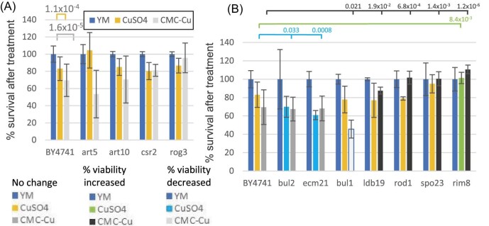
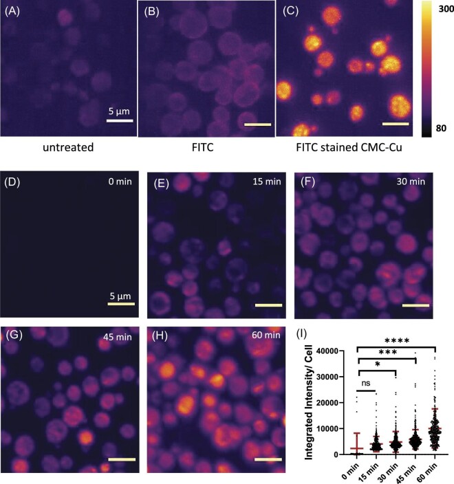
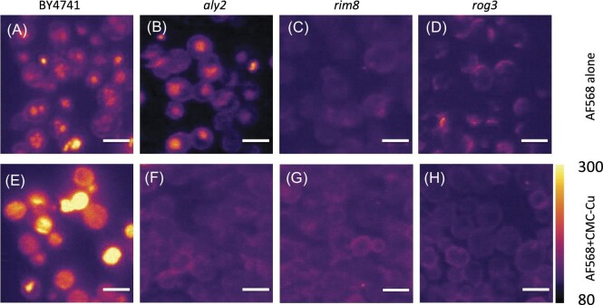
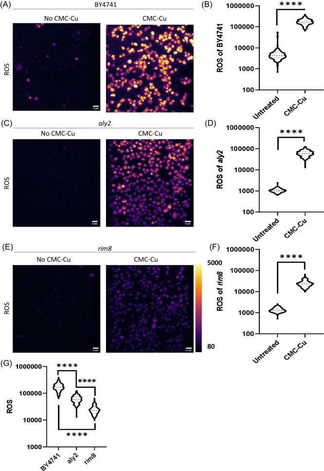
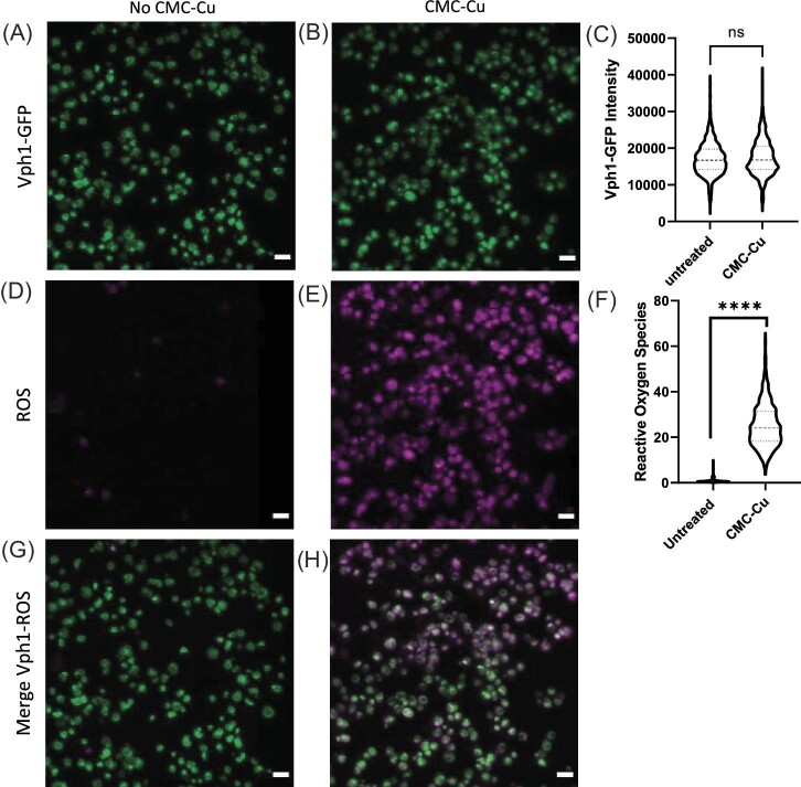
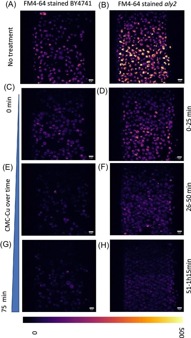
The increased use of antimicrobial compounds such as copper into nanoparticles changes how living cells interact with these novel materials. The increased use of antimicrobial nanomaterials combats infectious disease and food spoilage. Fungal infections are particularly difficult to treat because of the few druggable targets, and Saccharomyces cerevisiae provides an insightful model organism to test these new materials. However, because of the novel characteristics of these materials, it is unclear how these materials interact with living cells and if resistance to copper-based nanomaterials could occur. Copper nanoparticles built on carboxymethylcellulose microfibril strands with copper (CMC-Cu) are a promising nanomaterial when imported into yeast cells and induce cell death. The α-arrestins are cargo adaptors that select which molecules are imported into eukaryotic cells. We screened α-arrestins mutants and identified Aly2, Rim8, and Rog3 α-arrestins, which are necessary for the internalization of CMC-Cu nanoparticles. Internal reactive oxygen species in these mutants were lower and corresponded to the increased viability in the presence of CMC-Cu. Using lattice light-sheet microscopy on live cells, we determined that CMC-Cu were imported into yeast within 30 min of exposure. Initially, the cytoplasmic pH decreased but returned to basal level 90 min later. However, there was heterogeneity in response to CMC-Cu exposure, which could be due to the heterogeneity of the particles or differences in the metabolic states within the population. When yeast were exposed to sublethal concentrations of CMC-Cu no resistance occurred. Internalization of CMC-Cu increases the potency of these antimicrobial nanomaterials and is likely key to preventing fungi from evolving resistance.

Keywords: S. cerevisiae; copper nanoparticles; endocytosis; lattice light-sheet microscopy; reactive oxygen species; α-arrestins.


Conflict of interest statement

The authors declare no conflicts of interest.

Figures

Graphical Abstract
Graphical Abstract
Positively charged copper nanoparticles built on carboxymethylcellulose particles associate with the negatively charged cell wall of S. cerevisiae. CMC-Cu entry is mediated through numerous α-arrestins. Once assimilated in cells, the CMC-Cus increase internal ROS levels likely through Fenton reactions.
Fig. 1
Fig. 1
Viability of α-arrestin mutants treated with soluble copper or CMC-Cu. Mutants were grown to log-phase and then exposed to 400 μM soluble copper (orange) or 157 μM CMC-Cu (gray) for 90 min. Colony-forming units were normalized to BY4741 (wild-type) untreated in YM media (blue) and graphed as a percentage of viability. Mutants were then classified as to whether the response was different from wild-type. A. Mutants that had the same response as BY4741 to CuSO4 and CMC-Cu. B. Mutants that had changes in their copper or CMC-Cu viability. The direction of change was noted with colors increased copper viability (green), decreased copper viability (aqua), increased CMC-Cu viability (black), and decreased CMC-Cu viability (gray). The P-value of the change is noted above.
Fig. 2
Fig. 2
Maximum intensity projection of yeast A. BY4741 without treatment B. BY4741 were incubated with 2.5 μM FITC C. BY4741 were incubated 2.5 μM FITC stained CMC-Cu. D. BY4741 treated with CMC E. BY4741 treated with CMC-Cu, both CMC and CMC-Cu were stained by AF568 Hydrazine overnight, further BY4741 cells were incubated with CMC and CMC-Cu for 90 min. F. Violin plot of integrated intensity per cell for CMC (E) vs. CMC-Cu (D)treated yeast, using unpaired t-test with P-value **** ≤0.0001. AF568 stained CMC-Cu were incubated with BY4741 for G. 0 min, H. 15 min, I. 30 min, J. 45 min, and K. 60 min. L. The plot for integrated intensity per cell from 0 to 60 min. In this analysis, one-way ANOVA was used for the analysis with P-value higher than 0.05 means the value is not significant (ns), P-value less than or equal to ≤0.05, ≤0.01, ≤0.001, and ≤0.0001 mean significant difference which corresponds to *, **, ***, and ****, respectively.
Fig. 3
Fig. 3
Maximum intensity projection of yeast treated with AF568 alone and stained CMC-Cu A. BY4741 with AF568 alone. B.  aly2 mutant with AF568 alone. C.  rim8 mutant with AF568 alone. D.  rog3 mutant with AF568 alone. E. BY4741 with AF568 stained CMC-Cu. F.  aly2 mutant with AF568 stained CMC-Cu. G.  rim8 mutant with AF568 stained CMC-Cu. H.  rog3 mutant with AF568 stained CMC-Cu.
Fig. 4
Fig. 4
ROS of untreated vs. CMC-Cu treated of BY4741, aly2, and rim8 and intensity plot of ROS for untreated vs. CMC-Cu. All cells (BY4741, aly2, rim8) were incubated with CMC-Cu for 30 min and CellROx to measure ROS production. A. BY4741 untreated and CMC-Cu treated, B. Quantification of ROS in BY4741 CMC-Cu treated cells, C.  aly2 mutant untreated and CMC-Cu treated, D. Quantification of ROS in aly2 mutant CMC-Cu treated cells, E.  rim8 mutant untreated and CMC-Cu treated, F. Quantification of ROS in rim8 mutant CMC-Cu treated cells, G. ROS generated by CMC-Cu exposure of BY4741, aly2, and rim8 presented in the same graph. In this analysis, (Unpaired t) test was used for the analysis of Fig. B, D, and F, and one-way ANNOVA was used for the analysis of Fig. G with P-value higher than 0.05 means the value is not significant (ns), P-value less than or equal to ≤0.05, ≤0.01, ≤0.001, and ≤0.0001 mean significant difference which corresponds to *, **, ***, and ****, respectively.
Fig. 5
Fig. 5
Colocalization of vacuole and ROS production when yeast were treated with CMC-Cu. Cells were incubated with CMC-Cu for 30 min and added with 10 μM CellROx deep red and continuously incubated for another 60 min. Cells were imaged at 488 nm to image CMC-Cu and at 640 nm to detect reactive oxygen species. A. Vph1-GFP in green when untreated. B. Vph1-GFP in green in CMC-Cu treated yeast (90 min) C. Quantification of Vph1 intensity for untreated vs. CMC-Cu from panel A and B. D. ROS with CellROx staining in purple when untreated, E. ROS staining in purple when treated. F. Quantitation of ROS from panel C and D. G. Merged of ROS and Vph1-GFP without treatment, H. Merged of ROS and Vph1-GFP when exposed to CMC-Cu. Unpaired t-test P-value less than or equal to ≤0.0001 mean significant difference that corresponds to ****.
Fig. 6
Fig. 6
Visualization of vacuolar dynamics with CMC-Cu exposure. Yeast were stained with FM4-64 and exposed to CMC-Cu. Cells were stained with FM4-64 for 30 min then imaged for 75 min track. Cells were imaged before CMC-Cu treatment as a control sample/no treatment then CMC-Cu were added to the imaging chamber. Cells were imaged in three movies of 25 min (4.56 s/frame 200 frames) intervals at a different region of interest to prevent photobleaching. A. BY4741 untreated, B.  aly2 untreated, C. BY4741 exposed to CMC-Cu for 0–25 min, D.  aly2 exposed to CMC-Cu for 0–25 min, E. BY4741 exposed to CMC-Cu for 26–50 min, F.  aly2 exposed to CMC-Cu for 26–50 min, G. BY4741 exposed to CMC-Cu for 51–75 min, H.  aly2 exposed to CMC-Cu for 51–7.5 min.

Similar articles

References

    1. Habibi Y., Lucia L. A., Rojas O. J., Cellulose nanocrystals: chemistry, self-assembly, and applications, Chem. Rev., 2010, 110 (6), 3479–3500. 10.1021/cr900339w - DOI - PubMed
    1. Padalkar S., Capadona J. R., Rowan S. J., Weder C., Won Y. -. H., Stanciu L. A., Moon R. J., Natural biopolymers: novel templates for the synthesis of nanostructures, Langmuir, 2010, 26 (11), 8497–8502. 10.1021/la904439p - DOI - PubMed
    1. Shi Z., Zang S., Jiang F., Huang L., Lu D., Ma Y., Yang G., In Situ nano-assembly of bacterial cellulose-polyaniline composites, RSC Adv., 2012, 2 (3), 1040–1046. 10.1039/c1ra00719j - DOI
    1. Zhong T., Oporto G. S., Armstrong J., Jaczynski J., Tesfai A. T., Antimicrobial properties of the hybrid copper nanoparticles-carboxymethyl cellulose, Wood Fiber Sci, 2013, 45, 215–222.
    1. Cioffi N., Ditaranto N., Torsi L., Sabbatini L., Approaches to synthesis and characterization of spherical and anisotropic copper nanomaterials, Nanotechnol. Life Sci., 2010, 1, 3–69.

Publication types

MeSH terms