Review
. 2023 May;9(5):381-396.
doi: 10.1016/j.trecan.2023.02.001. Epub 2023 Feb 24.

Cell death, therapeutics, and the immune response in cancer

Affiliations
Review

Cell death, therapeutics, and the immune response in cancer

Kay Hänggi et al. Trends Cancer. 2023 May.

Abstract

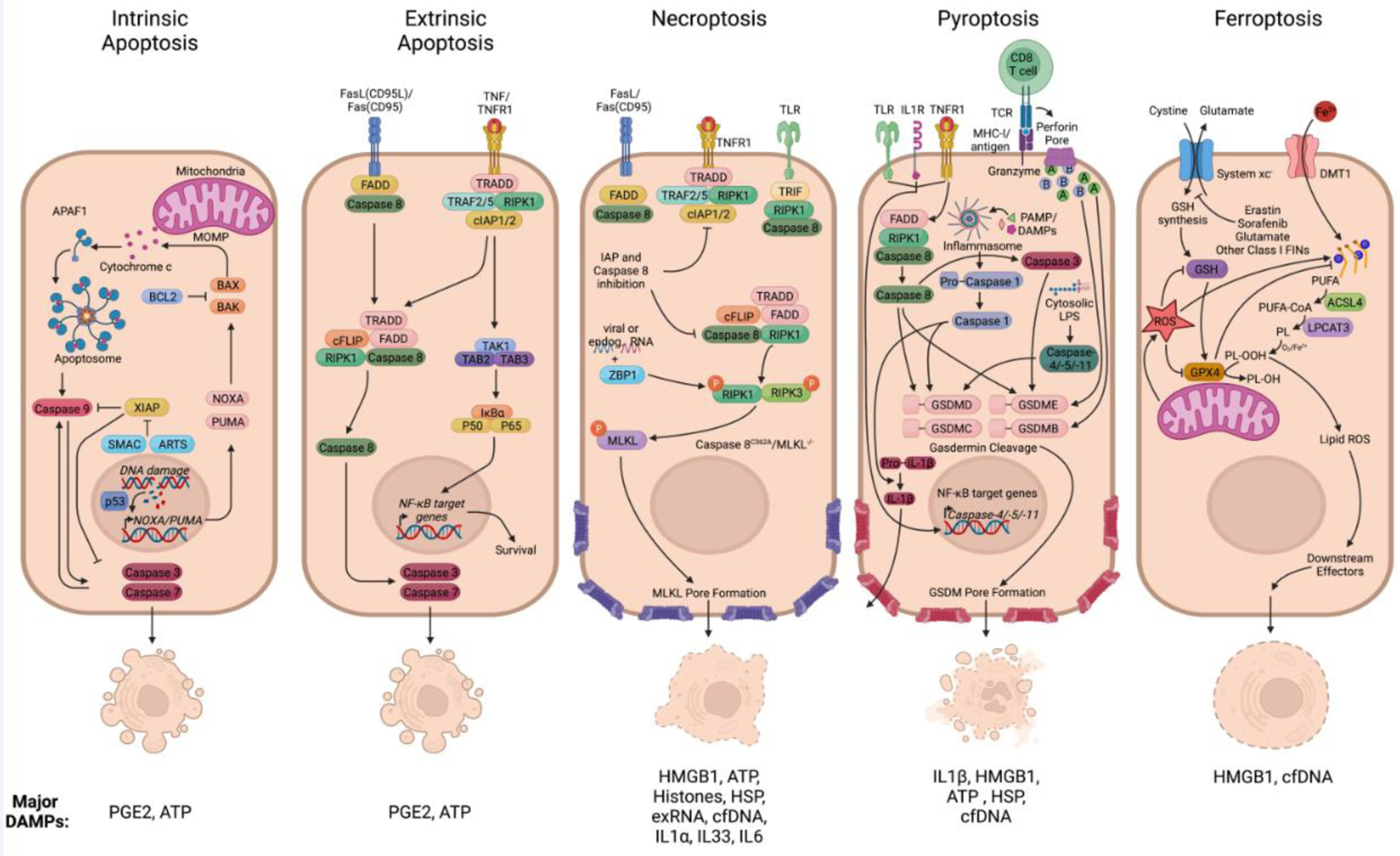
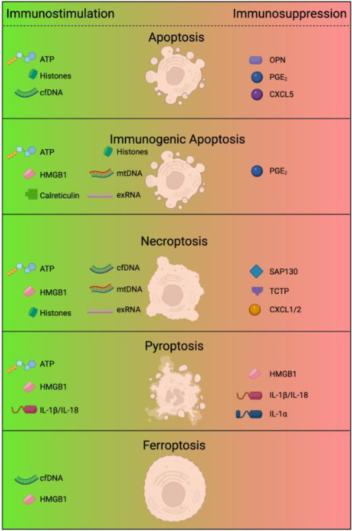
Induction of cell death is inexorably linked with cancer therapy, but this can also initiate wound-healing processes that have been linked to cancer progression and therapeutic resistance. Here we describe the contribution of apoptosis and the lytic cell death pathways in the response to therapy (including chemotherapy and immunotherapy). We also discuss how necroptosis, pyroptosis, and ferroptosis function to promote tumor immunogenicity, along with emerging findings that these same forms of death can paradoxically contribute to immune suppression and tumor progression. Understanding the duality of cell death in cancer may allow for the development of therapeutics that shift the balance towards regression.

Keywords: apoptosis; cell death; ferroptosis; immunogenic cell death; immunogenicity; immunotherapy; necroptosis; pyroptosis; tumor microenvironment.


Conflict of interest statement

Declaration of interests B.R. is a member of the scientific advisor board for Omios Biologics, LLC. B.R. has a secondary faculty appointment at the University of South Florida.

Figures

Figure 1.
Figure 1.. Overview of cell death modalities.
Active cell death processes are classified into sub forms according to the genes regulating each modality. (Panel 1) Caspase-dependent forms of non-lytic programmed cell death include intrinsic apoptosis that is initiated by MOMP and subsequent release of cytochrome c that can be controlled by BAX/BAK. This triggers the formation of the multiprotein complex termed the ‘apoptosome’ and activation of caspase-9, leading to downstream activation of executioner caspases-3 and -7. PGE2 and ATP are among the most common DAMPs released from apoptotic cells. While ATP is considered as a find-me signal for phagocytic cells, PGE2 negatively regulating death induced inflammation, promotes survival and angiogenesis and can act on surrounding stroma and the tumor microenvironment. (Panel 2) A variety of cell surface receptors, mainly from the TNF receptor superfamily such as CD95 or TNFR1, can lead to extrinsic apoptosis signaling via caspase-8 and RIPK1. Alternatively, these can lead to the induction of survival signaling via TAK1 mediated NF-kB activation driving survival gene expression. (Panel 3) Under apoptosis compromised conditions, such as depletion or pharmacological inhibition of caspase-8 and the IAPs, activation of RIPK1 can lead to necroptosis via RIPK3 and MLKL. The DAMPs described for necroptosis are HMGB1, ATP, Histones, HSP, exRNA, cfDNA, IL1α, IL33, and IL6. These generally have pro-inflammatory features; however, dose and context are critical in determining outcome. For example, IL-6 can promote and inhibit anti-tumor immunity. (Panel 4) Pyroptosis is induced through different cell surface receptors including TLRs, IL1R, and TNFR1. Essential for the execution of pyroptosis is the cleavage of GSDMs, leading to pore-formation, release of mature IL-1β, and the leakage of cellular content. This can be triggered by inflammasome activation and caspase-1, activated caspase-8 and caspase-3, or via granzymes from cytotoxic lymphocytes. Among the DAMPs release during pyroptosis (IL1β, HMGB1, HSP, cfDNA), IL1β is the strongest inducer of inflammation. (Panel 5) Ferroptosis occurs as a consequence of iron overload and accumulation of peroxidized lipids, caused either by GPX4 inhibition or glutathione deprivation. The few DAMPs described during ferroptotic cells have all been found to have pro-inflammatory functions. Figure created with BioRender. Figure 1 Abbreviations: BCL2 B-Cell Lymphoma 2 BAX BCL2 Associated X BAK BCL2 Antagonist/Killer 1 XIAP X-Linked Inhibitor Of Apoptosis SMAC Second Mitochondria-Derived Activator Of Caspase ARTS Apoptosis-Related Protein In The TGF-Beta Signaling PUMA P53-Upregulated Modulator Of Apoptosis p53 Tumor protein P53 PGE2 Prostaglandin E2 ATP adenosine 5′-triphosphate TNFR1 TNF receptor superfamily member 1A FADD Fas associated via death domain TRADD TNFRSF1A Associated Via Death Domain TRAF2/5 TNF Receptor Associated Factor 2/5 RIPK1 Receptor Interacting Serine/Threonine Kinase 1 cIAP1/2 Cellular Inhibitor Of Apoptosis 1/2 cFLIP CASP8 And FADD Like Apoptosis Regulator ZBP1 Z-DNA Binding Protein 1 RIPK3 Receptor Interacting Serine/Threonine Kinase 3 MLKL Mixed Lineage Kinase Domain Like Pseudokinase HMGB1 High Mobility Group Box 1 HSP heat shock proteins exRNA extracellular RNA cfDNA cell-free DNA IL1a Interleukine 1 alpha IL33 Interleukin-33 IL6 Interleukin-6 TLR Toll-like receptor MHC-I Major histocompatibility complex 1 TCR T cell receptor PAMP Pattern associated molecular patterns DAMP Danger associated molecular patterns LPS Lipopolysaccharides GSDMD/E/C/B Gasdermin D/E/C/B NF-kB Nuclear factor kappa B IL1b Interleukine 1 beta ROS Reactive oxigen species GSH Glutathione synthetase GPX4 Glutathione Peroxidase 4 PUFA Polyunsaturated fatty acids ACSL4 acyl-CoA synthetase long chain family member 4 LPCAT3 Lysophosphatidylcholine acyltransferase 3 CoA Coenzyme A PL Phospholipid
Figure 2.
Figure 2.. Cell death and release of immune regulatory factors.
The form of cell death determines the type of DAMPs, cytokines, or other factors released by cells. Depending on the cell death modality and the DAMPs released, this can have either immunostimulatory or immunosuppressive effects on the tumor microenvironment. Apoptotic death is often considered to be immunologically silent despite the release of ATP and other DAMPs, due to the balance with immunosuppressive factors such as OPN and PGE2, as well a consequence of macrophage efferocytosis. In contrast, the immunogenic potential and antitumor features of immunogenic apoptosis is attributed to the combined release of HMGB1, mtDNA, exRNA, and histones, along with calreticulin exposure on the cell surface. Necrotic-like forms of lytic death generally include the release of intracellular DAMPs, including ATP, TCTP, SAP130, Histones, HMGB1, mtDNA, cfDNA, and exRNA. However, different forms of cell necrotic death are not necessarily equivalent and the form, type, and quantity of DAMPs may vary. Just as importantly, different forms of cell death can promote the release of unique cytokines with immune regulatory function. Beyond the connection of IL-1β with pyroptosis, necroptosis has been shown to induce CXCL1 and CXCL2 expression, major chemokines involved in the recruitment of neutrophils. These factors could promote or inhibit anti-tumor immunity in a context-specific manner. Figure created with BioRender. Figure 2 Abbreviation list: ATP Adenosine 5′-triphosphate cfDNA Cell free DNA OPN Osteopontin PGE2 Prostaglandin E2 CXCL5 C-X-C motif chemokine ligand 5 mtDNA Mitochondrial DNA exRNA Extracellular RNA TCTP Translationally-Controlled Tumor Protein SAP130 Sin3A Associated Protein 130 CXCL1/2 C-X-C motif chemokine ligand 1/2 IL1α Interleukine-1 alpha IL1β Interleukine-1 beta

References

    1. Galluzzi L et al. (2018) Molecular mechanisms of cell death: recommendations of the Nomenclature Committee on Cell Death 2018. Cell Death & Differentiation 25, 486–541. - PMC - PubMed
    1. Green DR and Llambi F (2015) Cell Death Signaling. Cold Spring Harb Perspect Biol 7. - PMC - PubMed
    1. Kroemer G and Levine B (2008) Autophagic cell death: the story of a misnomer. Nat Rev Mol Cell Biol 9, 1004–1010 - PMC - PubMed
    1. Napoletano F et al. (2019) Intersections between Regulated Cell Death and Autophagy. Trends Cell Biol 29, 323–338 - PubMed
    1. Fuchs Y and Steller H (2015) Live to die another way: modes of programmed cell death and the signals emanating from dying cells. Nature Reviews Molecular Cell Biology 16, 329–344 - PMC - PubMed

Publication types