. 2023 Feb 25;14(2):159.
doi: 10.1038/s41419-023-05696-1.

NRP1 promotes prostate cancer progression via modulating EGFR-dependent AKT pathway activation

Affiliations

NRP1 promotes prostate cancer progression via modulating EGFR-dependent AKT pathway activation

Peng Zhang et al. Cell Death Dis. .

Abstract

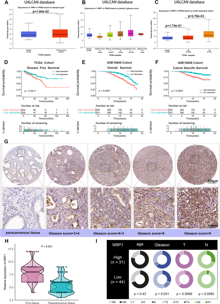
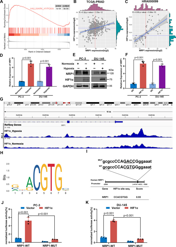
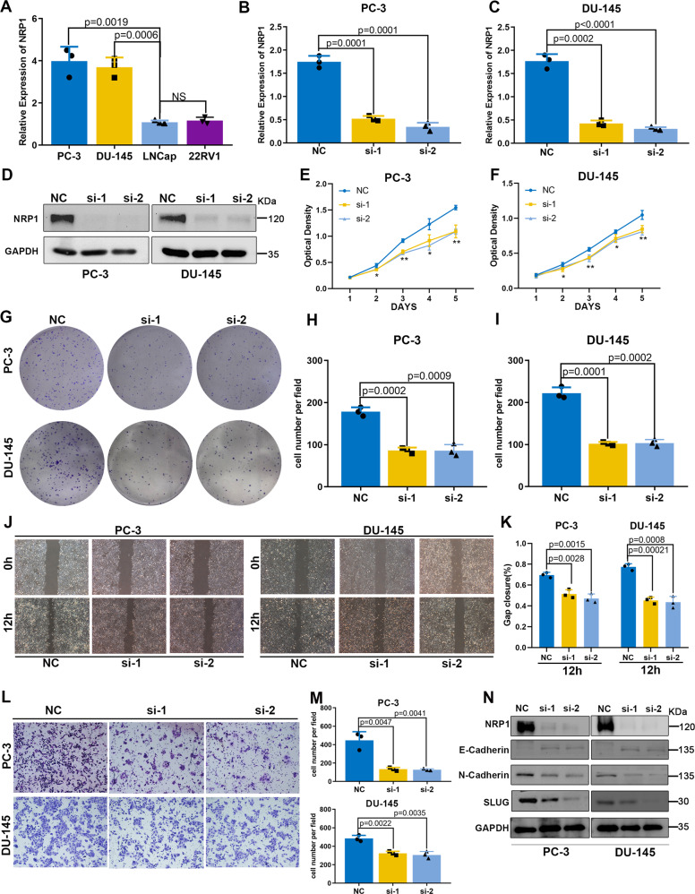
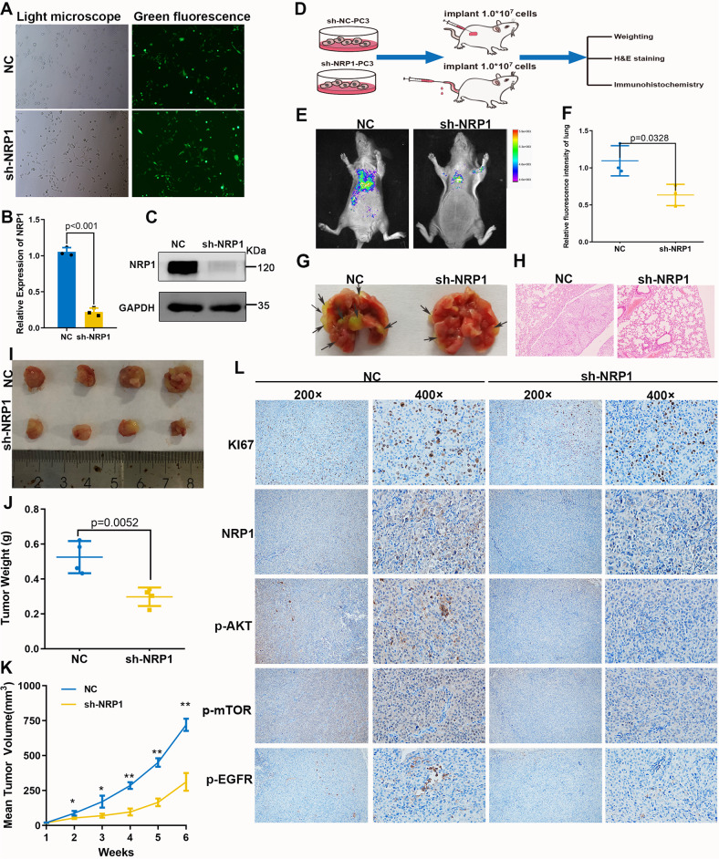
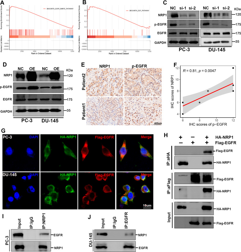
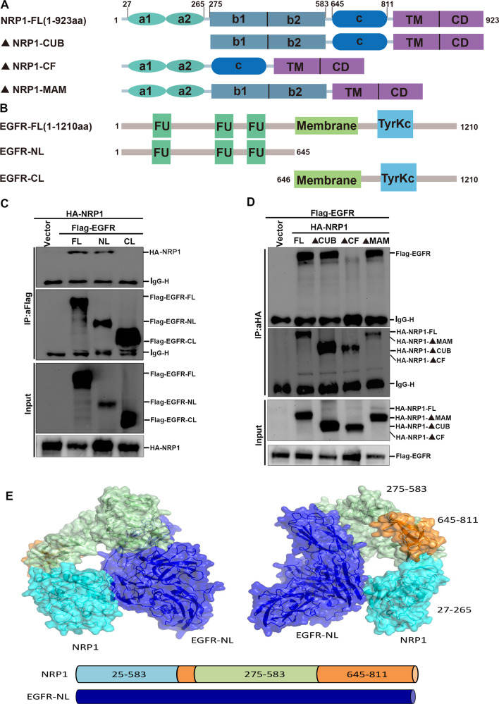
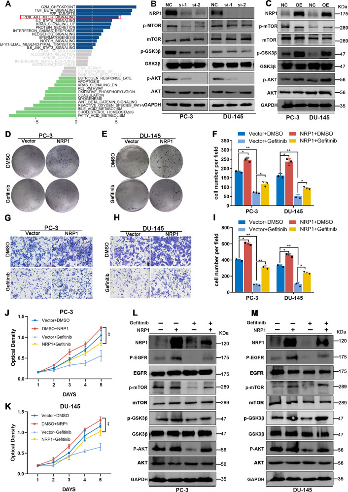
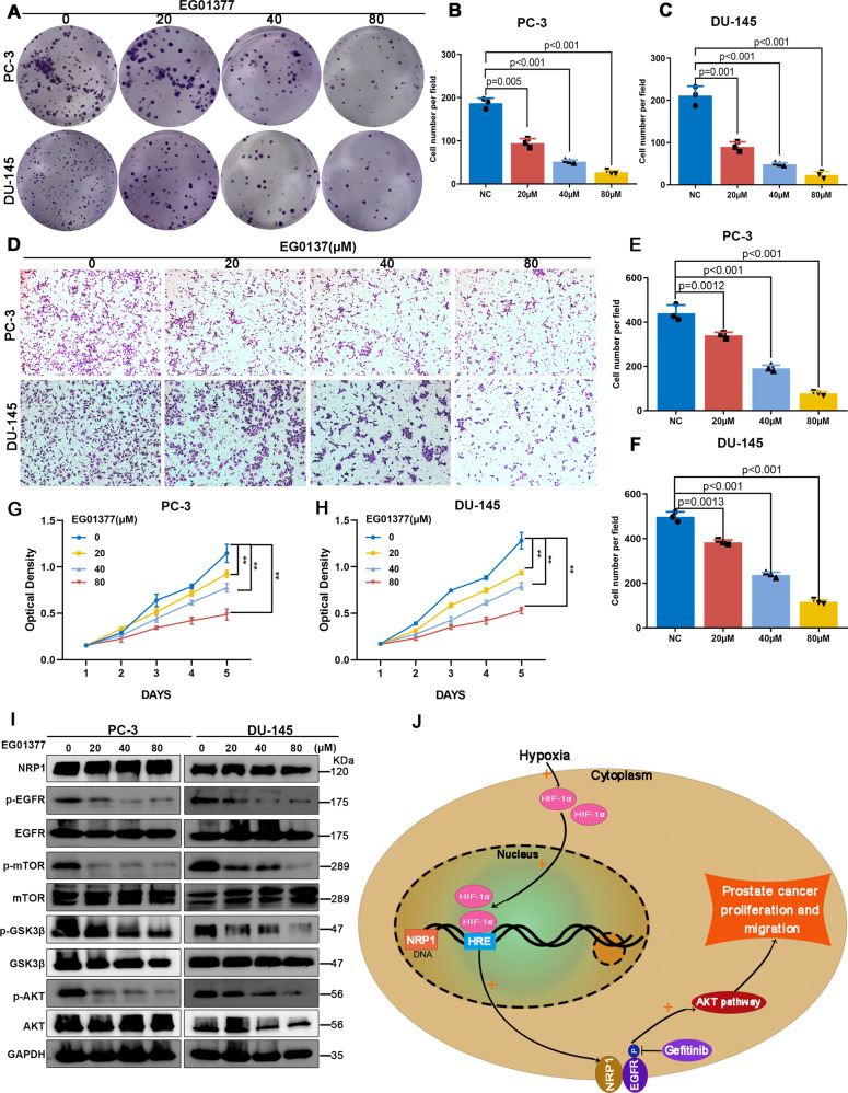
Prostate cancer (PCa) is the most common malignant tumor with a high global incidence in males. The mechanism underlying PCa progression is still not clear. This study observed that NRP1 was highly expressed in PCa and associated with poor prognosis in PCa patients. Functionally, NRP1 depletion attenuated the proliferation and migration ability of PCa cells in vitro and in vivo, while NRP1 overexpression promoted PCa cell proliferation and migration. Moreover, it was observed that NRP1 depletion induced G1 phase arrest in PCa cells. Mechanistically, HIF1α is bound to the specific promoter region of NRP1, thereby regulating its transcriptional activation. Subsequently, NRP1 interacted with EGFR, leading to EGFR phosphorylation. This study also provided evidence that the b1/b2 domain of NRP1 was responsible for the interaction with the extracellular domain of EGFR. Moreover, EGFR mediated NRP1-induced activation of the AKT signaling pathway, which promoted the malignant progression of PCa. In addition, the administration of NRP1 inhibitor EG01377 significantly inactivated the EGFR/AKT signaling axis, thereby suppressing PCa progression. In conclusion, the findings from this study highlighted the molecular mechanism underlying NRP1 expression in PCa and provide a potential predictor and therapeutic target for clinical prognosis and treatment of PCa.


Conflict of interest statement

The authors declare no competing interests.

Figures

Fig. 1
Fig. 1. NRP1 is overexpressed in PCa tissues and associated with unfavorable prognosis of PCa patients.
A Differential expression of NRP1 gene in PCa and adjacent tissues. B NRP1 expression is associated with higher Gleason scores in PCa. C NRP1 expression is associated with lymphatic metastasis in PCa. AC Complete data are obtained from the UALCAN database. DF Kaplan–Meier survival analysis of PCa patients from the TCGA and GSE10645 datasets. G IHC analysis of NRP1 expression in 75 PCa samples. Representative images are shown, with the scale bar of 400 μm and 100 μm, respectively. H Differential expression of NRP1 protein in 75 PCa tissues and paired paracancerous tissues (statistical significance assessed using a Mann–Whitney U test). I Comparison of age, Gleason score, T stage, and N stage between high NRP1 expression group and low NRP1 expression group (grouping based on the median of IHC staining score, and statistical significance assessed using the chi-square test).
Fig. 2
Fig. 2. HIF1α increases the expression of the NRP1 gene through transcriptional activation in PCa cells.
A GSEA analysis of the GSE70769 dataset indicated NRP1 is significantly associated with the hypoxia pathway. B, C HIF1α expression is correlated with NRP1 expression based on the TCGA-PRAD and HRA000099 datasets. D Expression of NRP1 mRNA level increases under hypoxic conditions. E Significant increase in protein levels of HIF1α and NRP1 in PCa cells under hypoxic conditions. F HIF1α overexpression promotes NRP1 mRNA expression. G GSE106305 ChIP dataset indicates HIF1α binding to the NRP1 promoter region shows a significant peak. H Schematic of canonical HIF1α motif binding sites provided by the JASPAR database. I Diagram represents the predicted HIF1α binding site on the NRP1 promoter, as well as wild-type/mutant NRP1 promoter plasmids. J, K Luciferase assay indicates that HIF1α significantly increases NRP1-WT promoter-driven luciferase activity, while the mutation of the binding site of HIF1α to NRP1 leads to significant attenuation of the luciferase activity in PC-3 and DU-145 cells, respectively. Values represent the mean ± SD of three independent experiments. Statistical significance was assessed using two-tailed t tests.
Fig. 3
Fig. 3. NRP1 depletion attenuates PCa cell proliferation and migration in vitro.
A mRNA expression level of NRP1 detected in four PCa cell lines (PC-3, DU-145, LNCap, 22RV1) by qRT-PCR. B, C qRT-PCR analysis evaluates the depletion efficiency of two NRP1-specific siRNA in PC-3 and DU-145 cells, and si-1 and si-2 represent favorable depletion efficiency. D Immunoblot analysis validates depletion efficiency of si-1 and si-2 in PC-3 and DU-145 cells. E, F MTT assay reveals that NRP1 depletion attenuates cell viability in PC-3 and DU-145 cells. GI Clone formation assay and the statistical chart represent NRP1 depletion attenuating clone formation ability in PC-3 and DU-145 cells. J, K Wound-healing assay and the statistical chart show that NRP1 depletion inhibits cell migration in PC-3 and DU-145 cells. L, M Transwell assay and the statistical chart demonstrate that NRP1 depletion inhibits cell migration in PC-3 and DU-145 cells. N Immunoblot assay shows EMT-related proteins in PC-3 and DU-145 cells after NRP1 depletion. Statistical significance was assessed using two-tailed t tests. Error bars represent the standard deviations of three independent experiments. *P < 0.01, **P < 0.001.
Fig. 4
Fig. 4. NRP1 promotes PCa cell proliferation and migration in vivo.
A Green fluorescence of PC-3 stable cells. B, C qRT-PCR and immunoblot assay verify NRP1-depletion efficiency in PC-3 stable cells. D Xenograft mice models and pulmonary metastasis models were established by subcutaneously injecting LV-NC cells or LV-sh-NRP1 cells. Mice were monitored continuously for 5 weeks in xenograft mice models and 8 weeks in the pulmonary metastasis model. Then the mice were sacrificed and the tumors were dissected. E Fluorescence intensity of pulmonary metastasis is detected by imaging apparatus for small animals in vivo, F sh-NRP1 group (n = 3) significantly inhibited lung metastasis of PCa cells compared with that in the NC group (n = 3). G Lung tissue specimens show pulmonary metastatic nodules, where the arrows denote the tumors transferred to the lungs. H H&E staining of lung tissues (scale bar = 100 μm). Statistical analysis of tumor volume and weight in two groups (n = 4 in each group). I Tumor image of xenograft mice models. J NRP1 depletion significantly decreases the weight of the tumor xenograft. K NRP1 depletion significantly inhibits tumor growth. L IHC staining detects the expression of Ki67, NRP1, p-AKT, p-mTOR, and p-EGFR. Data are shown as the mean ± SD. Statistical significance was assessed using a two-tailed t test. *P < 0.01, **P < 0.001.
Fig. 5
Fig. 5. NRP1 interacts with EGFR and induces EGFR phosphorylation.
A, B GSEA of TCGA-PRAD shows NRP1 is positively related to the EGFR pathway. C, D Immunoblot assay elucidates that NRP1 depletion attenuates EGFR phosphorylation level, while ectopic overexpression of NRP1 promotes EGFR phosphorylation level. E, F IHC assay and the correlation analysis represent that NRP1 protein is positively correlated with the expression of phosphorylated EGFR protein in PCa tissues (scale bar = 400 μm). G NRP1 (green) and EGFR (red) co-localization is examined by the immunofluorescence assay (scale bar = 15 μm) and the nucleus is indicated by DAPI (blue) staining. H Exogenous Co-IP assay represents NRP1 could interact with EGFR in 293T cells. I, J Endogenous Co-IP assay proves NRP1 could interact with EGFR in PC-3 and DU-145 cells. Statistical significance was assessed using a two-tailed t test.
Fig. 6
Fig. 6. b1/b2 domain of NRP1 interacts with the extracellular domain of EGFR to influence the activation of EGFR.
A, B Schematic diagram of NRP1 and EGFR primary structure and truncated plasmids construction. C Identification of the b1/b2 domain of NRP1 mediating its interaction with EGFR. D Identification of the extracellular domain of EGFR mediating its interaction with NRP1. E Molecular docking analysis shows the interaction domain between NRP1 and EGFR.
Fig. 7
Fig. 7. NRP1 promotes PCa cell proliferation through the EGFR/AKT signaling axis.
A GSVA result indicates the AKT pathway is significantly downregulated in the NRP1-depletion group compared to the control group based on RNA-Seq data of NRP1-depleted DU-145 cells and control cells. B, C Immunoblot assay elucidates that NRP1 depletion attenuates AKT, GSK3β, and mTOR phosphorylation levels, while ectopic overexpression of NRP1 increases AKT, GSK3β, and mTOR phosphorylation levels. DF Clone formation assay and the statistical chart represent EGFR inhibitor gefitinib attenuating clone formation ability induced by NRP1 overexpression in PC-3 and DU-145 cells. GI Transwell assay and the statistical chart represent gefitinib attenuates migration ability induced by NRP1 overexpression. J, K MTT assay elucidates gefitinib attenuates proliferation viability induced by NRP1 overexpression. L, M Immunoblot results elucidate that gefitinib attenuates AKT, GSK3β, and mTOR phosphorylation levels induced by NRP1 overexpression. Statistical significance was assessed using a two-tailed t test. Error bars represent the standard deviations of three independent experiments. *P < 0.01, **P < 0.001.
Fig. 8
Fig. 8. NRP1 inhibitor EG01377 attenuates PCa proliferation and migration via EGFR/AKT signaling axis.
AC Clone formation assay, DF transwell assay, and G, H MTT assay represent that NRP1 inhibitor EG01377 attenuates clone formation, proliferation, and migration ability in a concentration-dependent manner. I Immunoblot results elucidate that EG01377 attenuates AKT, GSK3β, and mTOR phosphorylation levels in a concentration-dependent manner. J A schematic model of HIF1α-NRP1-EGFR/AKT signaling axis promoting prostate cancer progression. Statistical significance was assessed using a two-tailed t test. Error bars represent the standard deviations of three independent experiments. **P < 0.001.

Similar articles

Cited by

References

    1. Sung H, Ferlay J, Siegel RL, Laversanne M, Soerjomataram I, Jemal A, et al. Global Cancer statistics 2020: GLOBOCAN estimates of incidence and mortality worldwide for 36 cancers in 185 countries. CA Cancer J Clin. 2021;71:209–49. doi: 10.3322/caac.21660. - DOI - PubMed
    1. Cornford P, van den Bergh RCN, Briers E, Van den Broeck T, Cumberbatch MG, De Santis M, et al. EAU-EANM-ESTRO-ESUR-SIOG guidelines on prostate cancer. Part II-2020 update: treatment of relapsing and metastatic prostate cancer. Eur Urol. 2021;79:263–82. doi: 10.1016/j.eururo.2020.09.046. - DOI - PubMed
    1. Kolodkin AL, Levengood DV, Rowe EG, Tai YT, Giger RJ, Ginty DD. Neuropilin is a semaphorin III receptor. Cell. 1997;90:753–62. doi: 10.1016/S0092-8674(00)80535-8. - DOI - PubMed
    1. Lu D, Shang G, He X, Bai XC, Zhang X. Architecture of the Sema3A/PlexinA4/Neuropilin tripartite complex. Nat Commun. 2021;12:3172. doi: 10.1038/s41467-021-23541-x. - DOI - PMC - PubMed
    1. Rizzolio S, Rabinowicz N, Rainero E, Lanzetti L, Serini G, Norman J, et al. Neuropilin-1-dependent regulation of EGF-receptor signaling. Cancer Res. 2012;72:5801–11. doi: 10.1158/0008-5472.CAN-12-0995. - DOI - PubMed

Publication types