. 2023 Feb 25;12(1):9.
doi: 10.1038/s41389-023-00457-3.

PELI1 and EGFR cooperate to promote breast cancer metastasis

Affiliations

PELI1 and EGFR cooperate to promote breast cancer metastasis

Jie Qi et al. Oncogenesis. .

Abstract

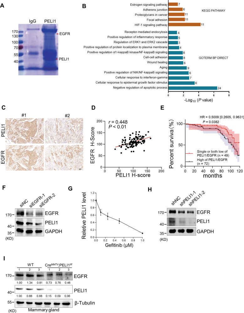
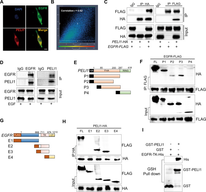
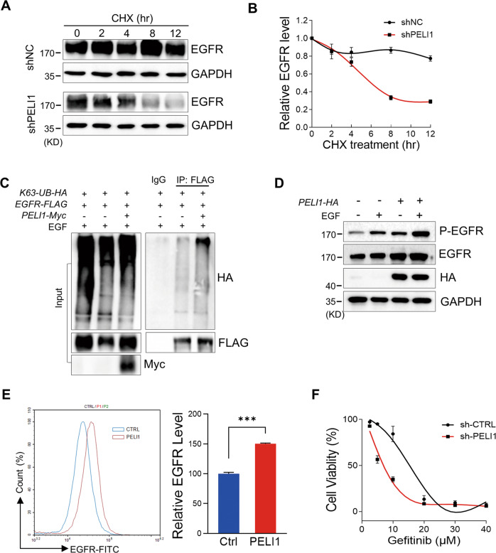
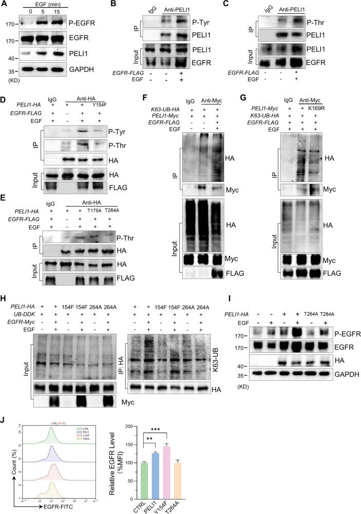
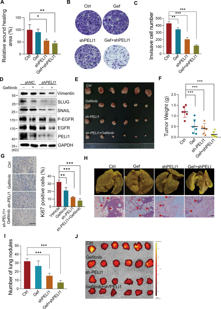
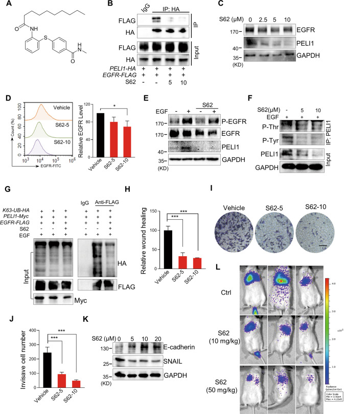
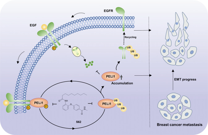
Pellino-1 (PELI1) is an E3 ubiquitin ligase acting as a key regulator for the inflammation and autoimmunity via the ubiquitination of the substrate proteins. There is increasing evidence to support that PELI1 functions as an oncoprotein in tumorigenesis and metastasis. However, the molecular mechanism underlying the high expression and oncogenic roles of PELI1 in cancers remains limited. Herein, we revealed a novel regulation mechanism by which PELI1 and EGFR cooperate to promote breast cancer metastasis. EGFR is positively correlated with PELI1 expression in breast cancers, and its activation led to the phosphorylation of PELI1 at Tyr154 and Thr264, which subsequently activated its E3 ubiquitin ligase. Simultaneously, PELI1 physically interacted with and enhanced the stability of EGFR via the K63-linked polyubiquitination in reverse. The co-inhibition of the PELI1-EGFR showed synergetic effect to repress breast cancer metastasis. Furthermore, we identified a compound S62 as a small molecule disruptor of PELI1/EGFR that effectively repressed breast cancer metastasis. Our study not only uncovered the emerging roles of PELI1/EGFR interaction in the progression of breast cancer, but also provided an effective strategy for the inhibition of metastasis in breast cancer.


Conflict of interest statement

The authors declare no competing interests.

Figures

Fig. 1
Fig. 1. The levels of PELI1 and EGFR were positively correlated in human breast cancer.
A MDA-MB-231 cells overexpressing PELI1 were lysed and immunopurified with normal IgG and anti-PELI1 antibody respectively. Then the complex was resolved by SDS–PAGE followed by Coomassie Blue staining. The distinct bands were analyzed by MS. Red arrows indicated the identified PELI1 and EGFR. B Representative biological processes and signaling pathways significantly enriched from proteins identified from Co-IP with PELI1 in MDA-MB-231 cells. GO and KEGG analysis were performed using DAVID bioinformatics database. The number of enriched proteins in relative terms was shown in each bar. C, D IHC analysis of PELI1 and EGFR in the tissues from breast cancer patients’ tissues microarray. Representative images of IHC staining (C) and the correlation rate (D) by Pearson’s test is shown (N = 121, scale bar, 50 μm). E Overall survival rates were determined by Kaplan–Meier analyses of indicated groups. Hazard ratio (HR) and P values (log rank P) are shown. F Western blotting analysis of PELI1 protein levels in MDA-MB-231 cells with EGFR knockdown. GAPDH was used as loading control. G ELISA analysis of the change of PELI1 in MDA-MB-231 cells with Gefitinib. H Western blotting analysis of EGFR protein levels in MDA-MB-231 cells with PELI1 knockdown. GAPDH was used as a loading control. I Sample immunoblotting showed the levels of PELI1 and EGFR proteins in mammary gland from indicated transgene mice. β-Tubulin was used as a loading control.
Fig. 2
Fig. 2. PELI1 directly interacted with EGFR.
A, B IF staining showed co-localization between PELI1and EGFR in MDA-MB-231 cells (scale bar, 10 μm). The co-localization correlation rate of PELI1 and EGFR in A is shown (B). C The interaction between PELI1 and EGFR was detected by Co-IP assay. HEK293T/17 cells were co-transfected with pCDNA3.1-HA-PELI1 and pLVX-FLAG-EGFR plasmids. The cells were harvested and subjected to immunoprecipitated with anti-HA and anti-FLAG antibodies respectively. Similar Co-IP analysis with normal IgG were performed as control. D Co-IP analysis of endogenous PELI1 and EGFR in MDA-MB-231 cells. The cell extracts were immunoprecipitated with anti-IgG, anti-EGFR and anti-PELI1 antibodies, respectively, upon EGF (100 ng/ml) stimulation. E The sketch map of the deletion mutant regions of PELI1. F Co-IP analysis of PELI1 mutants binding to EGFR. HEK293T/17 cells were co-transfected with PELI1 deletion mutants (HA tagged) and pLVX-FLAG-EGFR plasmids, and IP analysis was performed with anti-FLAG antibody. G The sketch map of the deletion mutant regions of EGFR. H Co-IP analysis of EGFR regions binding to PELI1. HEK293T/17 cells were co-transfected with EGFR deletion mutants (FLAG-tagged) and pCDNA3.1-HA-PELI1 plasmids, and IP analysis was performed with ani-HA antibody. I GST-pull down assay of the direct correlation between PELI1 and EGFR-TK.
Fig. 3
Fig. 3. PELI1 ubiquitinated EGFR and inhibited its degradation.
A, B Western blotting analysis of EGFR protein levels in MDA-MB-231 cells with PELI1 knockdown upon the treatment of CHX (10 μg/ml). EGFR levels were normalized to the change of GAPDH (N = 3). C Western blotting analysis of K63-linked polyubiquitination of EGFR immunoprecipitated from HEK293T/17 cells co-overexpressing PELI1 and EGFR upon EGF stimulation (100 ng/ml). D Western blotting analysis of EGFR proteins in MDA-MB-231 cells with or without PELI1 overexpression upon EGF (100 ng/ml) stimulation. E Flow cytometric analysis of membrane EGFR levels in MDA-MB-231 cells with PELI1 overexpression upon EGF (100 ng/ml) stimulation (N = 3). ***P < 0.001. F Effect of PELI1 knockdown on the cell viability of MDA-MB-231 cells was detected upon the treatment of Gefitinib.
Fig. 4
Fig. 4. EGFR phosphorylated PELI1 leading to its K63-linked auto-ubiquitination.
A Western blotting analysis of the levels of indicated proteins in response to EGF (100 ng/ml). B, C Western blotting analysis of the tyrosine (B) and threonine (C) phosphorylation of PELI1 with EGFR overexpression upon EGF stimulation (100 ng/ml) in MDA-MB-231 cells. D, E Western blotting analysis of the tyrosine or threonine phosphorylation of PELI1 immunoprecipitated from HEK293T/17 cells with overexpression of the indicated PELI1 mutants. F Western blotting analysis of the K63-mediated polyubiquitination of PELI1 immunoprecipitated from HEK293/17 cells co-overexpressing with EGFR and PELI1 upon EGF (100 ng/ml) stimulation. G Similar with F excluding overexpressing PELI1 mutant (K169F). H PELI1 was immunoprecipitated from HEK293T/17 cells transfected with PELI1, PELI1-Y154F or PELI1-T264A plasmids, followed by Western blotting analysis of the K63-mediated polyubiquitination. I Western blotting analysis of EGFR and phospho-EGFR in MDA-MB-231 cells overexpressed PELI1 or PELI1-T264A upon EGF (100 ng/ml) stimulation. J Flow cytometric analysis of membrane EGFR in MDA-MB-231 cells with overexpression of PELI1, PELI1-Y154F or PELI1-T264A (N = 3). **P < 0.01, ***P < 0.001. All P values were determined by unpaired two-tailed Student’s t test.
Fig. 5
Fig. 5. Inhibition of PELI1 and EGFR suppressed breast cancer.
AC Quantification of the migration (A) and invasion (B, C) of MDA-MB-231 cells transfected with PELI1-shRNA with or without Gefitinib (2 μM) treatment (N = 3, scale bar, 100 μm). D Western blotting analysis of the indicated proteins with or without PELI1 knockdown and Gefitinib (2 μM) treatment. EG Effects of PELI1 knockdown and Gefitinib treatment on the tumor incidence of MDA-MB-231 cells in nude mice. The mice were subcutaneously transplanted with MDA-MB-231/Con-shRNA and MDA-MB-231/PELI1-shRNA cells (5 × 106/mouse) and were treated with or without Gefitinib (50 mg/kg) orally every other day for 2 months. Representative images of tumors (E) and tumor weight (F) are represented (N = 6 per group). The tumors were made into paraffin sections and the Ki67-positive cells (G, scale bar, 50 μm) were quantified. H Effects of PELI1 knockdown and Gefitinib treatment on the lung-metastasis of MDA-MB-231 cells. NYG mice were injected with MDA-MB-231/Con-shRNA and MDA-MB-231/PELI1-shRNA cells (2 × 105/mouse) via tail vein, and were treated with or without Gefitinib (50 mg/kg) orally every other day for 1 month. The whole indicated lung tissues were stained with Bouin fluid and made into HE stained sections (scale bar, 50 μm). I Quantitative analysis of the metastatic lung nodules in H (N = 6 per group). J The GFP fluorescence intensity of lung tissues from H are shown. *P < 0.05, **P < 0.01, ***P < 0.001. All P values were determined by unpaired two-tailed Student’s t test.
Fig. 6
Fig. 6. The compound S62 interrupted the interaction between PELI1 and EGFR to suppress breast cancer metastasis.
A Chemical structure of S62. B HEK293T/17 cells with overexpression of PELI1 and EGFR were treated with or without S62 (5 or 10 μM) for 24 h. Co-IP assay was subsequently performed to detect the interaction between PELI1 and EGFR. C Western blotting analysis of EGFR and PELI1 in MBA-MB-231 cells with the treatment of S62 for 24 h. D Flow cytometric analysis of the membrane EGFR in MDA-MB-231 cells treated with or without S62 for 24 h (N = 3). E Western blotting analysis of EGFR, PELI1 and phosphorylation of EGFR with or without the treatment of S62 (10 μM) upon EGF stimulation (100 ng/ml). F Western blotting analysis of the tyrosine and threonine phosphorylation of PELI1 immunoprecipitated from MDA-MB-231 cells. The cells were treated as in E. G Western blotting analysis of the K63-mediated polyubiquitination of EGFR immunoprecipitated from HEK293T/17 cells with the treatment of S62 (10 μM). H Quantitative analysis of the migration of MDA-MB-231 cells with the treatment of S62 (N = 3). I Effect of S62 on the invasion of MDA-MB-231 cells. The representative images of invasive cells were shown (scale bar, 100 μm). J Quantitative analysis of the invasion of MDA-MB-231 cells in I (N = 3). K Western blotting analysis of E-cadherin and SNAIL in MDA-MB-231 cells with the treatment of S62. L The representative images of lung metastasis of breast cancer cells in NYG mice with S62 treatment. MDA-MB-231 transfected with lentivirus that stably expressed luciferase (2 × 105/mouse) were injected into NYG mice via the tail vein. The mice were treated with CMC-Na (0.5%) or S62 (10 or 50 mg/kg) every day for 2 weeks, and then were detected using the bioluminescence imaging. *P < 0.05, ***P < 0.001. All P values were determined by unpaired two-tailed Student’s t test.
Fig. 7
Fig. 7. Schematic diagram illustrates that PELI1 and EGFR cooperated to promote breast cancer metastasis.
EGFR acts as a phosphokinase of PELI1 to promote its phosphorylation, leading to the E3 ubiquitin ligase activation. Simultaneously, PELI1 enhanced the stability of EGFR via the K63-linked polyubiquitination. Compound S62 as a small molecule disruptor of PELI1/EGFR repressed breast cancer metastasis.

Similar articles

Cited by

References

    1. Sharma P, Barlow WE, Godwin AK, Parkes EE, Knight LA, Walker SM, et al. Validation of the DNA damage immune response signature in patients with triple-negative breast cancer from the SWOG 9313c trial. J Clin Oncol. 2019;37:3484–92. doi: 10.1200/JCO.19.00693. - DOI - PMC - PubMed
    1. Tan DS, Marchió C, Jones RL, Savage K, Smith IE, Dowsett M, et al. Triple negative breast cancer: molecular profiling and prognostic impact in adjuvant anthracycline-treated patients. Breast Cancer Res Treat. 2008;111:27–44. doi: 10.1007/s10549-007-9756-8. - DOI - PubMed
    1. Bray F, Ferlay J, Soerjomataram I, Siegel RL, Torre LA, Jemal A. Global cancer statistics 2018: GLOBOCAN estimates of incidence and mortality worldwide for 36 cancers in 185 countries. CA Cancer J Clin. 2018;68:394–424. doi: 10.3322/caac.21492. - DOI - PubMed
    1. Wu X, Tang P, Li S, Wang S, Liang Y, Zhong L, et al. A randomized and open-label phase II trial reports the efficacy of neoadjuvant lobaplatin in breast cancer. Nat Commun. 2018;9:832. doi: 10.1038/s41467-018-03210-2. - DOI - PMC - PubMed
    1. Park M, Kim D, Ko S, Kim A, Mo K, Yoon H. Breast cancer metastasis: mechanisms and therapeutic implications. Int J Mol Sci. 2022;23:6806. doi: 10.3390/ijms23126806. - DOI - PMC - PubMed