. 2023 Feb 25;14(1):1088.
doi: 10.1038/s41467-023-36780-x.

Stress-induced plasticity of a CRH/GABA projection disrupts reward behaviors in mice

Affiliations

Stress-induced plasticity of a CRH/GABA projection disrupts reward behaviors in mice

Matthew T Birnie et al. Nat Commun. .

Abstract

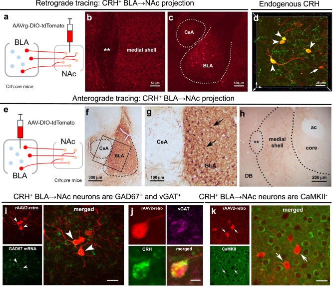
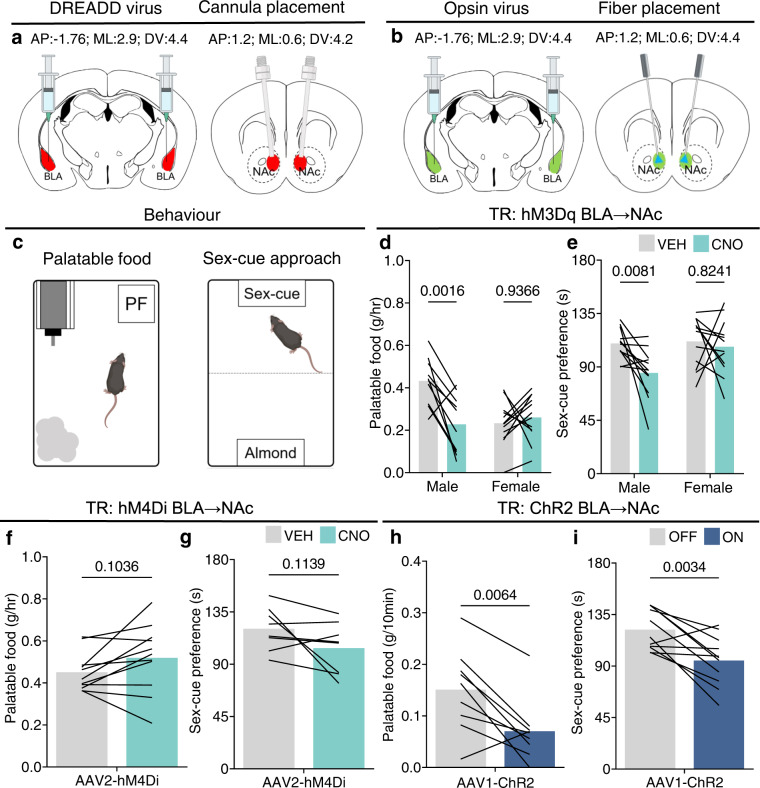
Disrupted operations of the reward circuit underlie major emotional disorders, including depression, which commonly arise following early life stress / adversity (ELA). However, how ELA enduringly impacts reward circuit functions remains unclear. We characterize a stress-sensitive projection connecting basolateral amygdala (BLA) and nucleus accumbens (NAc) that co-expresses GABA and the stress-reactive neuropeptide corticotropin-releasing hormone (CRH). We identify a crucial role for this projection in executing disrupted reward behaviors provoked by ELA: chemogenetic and optogenetic stimulation of the projection in control male mice suppresses several reward behaviors, recapitulating deficits resulting from ELA and demonstrating the pathway's contributions to normal reward behaviors. In adult ELA mice, inhibiting-but not stimulating-the projection, restores typical reward behaviors yet has little effect in controls, indicating ELA-induced maladaptive plasticity of this reward-circuit component. Thus, we discover a stress-sensitive, reward inhibiting BLA → NAc projection with unique molecular features, which may provide intervention targets for disabling mental illnesses.


Conflict of interest statement

The authors declare no competing interests.

Figures

Fig. 1
Fig. 1. A projection of CRH/GABA neurons in the medial BLA to the medial NAc shell.
ac Retrograde tracing of CRH+ neuronal inputs to medial NAc shell of CRH-ires-CRE mice. a Schematic of construct and injection location of AAV2‐retro‐CAG‐FLEX‐tdTomato‐WPRE virus that permits retrograde access to projection neurons providing afferent inputs to NAc. b Example confocal micrograph of locally infected CRH+ axon terminals in medial NAc shell. c Retrograde tracing identifies the medial BLA as a robust source of CRH+ NAc inputs. d 3D image (z-stack; 0.5 μm steps) confirmed localization in the BLA of AAV-retro infected cells (red) that co-express endogenous CRH (green); dual labeled neurons = yellow. eg Anterograde tracing of CRH+ axonal projections from BLA to medial NAc shell. e, The AAV1-DIO-tdTomato construct and the viral genetic experimental design. f Virus injection is confined to the BLA, g and absent from the central amygdala (CeA), shown by selective expression of tdTomato in BLA CRH+ neurons. h BLA-origin CRH+ axons and terminals in the medial NAc shell. ik Virus injection into the medial NAc shell retrogradely infected somata in the BLA. i Combined fluorescence in situ hybridization (FISH) and immunostaining with GAD67 mRNA in CRH+ cells in the BLA. Arrowheads point to co-localized GAD67 mRNA and virus-reporter labeling. j a BLA → NAc cell (red) co-expresses endogenous CRH (green) and vGAT (magenta), but k does not co-express the glutamatergic marker CaMKII. ** = Major Island of Calleja, ac  anterior commissure, DB  diagonal band. Scale bars in i and k = 10 µm. To confirm findings, virus injections, projection assessment, and immunohistochemistry were assessed in mice from at least two independent litters.
Fig. 2
Fig. 2. Optical stimulation of CRH+ BLA-origin axons in the NAc evokes exclusively IPSCs.
a Schematic of experimental design for electrophysiology recordings in the whole-cell patch-clamp configuration. Horizontal brain slices containing BLA and NAc from CRH-ires-CRE mice that were injected with Cre-dependent ChR2-EYFP in BLA. b Representative traces of optically evoked IPSCs (oIPSCs). These were blocked in the presence of GABAA receptor antagonist picrotoxin. c A neurobiotin (brown) filled neuron from which oIPSCs were recorded. Note spines (arrows) suggesting the cell is a medium spiny neuron (MSN). Arrowheads denote ChR2-expressing boutons from BLA-origin CRH+ axons (CRH; blue) on the soma. d oIPSCs amplitudes pre and post picrotoxin (n = 7 neurons, 5 mice). e Time-course plot of normalized oIPSCs amplitudes throughout the recording and following application of picrotoxin. Black triangles in e denote trace recordings in b and timepoint analysis in d and e. f Representative trace of a NAc cell showing no response from optically evoked EPSC (oEPSC) at −70 mV, obtained after verifying oIPSCs at 0 mV in the presence of picrotoxin. g Representative trace showing spontaneous EPSCs (sEPSCs) were still present; gray box shows magnified view recording. In d and e, bars represent mean. In e circles represent mean ± SEM. Two-sided paired t-tests in d and e. d oIPSCs (current) pre vs. post PTX: P = 0.0338. e oIPSCs (normalized) pre vs. post PTX: P = 0.0004. 3 V = third ventricle, Hip  Hippocampus, LV  lateral ventricle. Source data are provided as a Source Data file.
Fig. 3
Fig. 3. Stimulating the CRH/GABA BLA → NAc projection suppresses reward in typically reared male mice.
a, b Coordinate locations of a, DREADD injection and CNO infusion and b opsin injection and fiber placement. c schematic of the reward tasks. d, e Chemogenetically stimulating the CRH/GABA BLA → NAc projection with microinfusion of CNO in the medial NAc shell suppressed d, palatable food consumption (n = 10 male mice; 12 female mice) and e preference for a sex-cue (n = 12 male mice; 12 female mice) in males, but not females. f, g Inhibiting the CRH/GABA BLA → NAc projection in TR male mice did not increase f, palatable food consumption (n = 11 mice) or g, preference for a sex-cue (n = 8 mice). h, i stimulating the CRH/GABA ChR2-expressing BLA → NAc projection decreased h palatable food consumption (n = 9 mice) and i, approach time to a sex-cue (n = 11 mice). In di bars represent mean. Two-way ANOVA with repeated measures followed by post hoc tests (d, e). d hM3Dq BLA → NAc: Sex x Treatment—F = 5.691, DFn = 1, DFd = 40, P = 0.0219; post hoc with Tukey’s multiple comparison (Veh vs. CNO: Male—P = 0.0016; Female—P = 0.9366). e hM3Dq BLA → NAc: Treatment—F = 6.448, DFn = 1, DFd = 44, P = 0.0147; post hoc with Sidak’s multiple comparison (Veh vs. CNO: Male—P = 0.0081; Female—P = 0.8241). Two-sided paired t-tests (fi). f hM4Di BLA → NAc: P = 0.1036; g hM4Di BLA → NAc: P = 0.1139. h ChR2 BLA → NAc: P = 0.0064; i ChR2 BLA → NAc: P = 0.0034. PF  palatable food. Gray = vehicle/light off, teal = CNO, blue = light on. Source data are provided as a Source Data file.
Fig. 4
Fig. 4. Inhibiting the CRH/GABA BLA → NAc projection rescues reward deficits following early-life adversity.
a Timeline and environment (limited bedding and nesting) of ELA. bd ELA reduced palatable food consumption (n = 16 mice; TR = 10, ELA = 6), yet did not alter regular chow consumption (n = 20 mice; TR = 9, ELA = 11). ELA reduced preference for a sex-cue (n = 16 mice; TR = 8, ELA = 8) and preference for sucrose (n = 15 mice; TR = 7, ELA = 8). e Inhibiting the hM4Di+ CRH/GABA BLA → NAc projection with microinfusion of CNO in the medial NAc shell increased palatable food consumption (n = 9 mice) and f preference for a sex-cue (n = 8 mice). However, stimulating the hM3Dq+ CRH/GABA BLA → NAc did not influence g palatable food consumption (n = 9 mice) or h the preference for a sex-cue (n = 7 mice). In bd, bars represent mean ± SEM; in eh bars represent mean. Two-sided unpaired t-tests (b, d), two-sided unpaired U-test (c), two-sided paired t-tests (eh). b TR vs. ELA main: P = 0.0001, inset: P = 0.1263; c TR vs. ELA: P = 0.0281; d TR vs. ELA: P = 0.0053; e ELA hM4Di BLA → NAc: P = 0.0035; f ELA hM4Di BLA → NAc: P = 0.0170; g ELA hM3Dq BLA → NAc: P = 0.1375; h ELA hM3Dq BLA → NAc: P = 0.6267. Gray = typical reared, mauve = ELA/ELA + vehicle, pink = ELA + CNO. Source data are provided as a Source Data file.

References

    1. Berridge KC, Kringelbach ML. Pleasure systems in the brain. Neuron. 2015;86:646–664. doi: 10.1016/j.neuron.2015.02.018. - DOI - PMC - PubMed
    1. Yang H, et al. Nucleus accumbens subnuclei regulate motivated behavior via direct inhibition and disinhibition of VTA dopamine subpopulations. Neuron. 2018;97:434–449.e4. doi: 10.1016/j.neuron.2017.12.022. - DOI - PMC - PubMed
    1. Britt JP, et al. Synaptic and behavioral profile of multiple glutamatergic inputs to the nucleus accumbens. Neuron. 2012;76:790–803. doi: 10.1016/j.neuron.2012.09.040. - DOI - PMC - PubMed
    1. Beyeler A, et al. Organization of valence-encoding and projection-defined neurons in the basolateral amygdala. Cell Rep. 2018;22:905–918. doi: 10.1016/j.celrep.2017.12.097. - DOI - PMC - PubMed
    1. Ambroggi F, Ishikawa A, Fields HL, Nicola SM. Basolateral amygdala neurons facilitate reward-seeking behavior by exciting nucleus accumbens neurons. Neuron. 2008;59:648–661. doi: 10.1016/j.neuron.2008.07.004. - DOI - PMC - PubMed

Publication types

Substances