. 2023 Feb 25;8(1):11.
doi: 10.1038/s41536-023-00286-3.

Biomineralization-inspired mineralized hydrogel promotes the repair and regeneration of dentin/bone hard tissue

Affiliations

Biomineralization-inspired mineralized hydrogel promotes the repair and regeneration of dentin/bone hard tissue

Bo Wen et al. NPJ Regen Med. .

Abstract

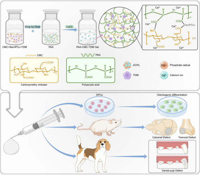
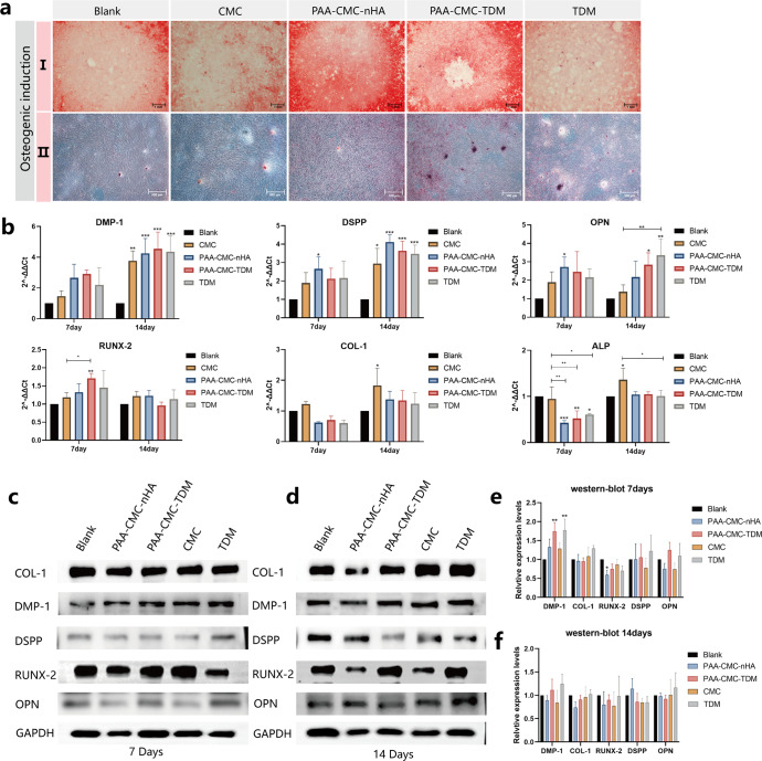
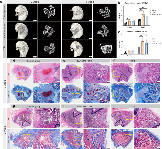
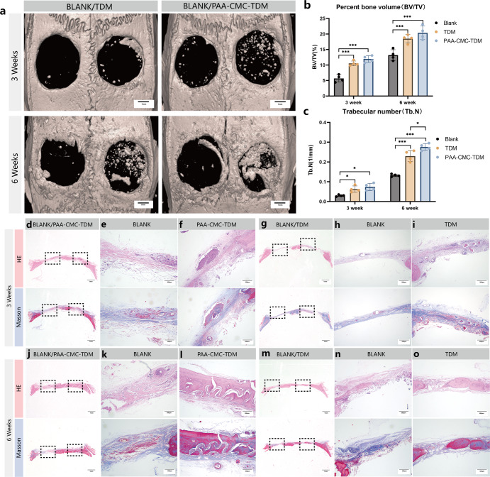
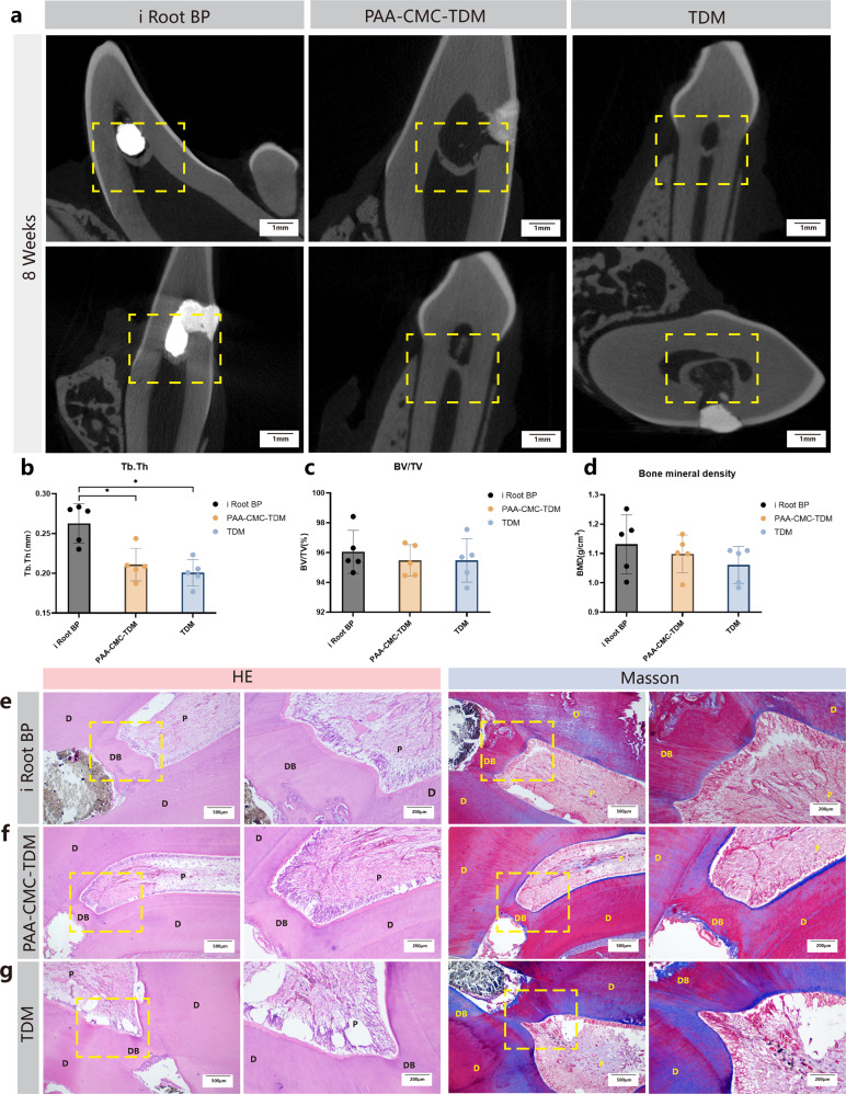
Maxillofacial hard tissue defects caused by trauma or infection often affect craniofacial function. Taking the natural hard tissue structure as a template, constructing an engineered tissue repair module is an important scheme to realize the functional regeneration and repair of maxillofacial hard tissue. Here, inspired by the biomineralization process, we constructed a composite mineral matrix hydrogel PAA-CMC-TDM containing amorphous calcium phosphates (ACPs), polyacrylic acid (PAA), carboxymethyl chitosan (CMC) and dentin matrix (TDM). The dynamic network composed of Ca2+·COO- coordination and ACPs made the hydrogel loaded with TDM, and exhibited self-repairing ability and injectability. The mechanical properties of PAA-CMC-TDM can be regulated, but the functional activity of TDM remains unaffected. Cytological studies and animal models of hard tissue defects show that the hydrogel can promote the odontogenesis or osteogenic differentiation of mesenchymal stem cells, adapt to irregular hard tissue defects, and promote in situ regeneration of defective tooth and bone tissues. In summary, this paper shows that the injectable TDM hydrogel based on biomimetic mineralization theory can induce hard tissue formation and promote dentin/bone regeneration.


Conflict of interest statement

The authors declare no competing interests.

Figures

Fig. 1
Fig. 1. Schematic diagram of PAA-CMC-TDM mineralized hydrogel for hard tissue regeneration and the experimental design.
The mineralized particles such as TDM or nHA are uniformly dispersed in the CMC/Na2HPO4 solution and mixed with the PAA solution by dripping. After the components are fully mixed, saturated calcium chloride is added to promote the formation of the hydrogel. The ability of bone regeneration was evaluated by cytological experiments in vitro, repair experiments of the femur and calvarial defects in rats and dental pulp defect model experiments in Beagle dogs.
Fig. 2
Fig. 2. Preparation and morphological characterization of materials.
ac Preparation process and hydrophilic evaluation of the PAA-CMC, PAA-CMC-nHA and PAA-CMC-TDM. The hydrogels showed favorable hydrophilia with a relatively low water contact angle. d The SEM surface and section observation of freeze-dried hydrogels. e TDM and PAA-CMC-TDM powder were mixed with water and molded, respectively, PAA-CMC-TDM powder could be formed smoothly and its shape was relatively complete. f Images of PAA-CMC-TDM hydrogel and TDM clumps sonicated for 0–15 min. g Self-healing process of PAA-CMC-TDM. The hydrogels were cut into two pieces and then reassembled at room temperature to form a completely new one. Hydrogel can be extruded through a syringe. Scale bar (d): 10 and 100 µm.
Fig. 3
Fig. 3. Study of the properties, swelling, and degradation of hydrogels after adjusting the material composition.
a The optical images and SEM surface observation of hydrogels of identical content CMC with different molecular weights. b The optical images and SEM surface observation of hydrogels of identical molecular weights CMC with different content. c The optical images and SEM surface observation of hydrogels of TDM with different content. d The optical images and SEM surface observation of hydrogels of nHA with different content. e I. Swelling rate of hydrogels containing different molecular weights and contents of CMC at the same amount of TDM. II. Swelling rate of the hydrogels containing different amounts of TDM or nHA with the same amount of CMC. f I. The water content of hydrogels containing different molecular weights and contents of CMC at the same amount of TDM. II. The water content of the hydrogels containing different amounts of TDM or nHA with the same amount of CMC. g Residual weight of hydrogels containing different molecular weights and contents of CMC in PBS with 2 U/mL collagenase at 37 °C for 12 days. h Residual weight of hydrogels containing different amounts of TDM or nHA in PBS with 2 U/mL collagenase at 37 °C for 12 days. Error bars indicate standard deviation (n = 3); *p < 0.05, **p < 0.01, and ***p < 0.001 (two-way ANOVA). Scale bar (ad): SEM, 10 µm.
Fig. 4
Fig. 4. Components and structure characterization of hydrogels.
a The TGA-DTG analysis curve of hydrogels PAA-ACP, PAA-CMC, PAA-CMC-nHA and PAA-CMC-TDM with temperature ranges from 50 to 800 °C. b The XRD patterns of the hydrogels and the mineral matrix fillers TDM and nHA. c ATR-FTIR spectra of PAA-ACP, PAA-CMC-ACP, PAA-CMC-nHA, PAA-CMC-TDM, CMC and PAA. d XPS spectrum and the high-resolution XPS spectra of Ca 2p, C 1s and P 1s of the fabricated PAA-CMC, PAA-CMC-nHA and PAA-CMC-TDM.
Fig. 5
Fig. 5. Rheological characterization of the mineralized hydrogel.
a Rheological strain sweeps of the hydrogel (strain = 0.01–100%, temperature: 20 °C, and frequency: 10 Hz). b Rheological frequency sweeps of the hydrogel (temperature: 20 °C and stress: 10 Pa, 0.1–50 Hz). c Step–strain sweeps of the hydrogel (temperature: 20 °C and frequency: 10 Hz, strain: 0.1%, 200 s, 100%, 200 s).
Fig. 6
Fig. 6. Effect of hydrogel on odontogenic differentiation of DPCs.
a Alizarin Red S staining of DPCs cultured in the control group (PBS), TDM, CMC and hydrogel groups. Red indicates mineral content. b RT-PCR of odontogenic differentiation marker genes DSPP, DMP-1, ALP, COL-I, OPN, and RUNX-2 after treatment with TDM, CMC and mineralized hydrogels for 7 days and 14 days. c, d Western blot analyses of odontogenic differentiation marker DSPP, DMP-1, ALP, COL-I, OPN, and RUNX-2 after treatment with TDM, CMC and mineralized hydrogels for 7 days (c) and 14 days (d). e Gray-scale analysis of western blot analysis results for 7 days. f Gray-scale analysis of western blot analysis results for 14 days. Error bars indicate standard deviation (n = 3); *p < 0.05, **p < 0.01, and ***p < 0.001 (two-way ANOVA). Scale bar (a-I):1 mm; (a-II): 500 µm.
Fig. 7
Fig. 7. HE staining and fluorescent immunohistological staining of PAA-CMC-TDM implanted subcutaneously in rats.
a, b H&E staining histologic sections of skin tissues surrounding PAA-CMC-TDM for 7 days (a) and 14 days (b). c, d Fluorescent immunohistological staining histologic sections of CD68 (Green) and CD163 (Red) of skin tissues surrounding PAA-CMC-TDM for 7 days (c) and 14 days (d). *The material. #The surrounding tissue. Scale bar (a): 1 mm, 500 µm, 200 µm, 100 µm; (c, d, CD68): 100 µm; (c, d, CD163): 100 µm; (c, d, CD68/CD163/DAPI): 100 and 50 µm.
Fig. 8
Fig. 8. In vivo bone regeneration on femoral defect after 3 and 6 weeks of implantation of mineralized hydrogels.
a Micro-CT assessment of bone regeneration in femoral defect at 3 and 6 weeks after implantation and 3D micro-CT reconstruction was carried out to observe the bone microstructure. b The quantitative analysis of bone volume/tissue volume (BV/TV) ratio by micro-CT. c The quantitative analysis of trabecular number (Tb.N, 1/mm) by micro-CT. df The HE staining and Masson’s trichrome staining evaluation of bone regeneration in defects at 3 weeks after implantation. gi The HE staining and Masson’s trichrome staining evaluation of bone regeneration in defects at 6 weeks after implantation (Control group, PAA-CMC-TDM group, TDM group). Error bars indicate standard deviation (n = 4); *p < 0.05, **p < 0.01, and ***p < 0.001 (two-way ANOVA). Scale bar (a): 1 mm; (d–i): 200 μm.
Fig. 9
Fig. 9. In vivo bone regeneration on calvarial defect after 3 and 6 weeks of implantation of mineralized hydrogels.
a Micro-CT assessment of bone regeneration in calvarial defect at 3 and 6 weeks after implantation, and 3D micro-CT reconstruction was carried out to observe the bone microstructure. b The quantitative analysis of bone volume/tissue volume (BV/TV) ratio by micro-CT. c The quantitative analysis of trabecular number (Tb.N, 1/mm) by micro-CT. di The HE staining and Masson’s trichrome staining evaluation of bone regeneration in calvarial defect at 3 weeks after implantation of PAA-CMC-TDM and TDM. jo The HE staining and Masson’s trichrome staining evaluation of bone regeneration in calvarial defect at 6 weeks after implantation of PAA-CMC-TDM and TDM. Error bars indicate standard deviation (n = 4); *p < 0.05, **p < 0.01, and ***p < 0.001 (two-way ANOVA). Scale bar (a): 1 mm; (d, j, g, m): 1 mm; (e, f, h, I, k, l, n, o): 500 μm.
Fig. 10
Fig. 10. The mineralized hydrogels as pulp capping material, evaluated in vivo in Beagle dogs.
a Micro-CT assessment of dentin bridge in dog tooth at 8 weeks after pulp capping with i Root BP plus, PAA-CMC-TDM and TDM. b The quantitative analysis of the thickness of the formed dentin bridge (Tb.Th) by micro-CT. c The quantitative analysis of dentin bridge mineralization ratio (BV/TV) by micro-CT. d The bone mineral density analysis of dentin bridge by micro-CT. e The HE staining and Masson’s trichrome staining evaluation of 8 weeks pulp capping with i Root BP plus in dog tooth. f The HE staining and Masson’s trichrome staining evaluation of 8 weeks pulp capping with PAA-CMC-TDM in dog tooth. g The HE staining and Masson’s trichrome staining evaluation of 8 weeks pulp capping with TDM in dog tooth. D dentin, P pulp tissue, DB dental bridge. Error bars indicate standard deviation (n = 5); *p < 0.05, **p < 0.01, and ***p < 0.001 (one-way ANOVA). Scale bar (a): 1 mm; (e, f, g, HE/Left): 500 μm; (e, f, g, HE/Right): 200 μm; (e, f, g, Masson/Left): 500 μm; (e, f, g, Masson/Right): 200 μm.

Similar articles

Cited by

References

    1. Cusack M, Freer A. Biomineralization: elemental and organic influence in carbonate systems. Chem. Rev. 2008;108:4433–4454. doi: 10.1021/cr078270o. - DOI - PubMed
    1. Green DW, Ben-Nissan B, Yoon KS, Milthorpe B, Jung HS. Natural and synthetic coral biomineralization for human bone revitalization. Trends Biotechnol. 2017;35:43–54. doi: 10.1016/j.tibtech.2016.10.003. - DOI - PubMed
    1. Liu J, Zhang SS, Zheng SG, Xu T, Si Y. Oral health status and oral health care model in China. Chin. J. Dent. Res. 2016;19:207–215. - PubMed
    1. Morotomi T, Washio A, Kitamura C. Current and future options for dental pulp therapy. Jpn Dent. Sci. Rev. 2019;55:5–11. doi: 10.1016/j.jdsr.2018.09.001. - DOI - PMC - PubMed
    1. Roohani I, Yeo GC, Mithieux SM, Weiss AS. Emerging concepts in bone repair and the premise of soft materials. Curr. Opin. Biotechnol. 2021;74:220–229. doi: 10.1016/j.copbio.2021.12.004. - DOI - PubMed