. 2023 Mar 28;42(3):112140.
doi: 10.1016/j.celrep.2023.112140. Epub 2023 Feb 25.

mRNA targeting eliminates the need for the signal recognition particle during membrane protein insertion in bacteria

Affiliations

mRNA targeting eliminates the need for the signal recognition particle during membrane protein insertion in bacteria

Pinku Sarmah et al. Cell Rep. .

Abstract

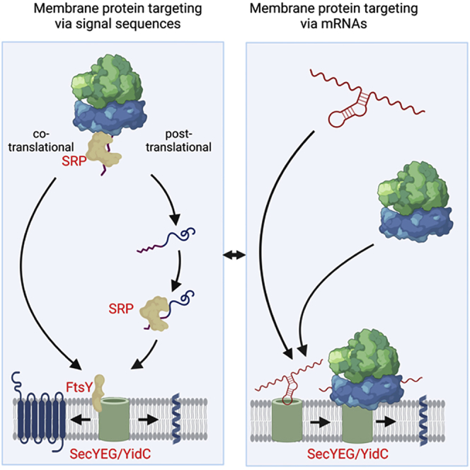
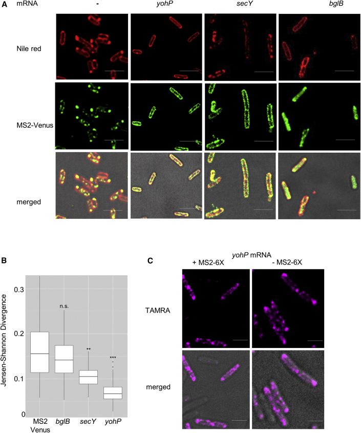
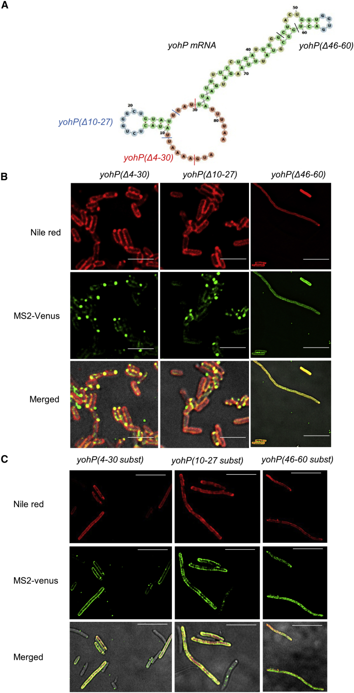
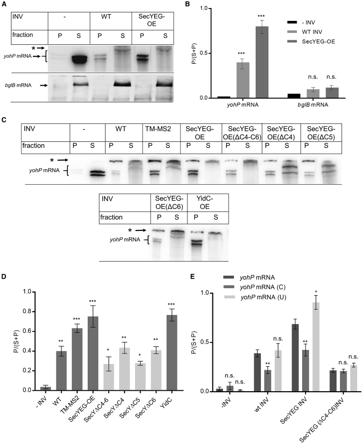
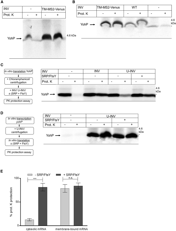
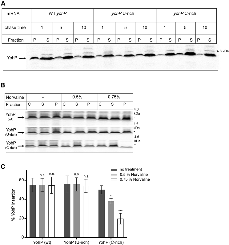
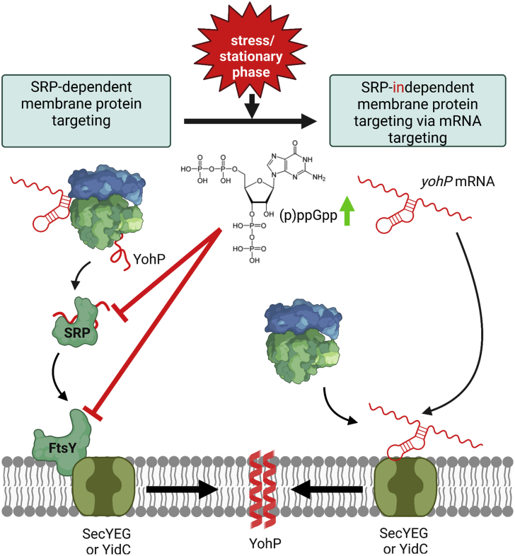
Signal-sequence-dependent protein targeting is essential for the spatiotemporal organization of eukaryotic and prokaryotic cells and is facilitated by dedicated protein targeting factors such as the signal recognition particle (SRP). However, targeting signals are not exclusively contained within proteins but can also be present within mRNAs. By in vivo and in vitro assays, we show that mRNA targeting is controlled by the nucleotide content and by secondary structures within mRNAs. mRNA binding to bacterial membranes occurs independently of soluble targeting factors but is dependent on the SecYEG translocon and YidC. Importantly, membrane insertion of proteins translated from membrane-bound mRNAs occurs independently of the SRP pathway, while the latter is strictly required for proteins translated from cytosolic mRNAs. In summary, our data indicate that mRNA targeting acts in parallel to the canonical SRP-dependent protein targeting and serves as an alternative strategy for safeguarding membrane protein insertion when the SRP pathway is compromised.

Keywords: (p)ppGpp; CP: Microbiology; FtsY; SecYEG translocon; YidC; alarmones; mRNA targeting; signal recognition particle; small membrane proteins; stringent response; translation.


Conflict of interest statement

Declaration of interests The authors declare no competing interests.

Figures

None
Graphical abstract
Figure 1
Figure 1
Translation-independent membrane enrichment of mRNAs encoding for membrane proteins (A) E. coli cells expressing just pBad24-MS2-Venus or together with a plasmid encoding yohP, secY, or bglB, each with a deleted ribosome binding site and a hexa-repeat MS2 stem loop sequence at the 3′ UTR. Imaging was performed with the Delta Vision Ultra microscope, and 3 μm Z-scans were recorded with an interval of 1 μm. The scale bar refers to 2.5 μm. (B) Jensen-Shannon divergence (JSD) plot for quantifying the correlation between Nile red distribution and mVenus distribution. The JSD value is based on scoring 60–80 individual cells. Statistical analyses were performed with the Satterthwaite-corrected unpaired two-sided Student’s t test using cells expressing just MS2-Venus but with no mRNA as reference. ∗∗p ≤ 0.01 and ∗∗∗p ≤ 0.001. n.s. denotes non-significant changes. (C) RNA-FISH of E. coli cells expressing yohP mRNA with or without the MS2 stem loop sequence. A set of 19 oligonucleotides against the yohP sequence were linked to the fluorescent probe TAMRA. Imaging was performed as in (A) with 0.1 s exposure time and 100% laser intensity. The scale bar refers to 2.5 μm. See also Figure S1.
Figure 2
Figure 2
Membrane localization of the yohP mRNA is dependent on both nucleotide sequence and secondary structure (A) Predicted secondary structure of the yohP mRNA and the strategy for monitoring the influence of nucleotide composition, length, and secondary structures on membrane binding. Structure was predicted by the RNAfold webserver (http://rna.tbi.univie.ac.at/cgi-bin/RNAWebSuite/RNAfold.cgi). (B) The wild-type yohP sequence was modified by increasing either the uracil (U-rich), cytosine (C-rich), guanine (G-rich), or adenine content (A-rich). Imaging was performed as described in Figure 1. (B) As in (A), but the mRNA variants with increased U-, C-, A- or G-content were designed with only minor influence on the predicted secondary structures of the yohP mRNAs (U-rich, C-rich, G-rich, A-rich). The scale bar refers to 2.5 μm. See also Figures S2 and S3.
Figure 3
Figure 3
Deletion or replacements of the predicted loops of the yohP mRNA impair membrane binding (A) Structure prediction of the yohP mRNA using the RNAfold webserver. The predicted loops and their deletions are indicated by colored lines. (B) Imaging of E. coli cells expressing pBad-24-MS2-Venus together with the indicated stem loop deletions, as described in Figure 1. (C) As is (B), but the sequence of the predicted stem loops were replaced with random sequences. The scale bar refers to 2.5 μm. See also Figures S2 and S3.
Figure 4
Figure 4
The SecYEG translocon and YidC constitute putative mRNA receptors at the E. coli membrane (A) The yohP and bglB mRNAs were in vitro transcribed and 32P labeled. After purification, the mRNA was incubated with either buffer or inner membrane vesicles (INVs) isolated from either wild-type (WT) E. coli cells or a SecYEG-overproducing strain (SecYEG-OE). After incubation, INVs and the bound mRNA were pelleted by centrifugation, and the membrane fraction (P) and the soluble fraction (S) were separated on a urea gel and analyzed by autoradiography. (B) Quantification of three independent experiments as shown in (A). For quantification, the radioactive signal in (P) was divided by the sum of the signals in (P) and (S). Shown are the mean values and the standard errors of the mean (SEMs). (C) As in (A) using INVs from different E. coli strains. TM-MS2 refers to INVs from a strain overproducing a membrane-tethered MS2 protein. INVs from E. coli strains overproducing SecYEG variants that lacked the three cytosolic loops C4–C6 of SecY or the individual loops were also analyzed. In addition, INVs from cells overproducing YidC were tested. The asterisk () corresponds to radioactive material that did not enter the gel. (D) Quantification of the data shown in (C) was performed as in (B). (E) Binding of WT, uracil-rich (U), and cytosine-rich (C) mRNAs to the indicated INVs. Quantification of three independent experiments was performed as in (B). Statistical analyses were performed as in Figure 1, using the amount of mRNA in (P) after incubation without INVs as reference. p ≤ 0.05, ∗∗p ≤ 0.01, and ∗∗∗p ≤ 0.001. n.s. denotes non-significant changes. See also Figures S4 and S5.
Figure 5
Figure 5
YohP translated from membrane-bound mRNAs does not require the SRP pathway for insertion (A) YohP mRNA was incubated with TM-MS2-containing INVs or INV buffer, and INV-bound mRNA was subsequently isolated by centrifugation. The pellet fraction was then incubated with an in vitro translation system containing 35S-labeled methionine and cysteine. After 30 min of incubation, proteinase K (PK) was added when indicated for monitoring YohP insertion into the membrane. (B) As in (A) but also with WT INVs as further control. (C) YohP was synthesized from cytosolic mRNAs, and post-translational membrane insertion of YohP was analyzed after in vitro synthesis and subsequent chloramphenicol treatment and centrifugation for stopping translation and for removing ribosomes. In-vitro-synthesized and 35S-labeled YohP was then incubated with INV buffer, INVs, or urea-treated INVs (U-INVs). When indicated, purified SRP and FtsY (20 ng/μL) were added, and membrane insertion of YohP was monitored by PK protection. (D) The yohP mRNA was in vitro transcribed and purified. Purified mRNA was then added to U-INVs, and U-INV-bound mRNA was re-isolated by centrifugation. The membrane-bound mRNA was translated in vitro in the presence or absence of SRP/FtsY (20 ng/μL). YohP insertion was monitored by PK protection. (E) Quantification of the YohP insertion into U-INVs derived from the data in (C) and (D). Shown are the mean values of three independent experiments and the SEMs. Statistical analyses were performed as in Figure 1 using the YohP insertion in the absence of SRP/FtsY as reference. ∗∗∗p ≤ 0.001. n.s. denotes non-significant changes. See also Figure S7.
Figure 6
Figure 6
mRNA targeting and signal sequence-dependent targeting via the SRP pathway act in parallel (A) In vivo pulse-chase experiments for monitoring membrane insertion of YohP. YohP variants with identical amino acid sequence were produced from three different T7-dependent expression plasmids containing either WT yohP or the uracil-rich or cytosine-rich sequences. After induction of T7-dependent mRNA production, endogenous E. coli RNA polymerase was blocked by rifampicin, and cells were labeled for 5 min with 35S-labeled methionine/cysteine and then chased with non-radioactive methionine/cysteine for 1, 5, or 10 min. Cells were then lysed by ultrasonic treatment and separated into the membrane fraction (P) and the cytosolic fraction (S). Samples were separated by SDS-PAGE and visualized by autoradiography. (B) As in (A), but cells were treated when indicated with norvaline for 1.5 h before mRNA production and rifampicin treatment. Cells were 5 min pulsed and 5 min chased before cell lysis, centrifugation, and further processing as in (A). (C) Quantification of three independent experiments. For quantification, the radioactive signal in the pellet fraction was divided by the sum of the radioactive signals in the pellet and soluble fraction and is displayed as the percentage of YohP insertion. Shown are the mean values and the SEMs. Statistical analyses were performed as in Figure 1 using YohP insertion in the absence of norvaline as reference. p ≤ 0.05 and ∗∗∗p ≤ 0.001. n.s. denotes non-significant changes. See also Figures 2, S2, and S7.
Figure 7
Figure 7
mRNA targeting as SRP-independent back-up strategy for membrane protein insertion in E. coli Left panel: membrane protein insertion in E. coli depends on SRP and its receptor FtsY, which target client proteins to either the SecYEG translocon or the YidC insertase for insertion. (1) In most cases, SRP targets its substrates co-translationally but can also act post-translationally for small membrane proteins, such as YohP., (2) SRP targets proteins to the SRP receptor FtsY, which is associated with either the SecYEG translocon or the YidC insertase. (3) Subsequently, YohP is inserted into the membrane and dimerizes (4). Right panel: during stress conditions or when cells enter stationary phase, the concentration of the alarmones (p)ppGpp increase, resulting in a cellular re-programming, which includes the inhibition of the SRP pathway by preventing SRP-FtsY complex formation. Under those conditions, mRNAs can bind directly to the SecYEG translocon or YidC (1), and ribosomes translate these membrane-bound mRNAs (2), allowing for SRP-independent membrane insertion (3). This SRP-independent pathway is likely particularly important for membrane proteins, which are upregulated during stress conditions or when cells enter stationary phase, such as YohP. The cartoon was generated using BioRender (https://biorender.com/).

Similar articles

Cited by

References

    1. Driessen A.J., Manting E.H., van der Does C. The structural basis of protein targeting and translocation in bacteria. Nat. Struct. Biol. 2001;8:492–498. doi: 10.1038/88549. - DOI - PubMed
    1. Müller M., Koch H.G., Beck K., Schäfer U. Protein traffic in bacteria: multiple routes from the ribosome to and across the membrane. Prog. Nucleic Acid Res. Mol. Biol. 2001;66:107–157. - PubMed
    1. Rapoport T.A., Li L., Park E. Structural and mechanistic insights into protein translocation. Annu. Rev. Cell Dev. Biol. 2017;33:369–390. doi: 10.1146/annurev-cellbio-100616-060439. - DOI - PubMed
    1. Zimmermann R., Eyrisch S., Ahmad M., Helms V. Protein translocation across the ER membrane. Biochim. Biophys. Acta. 2011;1808:912–924. - PubMed
    1. Oswald J., Njenga R., Natriashvili A., Sarmah P., Koch H.G. The dynamic SecYEG translocon. Front. Mol. Biosci. 2021;8:664241. doi: 10.3389/fmolb.2021.664241. - DOI - PMC - PubMed

Publication types

MeSH terms

LinkOut - more resources