. 2023 Mar 28;42(3):112161.
doi: 10.1016/j.celrep.2023.112161. Epub 2023 Feb 25.

Ribosomal DNA replication time coordinates completion of genome replication and anaphase in yeast

Affiliations

Ribosomal DNA replication time coordinates completion of genome replication and anaphase in yeast

Elizabeth X Kwan et al. Cell Rep. .

Abstract

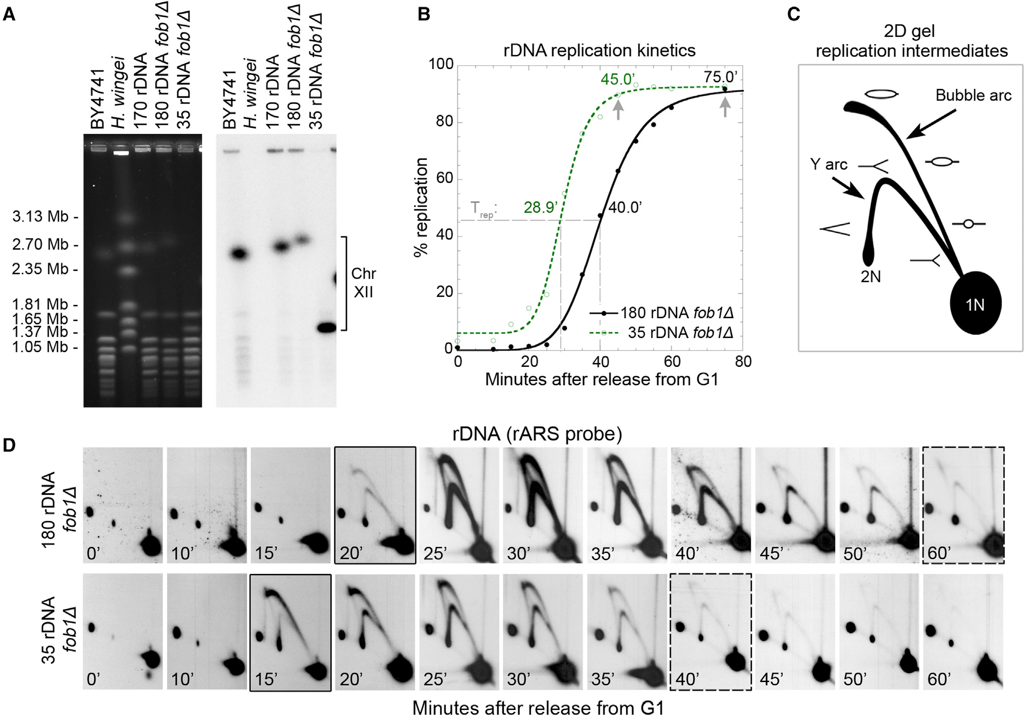
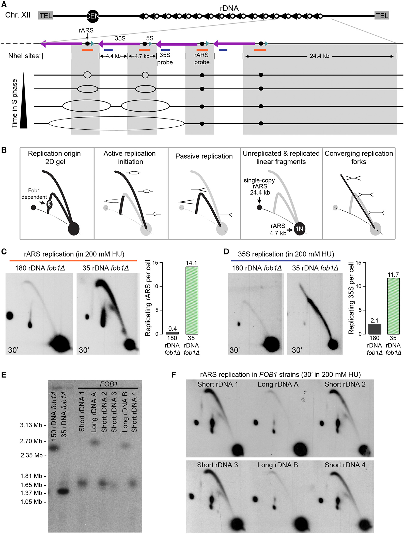
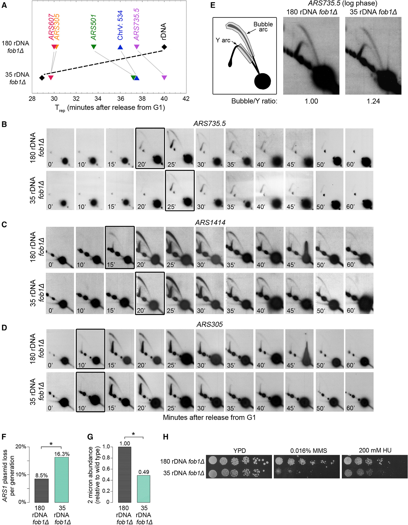
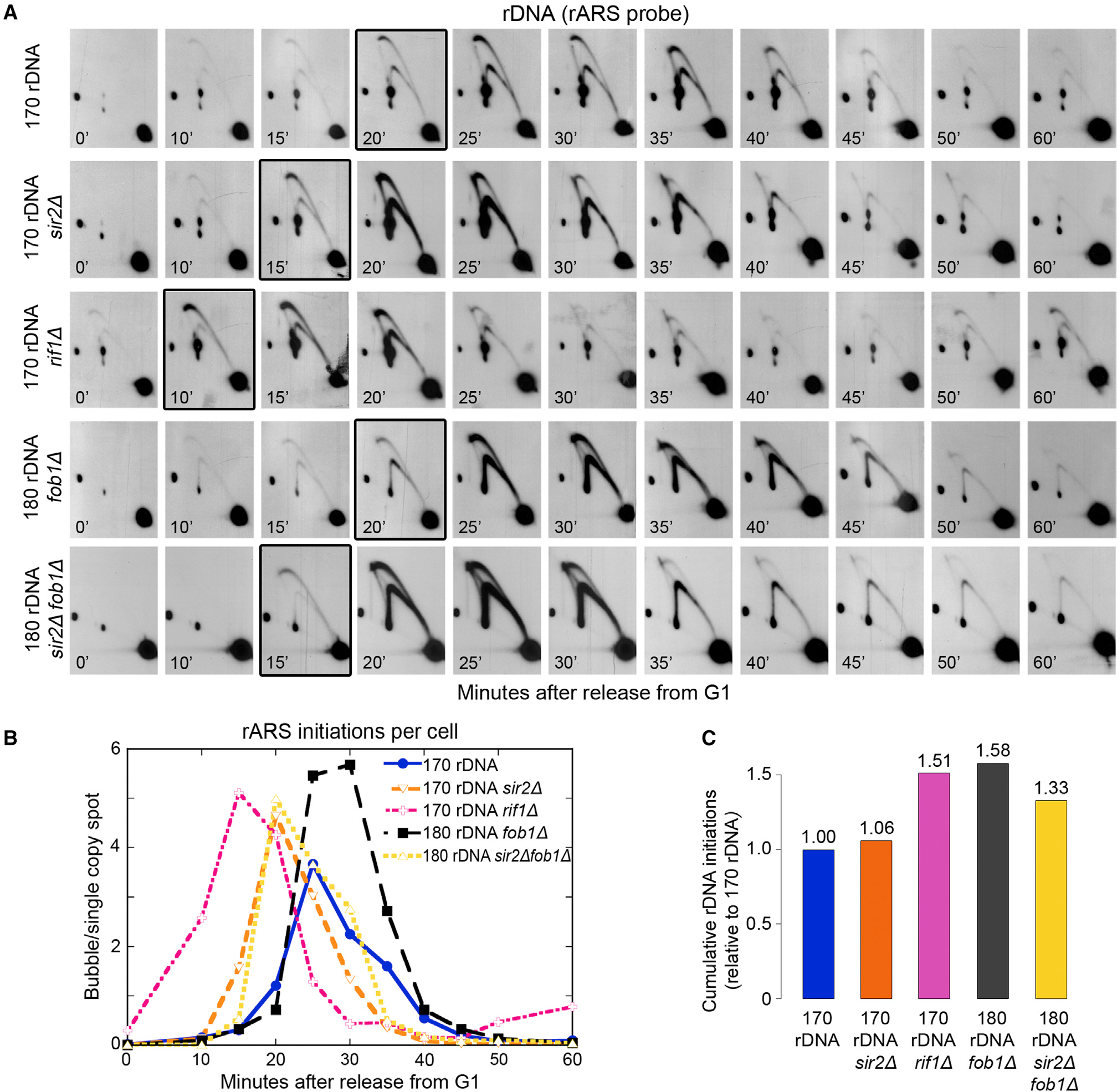
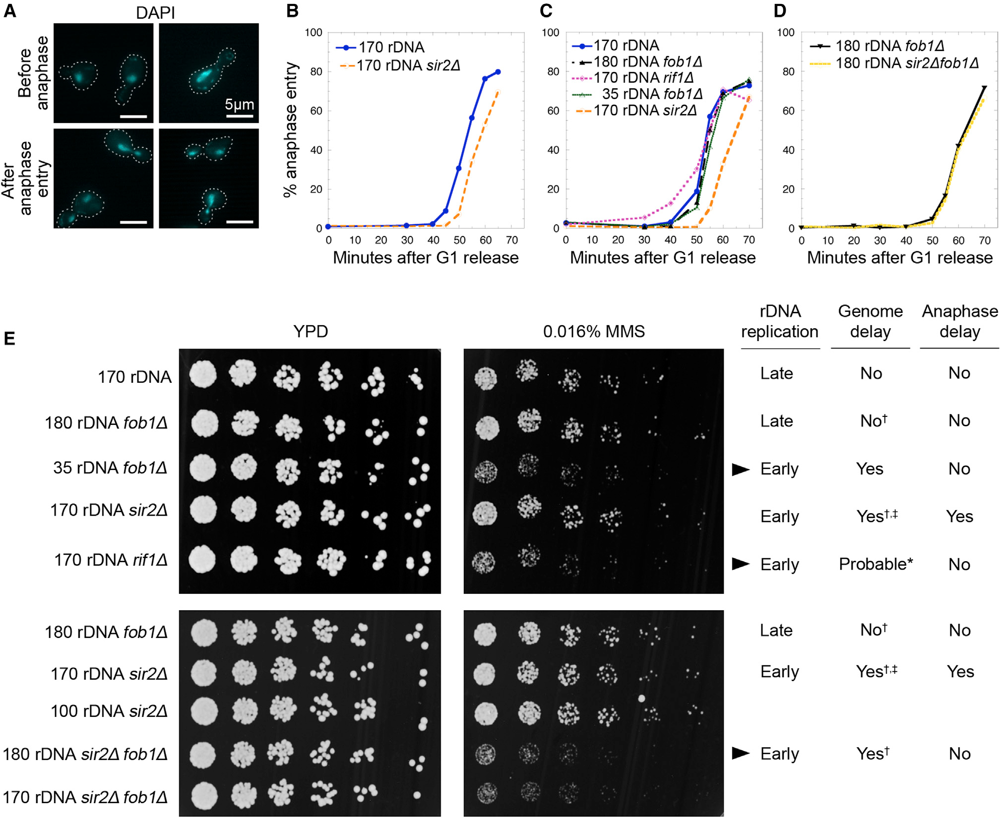
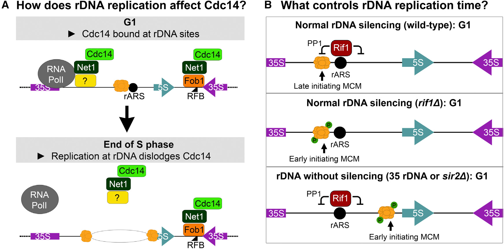
Timely completion of genome replication is a prerequisite for mitosis, genome integrity, and cell survival. A challenge to this timely completion comes from the need to replicate the hundreds of untranscribed copies of rDNA that organisms maintain in addition to the copies required for ribosome biogenesis. Replication of these rDNA arrays is relegated to late S phase despite their large size, repetitive nature, and essentiality. Here, we show that, in Saccharomyces cerevisiae, reducing the number of rDNA repeats leads to early rDNA replication, which results in delaying replication elsewhere in the genome. Moreover, cells with early-replicating rDNA arrays and delayed genome-wide replication aberrantly release the mitotic phosphatase Cdc14 from the nucleolus and enter anaphase prematurely. We propose that rDNA copy number determines the replication time of the rDNA locus and that the release of Cdc14 upon completion of rDNA replication is a signal for cell cycle progression.

Keywords: CP: Molecular biology; Cdc14; Fob1; anaphase; cell cycle; copy number variation; nucleolus; origin of replication; rDNA; replication timing; ribosomal DNA.


Conflict of interest statement

Declaration of interests C.Q. is a Cell Reports Advisory Board member, Ecology and Evolution.

Figures

Figure 1.
Figure 1.. rDNA arrays with fewer copies replicate early
(A) CHEF gel estimation of rDNA copy number using chromosome XII size. Ethidium bromide-stained gel (left) and the resulting Southern blot (right) hybridized with a single copy Chr. XII probe (CDC45). (B) Replication kinetic curves generated from density transfer slot blot analysis show that the minimal rDNA array replicates earlier than the 180-copy rDNA array. Dashed lines indicate time at half-maximal replication (Trep). Arrows indicate the estimated times at which rDNA replication has neared completion. (C) 2D gel diagram of positions for replication intermediates that contain an active origin (bubble arc) or are passively replicated (Y arc). (D) 2D gel analysis of replication across a synchronous S phase for the NheI fragment containing the rDNA ARS (rARS). One of three sets of biological replicates, all with similar results, is presented here. The black borders indicate times of first perceptible replication intermediates. The dashed borders indicate the times when rDNA replication is essentially completed.
Figure 2.
Figure 2.. Quantification of rDNA replication initiations in early S phase
(A) Schematic of rDNA locus organization, NheI restriction sites, Southern blot probe locations (orange and blue bars), and example of replication intermediates throughout S phase. The 24.4-kb NheI fragment on the telomere proximal edge of the rDNA array is present in a single copy per cell and hybridizes to the rARS probe (orange bar). (B) Diagram of replication intermediates resolved by 2D gel electrophoresis. (C and D) 2D gels of synchronized cells released into S phase in the presence of 200 mM HU, sequentially hybridized with the rARS probe (C) and the 35S probe (D). rARS replication bubbles per cell were quantified and normalized to the signal in the single-copy 24.4-kb linear spot. Estimation of the non-rARS 35S replication fork intermediates was normalized to the 4.4-kb 1N spot and adjusted for rDNA copy number. (E) Meiotic spores from a cross between a FOB1, 170-copy rDNA strain and a fob1Δ, 35-rDNA copy strain were analyzed by CHEF gel electrophoresis to measure rDNA repeat number. (F) 2D gels of DNA harvested as in (B) for the same six spores (from E) were analyzed for rARS initiation.
Figure 3.
Figure 3.. rDNA copy number reduction generates genome replication delays and defects
(A) Comparison of the Trep (time of half-maximal replication derived from a density transfer experiment) for five different genomic origins and an origin-free genomic region (Chr. V: 534) in strains with 180 vs. 35 rDNA copies. Additional biological replicates are presented in Figures S1F and S2G. (B–D) 2D gel analysis of replication across a synchronous S phase for time of initiation at the late-replicating genomic origins ARS735.5 and ARS1414 and the early-replicating origin ARS305. The boxed panels denote time of first perceptible bubble replication intermediates. (E) Relative origin efficiency for ARS735.5 was estimated by comparing the signal ratio of bubbles vs. Ys. (F) Measurement of plasmid loss rates of an ARS1-based plasmid in the 180-rDNA and 35-rDNA copy number strains. p = 0.03; one representative biological replicate of two is shown. (G) Natural abundance of the parasitic 2-micron plasmid in the 180-rDNA and 35-rDNA copy number strains. Paired two-tailed t test p = 0.007. (H) Spot assays to measure sensitivity to DNA damage from0.016%MMSand replication stress from200mMHUfor the 180-rDNAand 35-rDNAcopy number strains.
Figure 4.
Figure 4.. Characterization of altered rDNA replication time and rDNA origin efficiency in mutant strains
(A) Comparison of rDNA replication initiation time in synchronized cells using 2D gel electrophoresis for four mutants (sir2Δ, rif1Δ, fob1Δ, or sir2Δ fob1Δ) relative to wild-type cells—all with wild-type rDNA copy numbers. The boxed panels indicate the first appearance of more than 0.5 rARS bubble intermediates per cell. (B) Quantification of bubbles per cell, based on the ratio of bubbles to the 24.4-kb single-copy fragment for the five strains in (A). (C) Total number of rDNA origins fired in the three mutant strains in (A) compared with the wild-type strain.
Figure 5.
Figure 5.. Uncoupling of anaphase entry from delayed genome replication increases DNA damage sensitivity in strains with early rDNA replication
(A) Examples of DAPI-stained nuclei representing cells scored as either being “before anaphase” (no nuclear migration) or “after anaphase entry” (with nuclear migration into the daughter cell). Two representative images for each stage are presented, and cell outlines are indicated by white dotted lines. White scale bar indicates 5 μm. (B–D) Quantification of percentage of cells undergoing anaphase over time after release from G1 with n ≥ 200 cells for each timed sample in each replicate. One of two biological replicates is shown here; see Figure S6 for the other replicate. (E) Spot assays comparing growth on YPD vs. YPD +0.016% MMS. Each strain’s rDNA replication time (early or late), known genome replication delays (†; ‡; *,,), and anaphase entry delays (from B–D) are indicated. Arrowheads highlight strains for which anaphase entry is earlier than expected based on rDNA/genome replication times.
Figure 6.
Figure 6.. Strains with early rDNA replication have defects in Cdc14 localization
(A) C-terminally tagged Cdc14-GFP was visualized in G1-arrested cells with either 180 or 35 rDNA copies and compared with DAPI nuclear staining. (B) Quantification of the percentage of G1 cells with nucleolar localization of Cdc14-GFP or Utp13-GFP. (C) Utp13-GFP, a nucleolar protein that is involved in rRNA processing, was used to evaluate nucleolar structure in G1 arrested cells. (D and E) Examples of cells scored as being “before anaphase” or “after anaphase entry” using DAPI staining of nuclei, Utp13-mCherry, and Cdc14-GFP morphology. Quantification of cell fractions that (F and G) had lost nucleolar localization of Cdc14-GFP or (H and I) displayed nuclear migration indicative of anaphase entry. Two representative images are presented. Cell outlines are indicated by white dotted lines. White scale bar indicates 5 μm. For all samples, n ≥ 200.
Figure 7.
Figure 7.. Proposed models for early rDNA replication mechanisms and consequences
(A) In G1, Cdc14 is bound to the rDNA at the 35S rRNA transcription start site (TSS) and at the replication fork barrier (RFB). As rDNA begins replication in late S phase, Cdc14 is progressively released but persists at the RFB until replication forks stalled at the RFBs are resolved by oncoming forks and replication of the rDNA is completed. Thus, complete release of Cdc14 is coupled to completion of rDNA replication. (B) Regulation of rDNA initiation time in wild-type and mutant cells. In wild-type cells, late S phase replication is enforced by two factors: (1) repressive chromatin established by Sir2 confines the MCM helicase to the vicinity of the rARS where (2) Rif1 keeps the helicase in an inactive (unphosphorylated) form (top). In the absence of Rif1 (middle), the MCM helicase is phosphorylated early leading to early origin activation. In sir2Δ or 35-copy rDNA strains (bottom), the open chromatin conformation allows the loaded MCM helicase to translocate away from the repressive environment of Rif1 so that initiation occurs early.

Similar articles

Cited by

References

    1. Mohan J, and Ritossa FM (1970). Regulation of ribosomal RNA synthesis and its bearing on the bobbed phenotype in Drosophila melanogaster. Dev. Biol. 22, 495–512. 10.1016/0012-1606(70)90165-x. - DOI - PubMed
    1. Morton EA, Hall AN, Kwan E, Mok C, Queitsch K, Nandakumar V, Stamatoyannopoulos J, Brewer BJ, Waterston R, and Queitsch C (2020). Challenges and approaches to genotyping repetitive DNA. G3 10, 417–430. 10.1534/g3.119.400771. - DOI - PMC - PubMed
    1. Parks MM, Kurylo CM, Dass RA, Bojmar L, Lyden D, Vincent CT, and Blanchard SC (2018). Variant ribosomal RNA alleles are conserved and exhibit tissue-specific expression. Sci. Adv. 4, eaao0665. 10.1126/sciadv.aao0665. - DOI - PMC - PubMed
    1. Thompson O, Edgley M, Strasbourger P, Flibotte S, Ewing B,Adair R, Au V, Chaudhry I, Fernando L, Hutter H, et al. (2013). The million mutation project: a new approach to genetics in Caenorhabditis elegans. Genome Res. 23, 1749–1762. 10.1101/gr.157651.113. - DOI - PMC - PubMed
    1. Conconi A, Widmer RM, Koller T, and Sogo JM (1989). Two different chromatin structures coexist in ribosomal RNA genes throughout the cell cycle. Cell 57, 753–761. 10.1016/0092-8674(89)90790-3. - DOI - PubMed

Publication types

MeSH terms