

Measuring fascicle lengths of extrinsic and intrinsic thumb muscles using extended field-of-view ultrasound
- PMID: 36842405
- PMCID: PMC10849800
- DOI: 10.1016/j.jbiomech.2023.111512
Measuring fascicle lengths of extrinsic and intrinsic thumb muscles using extended field-of-view ultrasound
Abstract
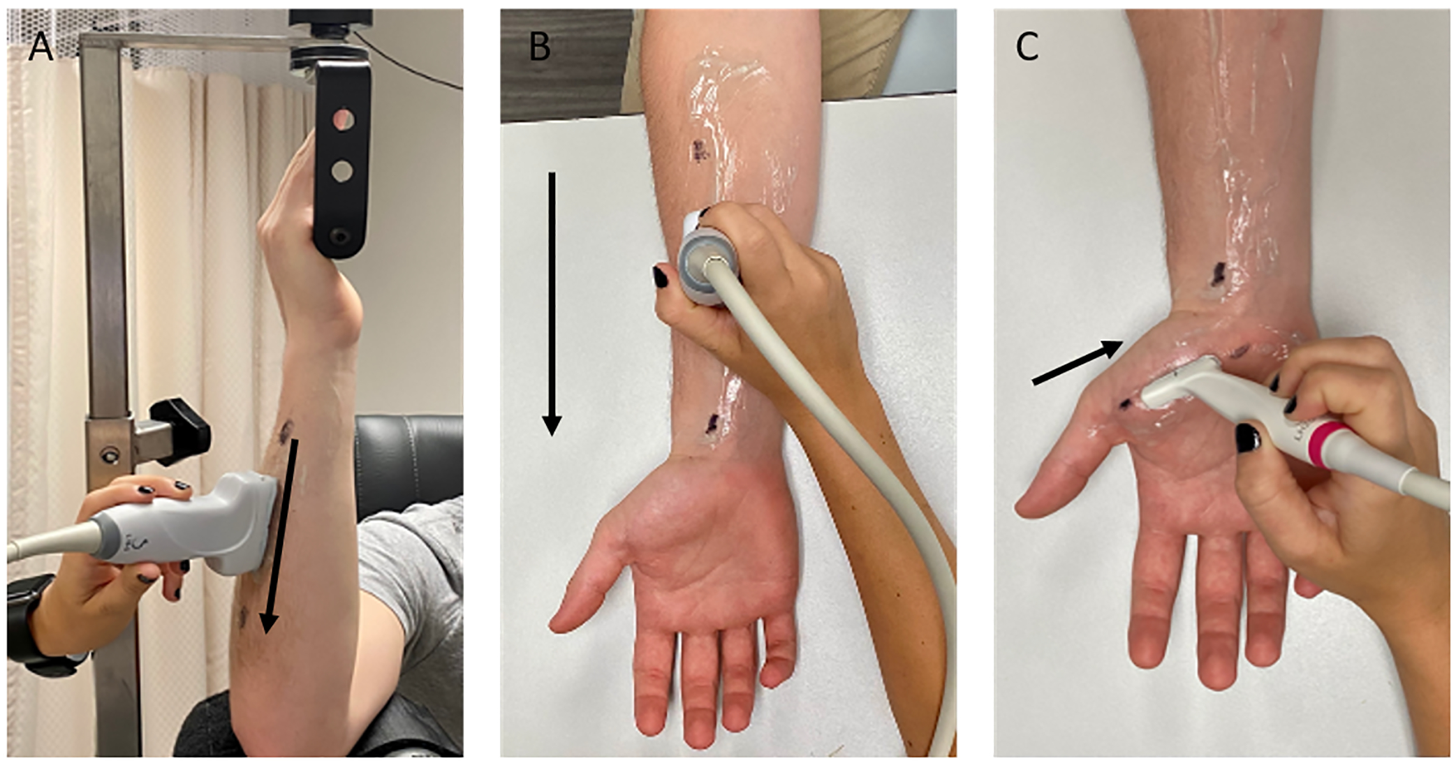
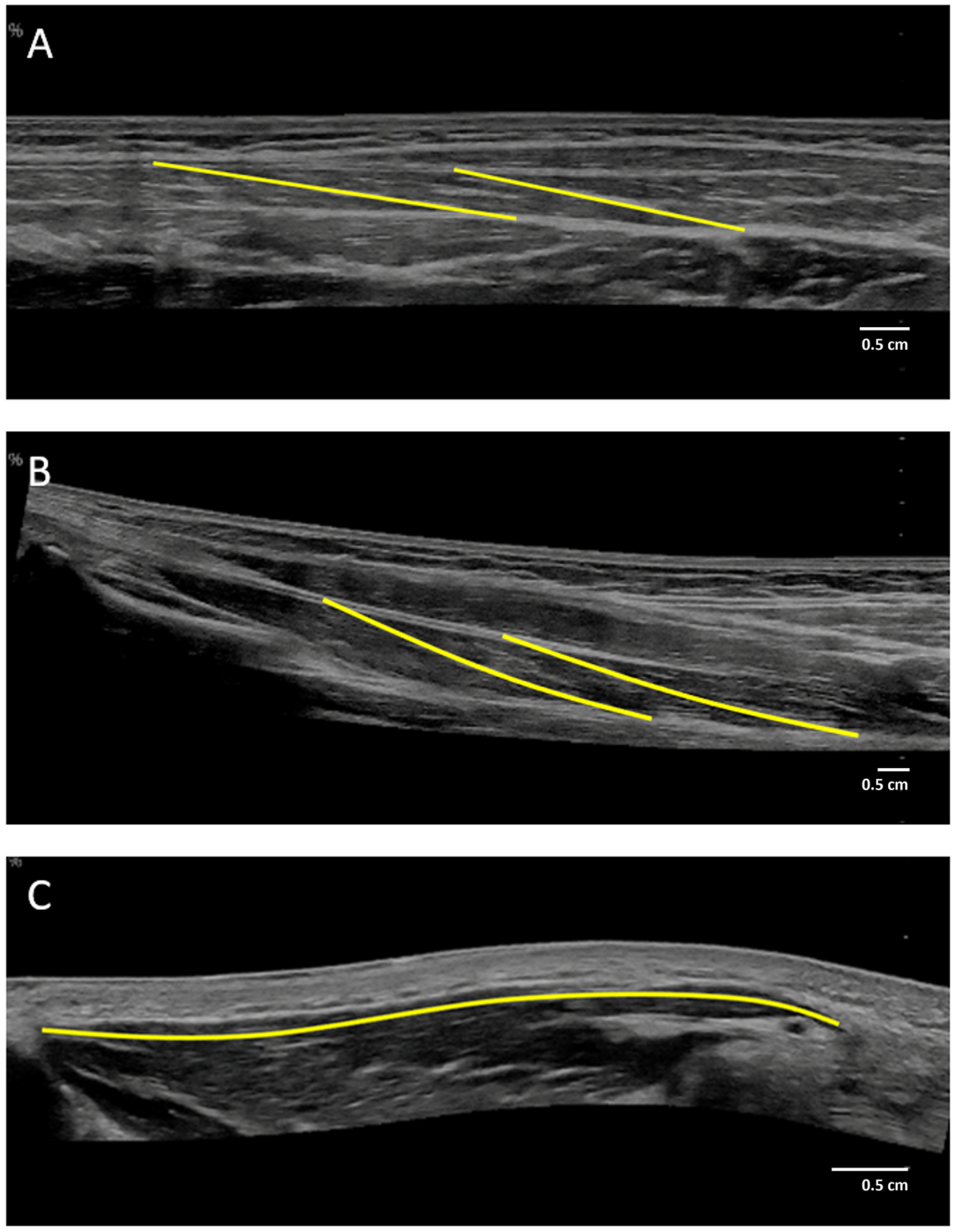
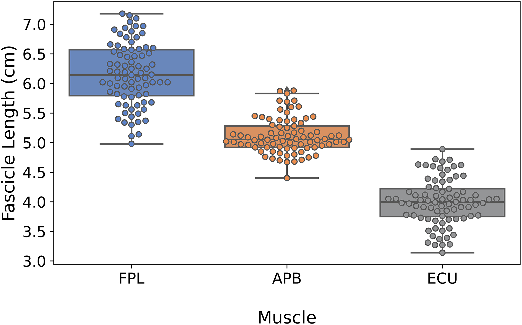
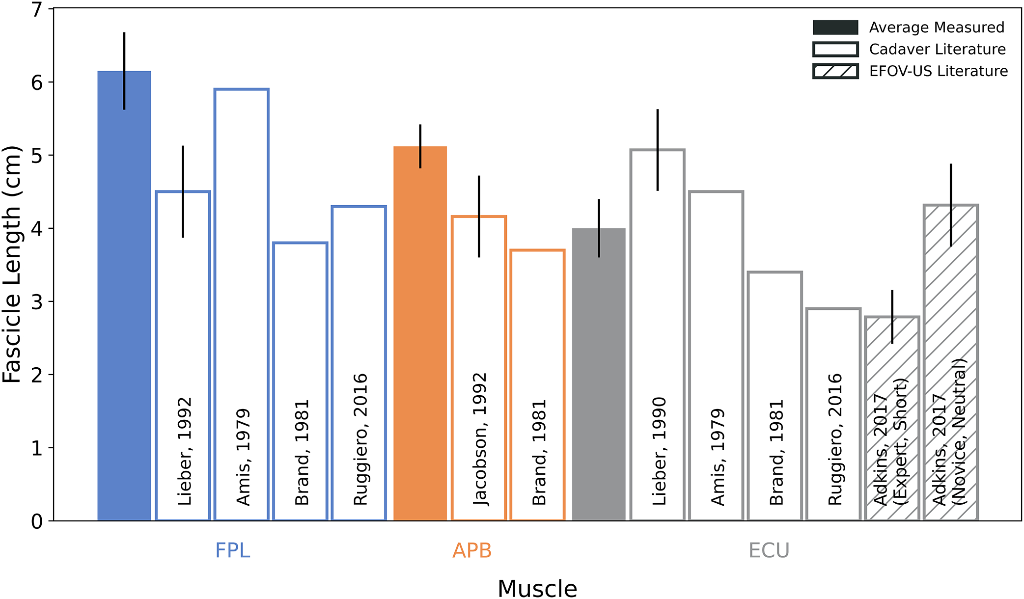
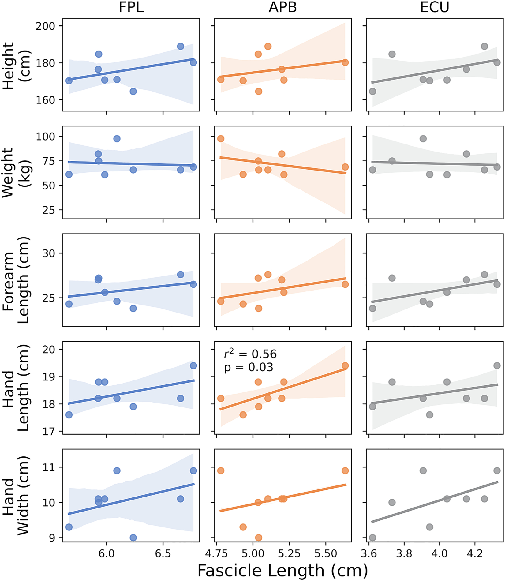
Complex motion of the human thumb is enabled by the balanced architectural design of the extrinsic and intrinsic thumb muscles. Given that recent imaging advances have not yet been applied to enhance our understanding of the in vivo properties of thumb muscles, the objective of this study was to test the reliability and validity of measuring thumb muscle fascicle lengths using extended field of view ultrasound (EFOV-US). Three muscles (FPL: flexor pollicis longus, APB: abductor pollicis brevis, and ECU: extensor carpi ulnaris) were imaged in eight healthy adults (4 female; age, 21.6 ± 1.3 years; height, 175.9 ± 8.3 cm)[mean ± SD]. Measured fascicle lengths were compared to cadaveric data (all muscles) and ultrasound data (ECU only). Additionally, to evaluate how fascicle lengths scale with anthropometric measurements, height, forearm length, hand length, and hand width were recorded. The EFOV-US method obtained precise fascicle length measurements [mean ± SD] for the FPL (6.2 ± 0.5 cm), APB (5.1 ± 0.3 cm), and ECU (4.0 ± 0.4 cm). However, our EFOV-US measurements were consistently different (p < 0.05) than prior cadaveric data, highlighting the need to better understand differences between in vivo and ex vivo fascicle length measurements. Fascicle length was significantly related to only hand length (r2 = 0.56, p = 0.03) for APB, highlighting that anthropometric scaling may not accurately estimate thumb muscle length. As the first study to apply EFOV-US to measure thumb muscle fascicle lengths, this study expands the utility of this imaging technology within the upper limb.
Keywords: Hand; Medical imaging; Muscle; Muscle architecture; Upper limb.
Copyright © 2023 Elsevier Ltd. All rights reserved.
Conflict of interest statement
Declaration of Competing Interest The authors declare that they have no known competing financial interests or personal relationships that could have appeared to influence the work reported in this paper.
Figures
Similar articles
-
Demonstration of extended field-of-view ultrasound's potential to increase the pool of muscles for which in vivo fascicle length is measurable.J Biomech. 2017 Oct 3;63:179-185. doi: 10.1016/j.jbiomech.2017.08.012. Epub 2017 Aug 23. J Biomech. 2017. PMID: 28882331 Free PMC article.
-
In vivo measurements of biceps brachii and triceps brachii fascicle lengths using extended field-of-view ultrasound.J Biomech. 2016 Jun 14;49(9):1948-1952. doi: 10.1016/j.jbiomech.2016.03.040. Epub 2016 Apr 1. J Biomech. 2016. PMID: 27083062 Free PMC article.
-
Obtaining Quality Extended Field-of-View Ultrasound Images of Skeletal Muscle to Measure Muscle Fascicle Length.J Vis Exp. 2020 Dec 14;(166):10.3791/61765. doi: 10.3791/61765. J Vis Exp. 2020. PMID: 33369599 Free PMC article.
-
Bilateral abductor pollicis longus muscle variation. Case report and review of the literature.Morphologie. 2004 Oct;88(282):160-3. doi: 10.1016/s1286-0115(04)98141-6. Morphologie. 2004. PMID: 15641655 Review.
-
Anatomy and function of the thenar muscles.Hand Clin. 2012 Feb;28(1):1-7. doi: 10.1016/j.hcl.2011.09.006. Hand Clin. 2012. PMID: 22117918 Review.
Cited by
-
Evaluating Extended Field of View Imaging for Measuring Rectal Tumor Lowest Boundary to Anal Verge Distance via Transrectal Biplane Ultrasound.Ultrasound Int Open. 2025 May 5;11:a25696939. doi: 10.1055/a-2569-6939. eCollection 2025. Ultrasound Int Open. 2025. PMID: 40396169 Free PMC article.
References
-
- Adkins Amy N., Franks Patrick W., and Murray Wendy M.. 2017. “Demonstration of Extended Field-of-View Ultrasound’s Potential to Increase the Pool of Muscles for Which in Vivo Fascicle Length Is Measurable.” Journal of Biomechanics 63 (October): 179–85. 10.1016/j.jbiomech.2017.08.012. - DOI - PMC - PubMed
-
- Amis AA, Dowson D, and Wright V. 1979. “Muscle Strengths and Musculoskeletal Geometry of the Upper Limb.” Engineering in Medicine 8 (1): 41–48. 10.1243/EMED_JOUR_1979_008_010_02. - DOI
Publication types
MeSH terms
Grants and funding
LinkOut - more resources
Full Text Sources
Research Materials
