Review
. 2023 Feb 9:14:957981.
doi: 10.3389/fendo.2023.957981. eCollection 2023.

Interactions between β-arrestin proteins and the cytoskeletal system, and their relevance to neurodegenerative disorders

Affiliations
Review

Interactions between β-arrestin proteins and the cytoskeletal system, and their relevance to neurodegenerative disorders

Tibor Szénási et al. Front Endocrinol (Lausanne). .

Abstract

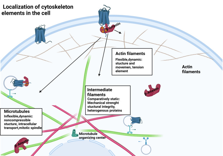
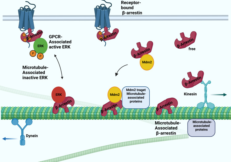
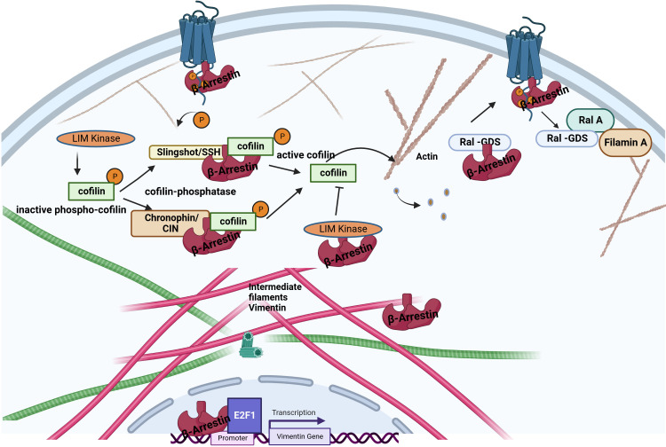
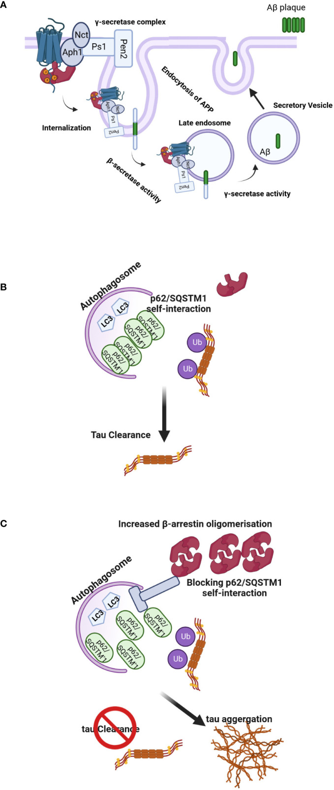
β-arrestins, which have multiple cellular functions, were initially described as proteins that desensitize rhodopsin and other G protein-coupled receptors. The cytoskeletal system plays a role in various cellular processes, including intracellular transport, cell division, organization of organelles, and cell cycle. The interactome of β-arrestins includes the major proteins of the three main cytoskeletal systems: tubulins for microtubules, actins for the actin filaments, and vimentin for intermediate filaments. β-arrestins bind to microtubules and regulate their activity by recruiting signaling proteins and interacting with assembly proteins that regulate the actin cytoskeleton and the intermediate filaments. Altered regulation of the cytoskeletal system plays an essential role in the development of Alzheimer's, Parkinson's and other neurodegenerative diseases. Thus, β-arrestins, which interact with the cytoskeleton, were implicated in the pathogenesis progression of these diseases and are potential targets for the treatment of neurodegenerative disorders in the future.

Keywords: Alzheimer’s disease; actin; arrestin; cytoskeleton; microtubule; neurodegeneration.


Conflict of interest statement

The authors declare that the research was conducted in the absence of any commercial or financial relationships that could be construed as a potential conflict of interest.

Figures

Figure 1
Figure 1
β-arrestin protein location within the cell. β-arrestins exist in distinct conformational states: free, receptor-bound, and microtubule-associated.
Figure 2
Figure 2
Possible interactions between arrestins and microtubular system. The β-arrestin proteins binding to microtubules involve the conformation change. They bind to the microtubule system and regulate their activity by recruiting ERK and Mdm2. The β-arrestin2 interacts with the kinesin-like protein KIF3A motor protein.
Figure 3
Figure 3
Interaction between β-arrestin proteins and other cytoskeletal elements. β-arrestins directly bind actin filaments and vimentin. Increased cofilin activity results from the scaffolding of cofilin by the upstream cofilin phosphatases chronophin and slingshot. The increased cofilin activity is also related to LIMK inhibition by arrestins. Older filaments are cut by cofilin, producing new filament seeds. β-arrestin2 binds to the guanine exchange factor for GTPase, Ral1(Ral-GDS). After receptor activation, RAL-GDS is released, which activates Ras-related protein Ral-A (RalA). RalA interacts with Filamin-A to promote actin rearrangement. β-arrestin1 can enhance vimentin gene expression in association with the transcription factor E2F1.
Figure 4
Figure 4
(A) Regulation of Aβ-related pathology by β-arrestin2. Activation of GPCRs by their ligands increases the affinity for β-arrestin2. Recruitment of β-arrestin2 to GPCRs ends up interacting with the γ-secretase complex via the Aph1 subunit. β-arrestin2 mediates the internalization of GPCRs and localizes γ-secretase to late endosomes, where the acidic environment often increases its activation. APP is cleaved by the β-secretase, and C-terminal APP fragments are produced, the direct substrates of the γ-secretase. These fragments are often subsequently cleaved by the γ-secretase to provide Aβ and APP intracellular domains. The produced Aβ is often discharged into intercellular space via secretory vesicles, resulting in extracellular amyloid plaque formation. (B, C) Model of β-arrestins-promoted tauopathy in AD and FTD. In healthy brains, monomeric β-arrestins regulate GPCR trafficking, and there is no excess of oligomeric β-arrestins, and thus misfolded tau is efficiently ubiquitylated and targeted for autophagy clearance. However, in FTD form-tau brains, β-arrestins oligomers are increased, inhibiting p62/SQSTM1-mediated autophagy, leading to failure of misfolded/aggregated tau to be efficiently cleared.

Similar articles

Cited by

References

    1. Cairns NJ, Lee VMY, Trojanowski JQ. The cytoskeleton in neurodegenerative diseases. J Pathol (2004) 204(4):438–49. doi: 10.1002/path.1650 - DOI - PMC - PubMed
    1. Pfister C, Chabre M, Plouet J, Tuyen VV, De Kozak Y, Faure JP, et al. . Retinal s antigen identified as the 48K protein regulating light-dependent phosphodiesterase in rods. Science (1985) 228:891–3. doi: 10.1126/science.2988124 - DOI - PubMed
    1. Caron MG, Barak LS. A brief history of the β-arrestins. Methods Mol Biol (2019) 1957:3–8. doi: 10.1007/978-1-4939-9158-7_1 - DOI - PMC - PubMed
    1. Jiang H, Galtes D, Wang J, Rockman HA. G Protein-coupled receptor signaling: transducers and effectors. Am J Physiol Cell Physiol (2022) 323(3):C731–48. doi: 10.1152/ajpcell.00210.2022 - DOI - PMC - PubMed
    1. Shukla AK, Westfield GH, Xiao K, Reis RI, Huang LY, Tripathi-Shukla P, et al. . Visualization of arrestin recruitment by a G-protein-coupled receptor. Nature (2014) 512(7513):218–22. doi: 10.1038/nature13430 - DOI - PMC - PubMed

Publication types