Review
. 2023 Jan 17;9(1):dvad002.
doi: 10.1093/eep/dvad002. eCollection 2023.

Transgenerational epigenetic inheritance of axonal regeneration after spinal cord injury

Affiliations
Review

Transgenerational epigenetic inheritance of axonal regeneration after spinal cord injury

Andy Madrid et al. Environ Epigenet. .

Abstract

Human epidemiological studies reveal that dietary and environmental alterations influence the health of the offspring and that the effect is not limited to the F1 or F2 generations. Non-Mendelian transgenerational inheritance of traits in response to environmental stimuli has been confirmed in non-mammalian organisms including plants and worms and are shown to be epigenetically mediated. However, transgenerational inheritance beyond the F2 generation remains controversial in mammals. Our lab previously discovered that the treatment of rodents (rats and mice) with folic acid significantly enhances the regeneration of injured axons following spinal cord injury in vivo and in vitro, and the effect is mediated by DNA methylation. The potential heritability of DNA methylation prompted us to investigate the following question: Is the enhanced axonal regeneration phenotype inherited transgenerationally without exposure to folic acid supplementation in the intervening generations? In the present review, we condense our findings showing that a beneficial trait (i.e., enhanced axonal regeneration after spinal cord injury) and accompanying molecular alterations (i.e., DNA methylation), triggered by an environmental exposure (i.e., folic acid supplementation) to F0 animals only, are inherited transgenerationally and beyond the F3 generation.

Keywords: CNS regeneration; DNA methylation; epigenetics; folate; folic acid; one carbon metabolism.


Conflict of interest statement

None declared.

Figures

Figure 1:
Figure 1:
Biphasic responses following increasing folic acid supplementation. (a) A spinal cord injury paradigm used to study the effects of folic acid supplementation on spinal axon regeneration in vivo. Animals were treated with intraperitoneal (i.p.) doses of folic acid, starting 3 days prior to the injury and given daily for 2 weeks. A sciatic nerve graft is implanted at the site of bilateral C3 dorsal column transection (arrow) to provide a permissive environment for the transected spinal axons to grow. At 2 weeks, a fluorescent tracer is placed at the free end of the graft (arrow). (b) At 48 hours, the lumbar DRG, in which the cell bodies of the transected axons reside, are harvested, sectioned, and inspected with fluorescence microscopy. The percent of fluorescent neurons is calculated. In this model, spinal axons on the side of the sciatic nerve harvest (conditioned side) are more likely to grow axons into the graft. (c) Percent axon regeneration follows a biphasic curve with increasing doses of folic acid, peaking at 80 µg/kg and returning to baseline levels by 800 µg/kg. (d, e) Global levels of DNA methylation measured in spinal cord tissue by a radioactive assay (note: the lower radioactive counts correspond to higher methylation levels) (d), and the expression of the de novo DNMTs DNMT3a and 3b, but not the maintenance DNMT1, measured in the same tissue by Western analysis (e), follows identical biphasic curves in response to folic acid supplementation (CPM: counts per minute; N: no spinal cord injury control; reproduced from previous publications [1, 3])
Figure 2:
Figure 2:
The folate and methylation pathway. Folic acid enters the cell through the FOLR1, which is upregulated with injury. The folic acid molecule is then converted into the active tetrahydrofolate (THF) form by DHFR. This allows the eventual production of nucleotides and certain amino acids, as well as the transfer of the methyl group into the methionine-methylation cycle. The latter occurs through the B12-dependent MS step. Subsequently, S-adenosylmethionine (SAM) is the substrate used by the methyltransferase enzymes for the methylation reactions. The inhibition of FOLR1, DHFR, MS, and DNMT suppresses CNS regeneration. In turn, the activation of DNMT enhances CNS regeneration (MTX: methotrexate; 3-ABA: 3-aminobenzamide; SAH: S-adenosylhomocysteine) (reproduced from previous publication) [3]
Figure 3:
Figure 3:
Ancestral folic acid enhances the regeneration of injured CNS axons in multiple generations of untreated progeny. (a) A mating pair of animals (F0) was treated with intraperitoneal folic acid (80 µg/kg) or vehicle distilled de-ionized water (DDI) starting 2 weeks before breeding and continuing in females until weaning and in males until pregnancy was assured. Four generations of progeny were bred without treatment. (b) Box plots show that the folic acid supplementation of progenitors enhances percent spinal axon regeneration in untreated F1–F4 male progeny (left boxes) compared to controls (right boxes). Single-generation control animals (S0) with direct exposure to folate supplementation exhibit ∼12% regeneration. Identical results were obtained using various routes of administration, breeding schemes, and two distinct genera (not shown) (reproduced from previous publication) [17]
Figure 4:
Figure 4:
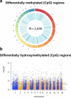
Ancestral folic acid results in transgenerational perturbations in DNA methylation and hydroxymethylation. (a) A circus plot depicts the representative locations of genome-wide DMRs identified when comparing spinal cord tissue derived from folic acid lineage F3 male offspring to F3 male vehicle control offspring. The outer circle represents the various chromosomes in the rat genome. The inner circle represents the relative location of the DMRs. (b) A Manhattan plot depicts the relative location of genome-wide regions tested for 5hmC abundance for the various chromosomes (x-axis) in the rat genome. The y-axis depicts the -log10 value of the P-value when comparing spinal cord tissue derived from folic acid lineage F3 male offspring to F3 male vehicle control offspring. Dots above the dashed line represent differentially hydroxymethylated regions (reproduced from previous publication) [17]

References

    1. Iskandar BJ, Nelson A, Resnick D et al. Folic acid supplementation enhances repair of the adult central nervous system. Ann Neurol 2004;56:221–7. - PubMed
    1. Selzer RR, Rosenblatt DS, Laxova R et al. Adverse effect of nitrous oxide in a child with 5,10-methylenetetrahydrofolate reductase deficiency. N Engl J Med 2003;349:45–50. - PubMed
    1. Iskandar BJ, Rizk E, Meier B et al. Folate regulation of axonal regeneration in the rodent central nervous system through DNA methylation. J Clin Invest 2010;120:1603–16. - PMC - PubMed
    1. Waterland RA, Jirtle RL. Transposable elements: targets for early nutritional effects on epigenetic gene regulation. Mol Cell Biol 2003;23:5293–300. - PMC - PubMed
    1. Anway MD, Memon MA, Uzumcu M et al. Transgenerational effect of the endocrine disruptor vinclozolin on male spermatogenesis. J Androl 2006;27:868–79. - PMC - PubMed